Thank you For watching.
Here are related videos and articles
Thank you For watching.
Leishmaniasis is a spectrum of protozoal diseases caused by Leishmania species, transmitted by the bite of infected female phlebotomine sandflies.
 |
| cutaneous leishmaniasis |
Disease manifestations depend on the species involved and the host immune response.
Major clinical forms:
Visceral leishmaniasis (VL / kala-azar)
Cutaneous leishmaniasis (CL)
Mucocutaneous leishmaniasis (MCL)
| Form | Causative Species | Geographic Distribution |
|---|---|---|
| Visceral | L. donovani, L. infantum (chagasi) | South Asia, East Africa, Latin America |
| Cutaneous | L. tropica, L. major, L. mexicana, L. braziliensis | Middle East, Africa, Americas |
| Mucocutaneous | L. braziliensis complex | Central & South America |
Endemic in >80 countries; affects poor, rural populations.
Vectors: Phlebotomus (Old World), Lutzomyia (New World).
Reservoirs: Humans (L. donovani), dogs, rodents.
Transmission: Sandfly bite, rarely congenital or via transfusion.
 |
| Phlebotomus |
Inoculation of promastigotes → engulfed by macrophages → transform into amastigotes → intracellular multiplication → spread to RES (liver, spleen, bone marrow).
Disease severity depends on cell-mediated immunity (CMI).
Strong CMI → localized CL.
Poor CMI → disseminated VL.
 |
| lifecycle of L donovani |
Incubation: 2 weeks–18 months.
") |
| Visceral Leishmaniasis (Kala-azar) |
Onset: Insidious.
Major triad:
Fever: Remittent, double-quotidian, or irregular.
Hepatosplenomegaly: Marked splenomegaly, moderate hepatomegaly.
Pancytopenia: due to hypersplenism and marrow infiltration.
Other features:
Weight loss, wasting, darkening of skin (“kala-azar” = black fever)
Lymphadenopathy (esp. African form)
Anemia, bleeding, infections (esp. bacterial superinfection)
Growth retardation and cachexia in chronic disease.
Occurs months–years after VL cure.
Hypopigmented macules, papules, nodules (face, arms, trunk).
Serves as a reservoir in endemic areas (notably India).
Lesion: Painless papule → ulcer with indurated margin (“oriental sore”).
Usually heals spontaneously in 3–6 months but leaves scar.
Chronic forms may resemble lupus vulgaris.
Extension from cutaneous lesion (nasal/oral mucosa).
Causes destructive ulcerations → severe disfigurement.
 |
| mucocutaneous leishmaniasis |
Amastigotes (Leishman–Donovan bodies) in:
Splenic aspirate (most sensitive, but risky)
Bone marrow aspirate (safe, moderately sensitive)
Lymph node or buffy coat smear
Giemsa-stained smears show:
Oval amastigotes (2–5 μm) with nucleus and kinetoplast inside macrophages.
Novy–MacNeal–Nicolle (NNN) medium → promastigote growth.
rK39 dipstick test: Highly sensitive & specific for VL (field use).
Direct agglutination test (DAT), IFA, ELISA also available.
PCR: Highly sensitive, species-specific; used in reference labs.
Pancytopenia, hypergammaglobulinemia, elevated ESR.
Liposomal Amphotericin B (preferred):
Total dose 10–21 mg/kg (varies by region/protocol)
Short-course regimens effective.
Alternative:
Amphotericin B deoxycholate: 1 mg/kg/day × 15–20 doses (toxic, nephrotoxic)
Miltefosine: 2.5 mg/kg/day (max 150 mg/day) × 28 days (oral)
Paromomycin (IM): 11 mg/kg/day × 21 days
Combination regimens (to prevent resistance):
Single-dose liposomal amphotericin B + short-course miltefosine or paromomycin.
Local therapy (cryotherapy, intralesional antimony) for small lesions.
Systemic therapy for multiple, mucosal, or immunocompromised cases:
Miltefosine, liposomal amphotericin B, or pentavalent antimonials.
Liposomal amphotericin B or pentavalent antimonials for ≥28 days.
Vector control: Insecticide spraying, bed nets.
Reservoir control: Treat dogs, cull infected reservoirs.
Personal protection: Repellents, protective clothing.
Early diagnosis and treatment reduce transmission.
Vaccine: None yet in routine use; trials ongoing.
Secondary bacterial infections
Severe anemia, hemorrhage
Disseminated infection in HIV patients
PKDL (in endemic regions like India/Nepal)
Excellent with prompt diagnosis and treatment.
Mortality >90% if untreated (mainly from secondary infections, cachexia).
Visceral leishmaniasis should be suspected in any febrile child with splenomegaly and pancytopenia in an endemic area.
rK39 test is the diagnostic test of choice in field settings.
Liposomal amphotericin B is the preferred therapy in both immunocompetent and immunocompromised children.
PKDL represents an important reservoir for ongoing transmission.
HIV co-infection complicates disease course and increases relapse risk.
Category: Inherited bone marrow failure syndrome (IBMFS)
Inheritance: Autosomal recessive (rarely X-linked)
Gene defects: >22 genes identified (FANCA, FANCC, FANCG most common) → defective DNA interstrand crosslink repair.
 |
| fanconi anemia notes |
Defect in DNA repair (Fanconi/BRCA pathway) → chromosomal breakage and hypersensitivity to DNA cross-linking agents (e.g., mitomycin C, diepoxybutane).
Progressive bone marrow failure (due to stem cell depletion) and genomic instability → predisposition to malignancies.
Multisystem developmental abnormalities due to impaired cell proliferation during embryogenesis.
Incidence: ~1 in 100,000–250,000 live births.
Carrier frequency: ~1 in 200.
Median age of diagnosis: 7–9 years.
~90% develop marrow failure by age 40.
Pancytopenia (usually first manifests with thrombocytopenia or macrocytic anemia).
Progressive bone marrow hypoplasia.
Increased fetal hemoglobin (HbF).
Myelodysplastic syndrome (MDS) or acute myeloid leukemia (AML) risk ↑ markedly.
Growth: Short stature, low birth weight.
Skeletal: Radial ray defects—absent/hypoplastic thumb, radius anomalies.
Skin: Café-au-lait spots, hypopigmentation, hyperpigmentation.
Head/Face: Microcephaly, triangular face, microphthalmia.
Genitourinary: Renal agenesis, horseshoe kidney, hypoplastic gonads, undescended testes, infertility.
Cardiac: Structural heart defects.
ENT: Hearing loss.
GI: Duodenal atresia, anal anomalies (occasionally).
Hypothyroidism, glucose intolerance, gonadal failure, low IGF-1.
AML, MDS, and solid tumors (esp. head & neck SCC, gynecologic SCC, liver tumors) due to chromosomal instability.
| Test | Finding/Use |
|---|---|
| CBC | Pancytopenia, macrocytosis, increased HbF |
| Bone marrow biopsy | Hypocellular marrow with fatty replacement |
| Chromosomal breakage test | Diagnostic — increased breaks after exposure to diepoxybutane (DEB) or mitomycin C |
| Molecular genetic testing | Confirms FANCA–FANC gene mutations |
| Flow cytometry for CD34 | Decreased hematopoietic stem cells |
| Ultrasound abdomen | Renal anomalies |
| Endocrine profile | Hypothyroidism, gonadal failure screening |
Acquired aplastic anemia
Dyskeratosis congenita
Shwachman-Diamond syndrome
Diamond–Blackfan anemia
Regular CBC monitoring.
Transfusion support (RBCs, platelets) — minimize iron overload.
Iron chelation therapy if ferritin ↑.
Androgens (e.g., oxymetholone, danazol) → stimulate erythropoiesis (transient benefit).
G-CSF for neutropenia (short-term).
Avoid DNA-damaging agents (chemotherapy, radiation).
Allogeneic hematopoietic stem cell transplantation (HSCT) — only curative therapy for marrow failure.
Ideal: HLA-matched sibling donor.
Conditioning regimens: low-intensity to minimize toxicity (avoid alkylators, irradiation).
Annual oral, gynecologic, and dermatologic exams.
CBC every 3–6 months.
Avoid smoking, alcohol, and UV exposure.
Hormonal replacement as indicated (thyroid, sex steroids, GH).
Orthopedic/surgical correction for congenital anomalies.
Median survival (without HSCT): ~20–30 years.
With HSCT: markedly improved, though risk of secondary malignancy persists.
Lifelong surveillance for cancer and organ dysfunction required.
Classic triad: Bone marrow failure + congenital anomalies + cancer predisposition.
Diagnostic hallmark: Chromosomal breakage test positive with DEB/Mitomycin C.
Curative therapy: HSCT.
Common mutation: FANCA.
AML/MDS risk: markedly increased.
Androgens improve counts transiently but cause virilization/hepatotoxicity.
हामी प्रायः रगत परीक्षणका लागि हातको नसाबाट (vein) रगत निकालिन्छ भन्ने कुरा जान्दछौं। तर कहिलेकाहीँ स्वास्थ्यकर्मीले नाडीबाट (artery) पनि रगत निकाल्छन्। यो सामान्य रगत परीक्षणभन्दा फरक र विशिष्ट उद्देश्यका लागि गरिन्छ।
 |
| ABG sampling technique why and when |
नाडीबाट रगत निकाल्ने मुख्य उद्देश्य Arterial Blood Gas (ABG) test हो।
यो परीक्षणले शरीरमा रहेका अक्सिजन (O₂), कार्बन डाइअक्साइड (CO₂) र रगतको अम्ल–क्षार (pH) सन्तुलन कस्तो छ भन्ने देखाउँछ।
यो जानकारी फोक्सो र मुटुको कार्य कस्तो छ भन्ने बुझ्न अत्यन्त जरुरी हुन्छ।
जब बिरामीलाई अक्सिजन कमी (hypoxia) को शंका हुन्छ।
सास फेर्न गाह्रो भएको अवस्थामा (जस्तै– दमा, COPD, pneumonia, ARDS)।
भेन्टिलेटरमा राखिएका बिरामीहरूमा, अक्सिजनको मात्रा ठिक छ कि छैन भनेर हेर्न।
गम्भीर रोगीहरूमा, अम्ल–क्षार सन्तुलन (acid–base balance) पत्ता लगाउन।
सर्जरीपछि वा गम्भीर संक्रमण (sepsis) भएका बिरामीहरूमा।
सबैभन्दा धेरै प्रयोग हुने नाडीहरू:
Radial artery (कलाईको नाडी) – सबैभन्दा सामान्य र सुरक्षित।
Femoral artery (जाँघको नाडी) – आपतकालमा प्रयोग।
Brachial artery (काँधतर्फको नाडी) – कहिलेकाहीँ प्रयोग।
ABG गर्नुअघि प्रायः Allen’s test गरिन्छ, जसले हातको रक्तप्रवाह सुरक्षित छ कि छैन भन्ने पक्का गर्छ।
बिरामीलाई आराम दिन्छ।
छालालाई सफा गरिन्छ (antiseptic)।
नाडीको धड्कन भेटाएर सुई प्रयोग गरी सिधै नाडीभित्र सुई प्रवेश गरिन्छ।
रगत सिधै syringe मा स्वचालित रूपमा भरिन्छ, किनकि नाडीको दबाब (pressure) बढी हुन्छ।
त्यसपछि तुरुन्तै syringe लाई बर्फमा राखी ल्याबमा पठाइन्छ ताकि ग्यासहरू नबदलिऊन्।
नसाको रगतले शरीरको अक्सिजन र कार्बन डाइअक्साइडको सन्तुलन सही रूपमा देखाउँदैन, किनभने त्यो पहिले नै ऊतकहरूबाट फर्किएको हुन्छ।
तर नाडीको रगत भने फोक्सोबाट निस्किएको ताजा अक्सिजनयुक्त रगत हो, जसले शरीरको साँच्चिकै ग्यास स्थिति जनाउँछ।
त्यसैले फोक्सो, सासफेर्ने प्रणाली वा अक्सिजन थेरापी मूल्याङ्कन गर्न नाडीबाट रगत आवश्यक पर्छ।
सामान्यतया सुरक्षित भए पनि केही साइड इफेक्ट हुन सक्छन् —
नाडीमा दबाबको कारण दुखाइ वा निलो दाग (bruise)
कहिलेकाहीँ रगत बग्ने वा clot बन्ने समस्या
धेरै पटक सुई लगाउँदा नाडीको क्षति वा हात सुन्निनु
त्यसैले यो परीक्षण प्रशिक्षित स्वास्थ्यकर्मी (जस्तै चिकित्सक वा नर्स) ले मात्र गर्नुपर्छ।
नाडीबाट रगत निकाल्नु साधारण परीक्षण होइन, तर अत्यन्त महत्त्वपूर्ण चिकित्सकीय प्रक्रिया हो जसले शरीरको अक्सिजन, कार्बन डाइअक्साइड र अम्ल–क्षार सन्तुलनबारे सटीक जानकारी दिन्छ।
यसले चिकित्सकलाई बिरामीको सासफेर्ने स्थिति बुझ्न, भेन्टिलेटर मिलाउन, र उपचारको प्रभाव मूल्याङ्कन गर्न मद्दत गर्छ।
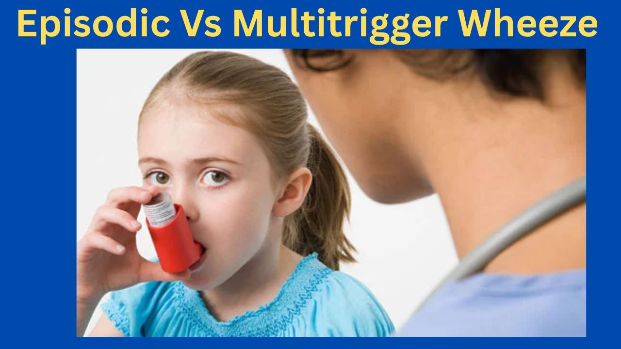
Episodic (Viral) Wheeze vs. Multiple Trigger Wheeze
A Clinically Oriented Review for the Practicing Pediatrician
Based on Nelson Textbook of Pediatrics (21st ed.) | Kendig’s Disorders of the Respiratory Tract in Children (9th ed.) | AAP & IAP-NAPCON Official Resources
Wheezing in preschool children (0–5 years) is one of the most common reasons for pediatric consultation and hospital admission worldwide. It is now well established that ‘preschool wheeze’ is not a single disease but a heterogeneous group of phenotypes with distinct pathophysiology, natural history, and responses to therapy. The two most clinically useful and validated phenotypes—recognized in both the Nelson Textbook of Pediatrics and major international guidelines—are:
This classification, initially proposed by Brand et al. and incorporated into the PRACTALL Consensus Report (2008) of the European Academy of Allergy and Clinical Immunology (EAACI) and the American Academy of Allergy, Asthma and Immunology (AAAAI), is now endorsed by the American Academy of Pediatrics (AAP) and the Indian Academy of Pediatrics (IAP) / National Asthma Consensus Group (NACG).
According to Nelson Textbook of Pediatrics (21st edition, Chapter 169: Wheezing in Infants and Children), approximately 30–40% of all children will experience at least one wheezing episode in the first three years of life, yet fewer than one-third of these will develop persistent asthma. Data from the Tucson Children’s Respiratory Study (TCRS), cited prominently in Nelson, delineates three early wheezing trajectories:
The IAP NAPCON 2019 Consensus Statement on Childhood Asthma notes that in South Asian children, including India and Nepal, the prevalence of preschool wheeze is significant, often complicated by high pollution exposure and early sensitization to house dust mite and cockroach allergens, features that shift the phenotype toward MTW.
As described in Nelson (Chapter 169) and Kendig’s Disorders of the Respiratory Tract in Children (9th edition, Chapter 38), EVW is predominantly mediated by:
MTW pathophysiology, as detailed in both Nelson and Kendig’s, resembles that of classic atopic asthma:
Nelson (21st ed., Chapter 169) and AAP Clinical Practice Guidelines for Asthma (2020 Update) recommend a detailed history focusing on:
The modified API (mAPI), described in Nelson and endorsed by the AAP, is a validated tool to identify preschool wheezers likely to develop persistent asthma (MTW phenotype). A positive mAPI in a child with ≥3 wheezing episodes in the past year has a positive predictive value of ~80% for asthma at school age.
Major criteria: (1) Parental asthma; (2) Physician-diagnosed atopic dermatitis; (3) Aeroallergen sensitization.
Minor criteria: (1) Food allergen sensitization; (2) ≥4% peripheral eosinophilia; (3) Wheezing apart from colds.
A positive API (1 major OR 2 minor) in a high-frequency wheezer predicts MTW/asthma phenotype and guides more aggressive preventive therapy.
Physical findings are largely similar during acute episodes in both phenotypes. However, clinicians should look for:
Kendig’s (9th ed., Chapter 38) and AAP Guidelines recommend the following investigations based on clinical context:
Table 1 summarizes the key distinguishing features of the two preschool wheeze phenotypes.
Table 1. Episodic Viral Wheeze vs. Multiple Trigger Wheeze — Comparative Features
| Feature | Episodic Viral Wheeze (EVW) | Multiple Trigger Wheeze (MTW) |
| Trigger pattern | Only viral URTIs; symptom-free between episodes | Viral + aeroallergens, exercise, cold air, smoke; persistent/interval symptoms |
| Typical age | Predominantly <3 years (preschool) | Any preschool age; more likely to persist into school age |
| Atopic features | Usually absent; non-atopic phenotype | Often present: eczema, allergic rhinitis, sensitization |
| Family history | Less prominent | Positive asthma/atopy family history common |
| Lung function (interval) | Normal between episodes | May show airflow limitation between episodes |
| Airway inflammation | Predominantly neutrophilic; transient | Eosinophilic; chronic even between episodes |
| Response to ICS | Limited/inconsistent benefit in trials | Better response; ICS often indicated |
| LABA benefit | Not established | May be considered as add-on (age-appropriate) |
| Montelukast | Modest benefit in some studies (episodic use) | Regular use may help; part of step-up therapy |
| Prognosis | Many remit by school age | Higher risk of persisting asthma |
Source: Nelson Textbook of Pediatrics 21e (Chapter 169); Kendig’s 9e (Chapter 38); Brand et al., PRACTALL Consensus Report 2008; AAP; IAP-NAPCON 2019.
Both Nelson and Kendig’s emphasize that preschool wheeze is not always asthma or EVW/MTW. The following should be actively excluded:
Per AAP Clinical Practice Guidelines (2020) and Nelson (Chapter 169), acute management is phenotype-independent and follows standard bronchodilator therapy:
This is where the phenotype distinction critically guides management:
Per Nelson, Kendig’s, and AAP Guidelines:
Per Nelson, Kendig’s, AAP (2020), and IAP-NAPCON (2019):
Table 2. Stepwise Treatment Approach for EVW and MTW
| Step | EVW Management | MTW Management |
| Acute | SABA (salbutamol) via spacer/nebulizer; oral prednisolone for moderate-severe | SABA; oral/systemic corticosteroids; consider early ICS step-up |
| Preventer | Not routinely indicated; trial ICS only if frequent/severe episodes (≥3/year) | Low-dose ICS (e.g., budesonide 100–200 mcg/day) as first-line preventer |
| Step-up | Episodic ICS at onset of URTI (intermittent therapy); montelukast episodic use | Increase ICS dose; add montelukast or LABA (≥5 yr); consider specialist referral |
| Monitoring | Symptom diary; reassess trigger pattern at each visit | Spirometry (if age-appropriate); allergy testing; adherence review |
Adapted from: Nelson Textbook of Pediatrics 21e; AAP Clinical Practice Guidelines (2020); IAP-NAPCON Consensus Statement 2019.
Per AAP and IAP-NAPCON recommendations:
Nebulizers are not superior to pMDI+spacer for acute bronchodilation and carry infection transmission risk in healthcare settings. Both AAP and IAP recommend prioritizing spacer-based delivery.
Nelson, AAP (2020 Expert Panel Report 3 Update), and IAP-NAPCON recommend the following monitoring framework:
The TCRS and birth cohort studies cited in Nelson provide the most robust data on prognosis:
Many children present with features of both EVW and MTW, especially between ages 2–4 years. Nelson recommends using the mAPI as a practical decision aid in such cases. If the mAPI is positive, treat as MTW (initiate regular ICS); if negative, manage as EVW (episodic/as-needed therapy).
Wheezing in infants under 12 months is most commonly due to bronchiolitis (RSV) and should not be classified as EVW or MTW. Per AAP Clinical Practice Guideline for the Diagnosis, Management, and Prevention of Bronchiolitis (2014, reaffirmed 2020), bronchodilators are not recommended for infants with bronchiolitis. ICS and systemic steroids are similarly not recommended in this age group for acute bronchiolitis.
The AAP has issued guidance noting that SARS-CoV-2 infection in young children may trigger wheezing episodes similar to other viral URTI triggers in EVW. Standard asthma action plans should include COVID-19 as a potential EVW trigger; ICS should not be stopped during COVID-19 illness in MTW/asthma patients.
Both AAP and IAP recommend annual influenza vaccination for all children with recurrent wheezing (EVW or MTW), as influenza is a significant trigger for severe exacerbations. Pneumococcal vaccination per national immunization schedules is also recommended.
AAP and IAP emphasize that education is a cornerstone of management:
Primary Textbook References:
AAP Official Resources:
IAP Official Resources:
Landmark Studies and Consensus Documents (cited in Nelson/Kendig’s):
 |
| splenomeglay illustration |
Splenomegaly is enlargement of the spleen beyond its normal size (normally not palpable below the left costal margin).
Normal weight: ~150–200 g
Normal length: ~11 cm
Massive splenomegaly: Spleen palpable below the umbilicus or crossing the midline.
Functions: Filtration of old RBCs, immune surveillance, hematopoiesis (fetal), platelet and RBC reservoir.
Normal spleen not palpable; becomes palpable when enlarged ≥2–3×.
| Type | Spleen size | Examples |
|---|---|---|
| Mild (2–3 cm) | Slight enlargement | Viral infections, hemolysis |
| Moderate (3–8 cm) | Reaches midway to umbilicus | Malaria, portal hypertension |
| Massive (>8 cm / crosses midline) | Large spleen | CML, myelofibrosis, Kala-azar |
Increased workload (reticuloendothelial hyperplasia)
→ Infections, hemolysis
Congestive (venous pooling)
→ Portal hypertension, splenic vein thrombosis
Infiltrative / Neoplastic
→ Leukemia, lymphoma, storage diseases
Immune / Inflammatory
→ SLE, rheumatoid arthritis (Felty’s syndrome)
Extramedullary hematopoiesis
→ Myelofibrosis, severe thalassemia
Acute infections:
Infective mononucleosis (EBV)
Viral hepatitis
Typhoid fever
Infective endocarditis
Sepsis (esp. in children)
Chronic infections:
Malaria
Kala-azar (Visceral leishmaniasis)
Tuberculosis
Schistosomiasis
Brucellosis
Hemolytic anemias
Thalassemia major/intermedia
Hereditary spherocytosis
Sickle cell disease (early phase)
Autoimmune hemolytic anemia
Leukemias & Lymphomas
Chronic myeloid leukemia (CML) → massive splenomegaly
Chronic lymphocytic leukemia (CLL)
Hairy cell leukemia
Hodgkin / Non-Hodgkin lymphoma
Myeloproliferative / Myelofibrotic disorders
Portal hypertension (cirrhosis, extrahepatic portal vein obstruction)
Splenic vein thrombosis
Right heart failure, constrictive pericarditis
Gaucher’s disease
Niemann–Pick disease
Amyloidosis
Sarcoidosis
Systemic lupus erythematosus (SLE)
Rheumatoid arthritis (Felty’s syndrome)
Autoimmune hepatitis
Cysts, abscess, hydatid disease
Primary splenic tumor (hemangioma, angiosarcoma)
Secondary metastasis (rare)
C – Chronic myeloid leukemia
H – Hairy cell leukemia
A – Agnogenic myeloid metaplasia (myelofibrosis)
M – Malaria (chronic)
P – Portal hypertension / Kala-azar
S – Storage diseases (Gaucher, Niemann-Pick)
 |
| massive splenomegaly in CT scan |
Fullness or dragging sensation in LUQ
Early satiety
Pain due to infarction or capsule stretch
Hypersplenism → Anemia, leukopenia, thrombocytopenia
Palpable firm or hard spleen below costal margin

CBC & Peripheral smear: cytopenias, abnormal cells
LFT, RFT
Viral markers (EBV, hepatitis, HIV)
Bone marrow examination
Ultrasound / CT abdomen: spleen size, portal system, lymphadenopathy
Serology: malaria, kala-azar (rk39), brucella
Liver biopsy / portal venography if portal cause suspected
Hypersplenism → cytopenias
Splenic rupture (trauma or spontaneously in infections)
Splenic infarction
Portal hypertension
Treat underlying cause (infection, hematologic disorder, etc.)
Avoid trauma / contact sports
Splenectomy – indicated in:
Hypersplenism with cytopenias unresponsive to therapy
Hereditary spherocytosis
Immune thrombocytopenic purpura (refractory)
Splenic abscess, cyst, rupture
Vaccinations before splenectomy: Pneumococcal, Hib, Meningococcal
Always examine in right lateral position
Start palpation from right iliac fossa towards LUQ
Note size, consistency, tenderness, notching, relation to costal margin
| Mechanism | Common Causes |
|---|---|
| Infective | Malaria, Kala-azar, EBV |
| Hemolytic | Thalassemia, HS, AIHA |
| Neoplastic | CML, Lymphoma |
| Congestive | Cirrhosis, Portal HTN |
| Storage | Gaucher, Niemann-Pick |
| Autoimmune | SLE, Felty’s |
| Miscellaneous | Cyst, Abscess |
 |
| checking oedema in nephrotic syndrome |
Nelson Textbook of Pediatrics (22nd ed., Ch. 494, p. 2570)
Prednisolone 2 mg/kg/day (maximum 60 mg/day) for 6 weeks, followed by
1.5 mg/kg on alternate days (maximum 40 mg) for next 6 weeks.
Rationale:
Earlier protocols (e.g., 4+4 week or 8-week total) are less effective.
Prolonged 12-week regimen (6+6) gives fewer relapses.
A 20 kg child presents with first episode NS.
Daily dose: 2 mg/kg = 40 mg daily × 6 weeks.
Then alternate-day: 1.5 mg/kg = 30 mg on alternate days × 6 weeks.
Total course: 12 weeks.
Avoid: tapering below alternate day dose before completion of 12 weeks — increases relapse.
<2 relapses in 6 months or <3 in 1 year.
Dose:
Prednisolone 2 mg/kg/day until remission (urine protein nil/trace × 3 days),
then 1.5 mg/kg on alternate days for 4 weeks, then stop.
Example:
Child relapses after 5 months remission → give daily 2 mg/kg till protein nil ×3 days → shift to 1.5 mg/kg AD ×4 weeks → stop.
≥2 relapses in 6 months or ≥4 in 12 months.
Dose:
Same as above for each relapse, but consider tapering or steroid-sparing agent.
Maintenance (if steroid-only used):
Alternate day 0.5–0.7 mg/kg prednisolone for 3–6 months.
Example:
If child relapses every 2 months — after inducing remission, maintain on 0.5 mg/kg AD for 6 months to break cycle.
Relapse during tapering or within 2 weeks of stopping steroids.
Strategy 1: Low-dose alternate-day steroids
Maintain remission with 0.3–0.5 mg/kg AD for 6–12 months.
Strategy 2: Add steroid-sparing agent
Cyclophosphamide, levamisole, MMF, or calcineurin inhibitor depending on toxicity and previous exposure.
Example:
A 7-year-old develops relapse each time dose falls below 0.5 mg/kg AD → maintain at 0.5 mg/kg AD × 6 months; if Cushingoid, add levamisole.
No remission after 6 weeks of daily 2 mg/kg prednisolone.
Confirm compliance, dose accuracy, and rule out secondary NS before labeling SRNS.
Protocol:
Continue same dose for total 6–8 weeks before biopsy and calcineurin inhibitor introduction.
Example:
A 6-year-old on pred 2 mg/kg × 6 weeks still 3+ protein — if compliance ensured, classify as SRNS, proceed to biopsy.
If urine protein reduces but not nil after 6 weeks →
continue full dose 2 mg/kg/day for additional 2 weeks before deciding resistance.
Switch to 2 mg/kg/day until remission × 3 days,
then back to alternate-day baseline dose for 4 weeks.
Do not increase dose; continue same daily dose until infection settles.
After remission, taper normally.
Use IV methylprednisolone (10–15 mg/kg/day × 3 days) if poor oral absorption suspected, then switch to oral 2 mg/kg/day.
Confirm no hypovolemia before diuretics.
Usually genetic; steroid trial limited: 2 mg/kg/day × 6 weeks, but if no response by 4 weeks, stop (to avoid toxicity).
Dosing guided by underlying disease.
Lupus NS: 2 mg/kg/day (max 60 mg) × 4 weeks + taper; or IV methylpred pulses.
Always taper after alternate-day phase, not during daily phase.
After 6+6 weeks:
Reduce to 1 mg/kg AD × 2 weeks
Then 0.5 mg/kg AD × 2 weeks
Then stop.
| Mistake | Consequence |
|---|---|
| Stopping abruptly after remission | Rapid relapse |
| Reducing to daily low-dose steroid | Loss of HPA rhythm |
| Using every 3rd day dosing | Relapse risk ↑ |
| Complication | Prevention |
|---|---|
| Cushingoid features | Prefer alternate-day dosing after remission |
| Growth retardation | AD dosing, Vitamin D & calcium |
| Infections | Live vaccines contraindicated during high-dose |
| Hypertension | Salt restriction, monitor BP weekly |
| Cataract | Yearly ophthalmic review |
| Indication | Next Step |
|---|---|
| ≥2 toxic relapses or dependence | Levamisole 2.5 mg/kg AD |
| SDNS with toxicity | Cyclophosphamide 2 mg/kg/day × 12 weeks |
| FRNS with poor tolerance | MMF 600 mg/m² BD |
| Calcineurin inhibitor use | Tacrolimus 0.05–0.1 mg/kg/day in 2 doses |
| Clinical Scenario | Correct Steroid Plan | Explanation |
|---|---|---|
| Relapse during alternate-day 0.5 mg/kg | Switch to 2 mg/kg/day until remission; resume baseline dose 4 weeks | AD dose insufficient; needs induction again |
| 3rd relapse in 3 months, cushingoid | Induce remission, then add levamisole; maintain on 0.3 mg/kg AD | To reduce toxicity |
| First episode remission after 4 weeks | Continue daily to complete 6 weeks; then AD 6 weeks | Early remission doesn’t mean early taper |
| Proteinuria returns within 7 days of stopping steroids | Steroid-dependent → restart 2 mg/kg/day → maintain 0.5 mg/kg AD × 6 months | Defines dependence |
| SRNS after 8 weeks | Proceed biopsy, add tacrolimus + low-dose pred 0.5 mg/kg AD | Steroid resistance confirmed |
| Child unable to take orally due to vomiting | IV methylpred 10 mg/kg/day × 3 days → switch to oral | Ensures systemic delivery |
| Child develops varicella while on 2 mg/kg/day | Stop steroids temporarily; IV acyclovir; restart after lesion crusting | Prevent fatal dissemination |
Bioavailability: Prednisolone preferred (not deflazacort for initial induction).
Equivalent doses: 5 mg prednisolone = 4 mg methylpred = 0.75 mg dexamethasone.
Morning dosing preferable to preserve circadian rhythm.
Kliegman RM, Nelson Textbook of Pediatrics, 22nd ed., Elsevier, 2023.
Indian Pediatrics Nephrology Group, Consensus Statement on Management of Nephrotic Syndrome, 2021.
IPNA Clinical Practice Recommendations for Idiopathic NS, 2020.
Avner ED et al., Pediatric Nephrology, 8th ed. (RPS, 2022).
Table of Contents(toc)
When a young child comes in with cough, difficulty breathing, and fever, one of the most important — and sometimes confusing — clinical questions is:

Is this bronchiolitis, pneumonia, or something else entirely?
According to Nelson, bronchiolitis is primarily a disease of infants, typically below 2 years of age, with the peak incidence between 2–6 months. It usually appears during the winter and early spring months, corresponding to RSV season.
Pneumonia, on the other hand, can occur in all age groups. Viral pneumonias are more common in infants and preschoolers, while bacterial pneumonias increase with age. There’s no strict seasonal restriction, though viral etiologies may peak in winter.
Bronchiolitis:
Caused most commonly by Respiratory Syncytial Virus (RSV) — responsible for the majority of cases in infants. Other causes include parainfluenza, influenza, human metapneumovirus, and adenovirus.
Pneumonia:
Viral — RSV, influenza, parainfluenza, adenovirus.
Bacterial — Streptococcus pneumoniae, Haemophilus influenzae, Staphylococcus aureus, and Mycoplasma pneumoniae (in older children).
In short, RSV = bronchiolitis, while bacterial pathogens = pneumonia is a good starting point, though overlap exists.
Nelson emphasizes that the site of pathology differentiates the two:
Bronchiolitis: Inflammation and edema of small airways (bronchioles) → obstruction → air trapping, atelectasis, and wheeze.
Pneumonia: Involves alveoli → consolidation, impaired gas exchange, and reduced compliance.
So, in bronchiolitis, the problem is in airflow, whereas in pneumonia, it’s in oxygen exchange.
| Feature | Bronchiolitis | Pneumonia |
|---|---|---|
| Age | <2 years (especially infants) | All ages |
| Onset | Gradual, following coryzal symptoms | Sudden or gradual, depending on cause |
| Fever | Low-grade or absent | Often high (especially bacterial) |
| Cough | Prominent, paroxysmal | Productive or dry |
| Wheeze | Characteristic; diffuse | Usually absent (except in viral) |
| Crepitations | Fine, diffuse, bilateral | Localized (lobar) or diffuse (interstitial) |
| Respiratory rate | Elevated, often >60/min in infants | Elevated; tachypnea proportional to severity |
| Feeding difficulty | Common due to distress | May occur if severe |
| Oxygen saturation | May be low due to air trapping | Often low due to consolidation |
In bronchiolitis, wheezing and hyperinflation dominate; in pneumonia, crackles and focal findings dominate.
Nelson advises that radiologic findings should not be used in isolation to differentiate.
However, classic patterns help:
Bronchiolitis: Hyperinflated lungs, flattened diaphragm, peribronchial thickening, patchy atelectasis — no focal consolidation.
Pneumonia: Lobar or segmental consolidation, air bronchograms, or patchy infiltrates.
Another practical clue from Nelson:
Bronchiolitis: Poor response to antibiotics; supportive care is the mainstay (hydration, oxygen if hypoxemic).
Pneumonia: Marked improvement with appropriate antibiotics if bacterial.
Nelson lists several conditions that mimic bronchiolitis or pneumonia:
Asthma (viral-induced wheeze):
Often recurrent episodes.
Family/personal history of atopy or asthma.
Responds well to bronchodilators, unlike classic bronchiolitis.
Pertussis:
Paroxysmal cough, inspiratory “whoop,” vomiting after coughing.
Minimal wheeze, may have leukocytosis with lymphocytosis.
Foreign Body Aspiration:
Sudden onset, unilateral decreased air entry, localized hyperinflation or collapse.
Congestive Heart Failure:
Tachypnea, hepatomegaly, but no true wheezing unless pulmonary edema present.
Cardiomegaly on chest X-ray.
Aspiration Pneumonitis / GER-related:
History of vomiting, feeding difficulty, neurological disease.
Recurrent or persistent infiltrates in dependent lung areas.
Bronchiolitis:
Supportive: Oxygen, hydration, nasal suctioning.
Avoid routine bronchodilators, steroids, antibiotics.
Hospitalization: If severe distress, apnea, poor feeding, or SpO₂ < 90%.
Pneumonia:
Empiric antibiotics based on age and likely pathogen.
Supportive care: Oxygen, fluids, antipyretics.
“Bronchiolitis should be suspected in infants with their first episode of wheezing following a viral prodrome, whereas pneumonia should be suspected in the presence of fever, focal crackles, and signs of consolidation.”
In practice, overlap exists — especially when viral pneumonia blurs the line — but understanding age, pattern, and auscultatory findings helps steer the diagnosis right.
| Parameter | Bronchiolitis | Pneumonia |
|---|---|---|
| Site | Bronchioles | Alveoli |
| Age | <2 years | All ages |
| Etiology | RSV (most common) | Bacterial or viral |
| Fever | Mild or absent | Usually high |
| Wheeze | Prominent | Usually absent |
| Cough | Paroxysmal | Productive/dry |
| CXR | Hyperinflation | Consolidation |
| Treatment | Supportive | Antibiotics (if bacterial) |
Nelson Textbook of Pediatrics, 21st Edition, Chapters 390 (Bronchiolitis) and 391 (Pneumonia).
Nelson Essentials of Pediatrics, 9th Edition, Section: Respiratory Disorders in Children.
 |
| a child with umbilical vein catheter insitu cc by 4 wikimedia |
Purpose:
For vascular access in neonates (especially preterm or critically ill).
Used for fluid, blood, medication administration, exchange transfusion, and central venous pressure (CVP) monitoring.
Emergency vascular access in neonates
Exchange transfusion
Administration of IV fluids, parenteral nutrition, inotropes, or antibiotics
Blood sampling or transfusion
Monitoring of central venous pressure
Omphalitis or periumbilical infection
Peritonitis
Necrotizing enterocolitis (NEC)
Umbilical or portal vein thrombosis
Imperforate or absent umbilical vein
Umbilical vein: single, large, thin-walled vessel at 12 o’clock position in the umbilical stump.
Leads to left portal vein → ductus venosus → inferior vena cava.
Two smaller umbilical arteries at 4 and 8 o’clock positions.
Sterile gloves, drapes, antiseptic solution
Umbilical catheter (3.5 Fr for <1.5 kg, 5 Fr for >1.5 kg)
Sterile scissors, forceps, and sutures
3-way stopcock and syringes
Normal saline for flush
Adhesive tape and umbilical tie
Sterile dressing
1. Preparation
Maintain aseptic technique.
Place baby under radiant warmer.
Monitor heart rate, SpO₂, and temperature.
Restrain limbs gently.
2. Identify Vessels
Clean umbilical stump with antiseptic.
Trim cord to ~1–2 cm from skin margin.
Identify one large thin-walled umbilical vein (12 o’clock) and two smaller thick-walled arteries (4 and 8 o’clock).
3. Catheter Measurement
Measure insertion length:
Formula (Shukla’s):
[
Length (cm) = (3 × weight [kg]) + 9 text{ cm (for term)}
]
or
[
Length (cm) = (1.5 × birthweight [kg]) + 5.6 text{ cm (for preterm)}
]
Aim: tip at IVC–right atrial junction (high position).
4. Catheter Insertion
Tie umbilical tape loosely at the base of the cord.
Gently dilate the vein with forceps.
Insert catheter filled with saline (to prevent air embolism).
Advance slowly until free blood return is obtained.
For emergency use, low position (2–4 cm) acceptable until radiographic confirmation.
5. Confirmation of Position
Aspirate blood freely (should not be pulsatile).
X-ray (AP chest–abdomen) to confirm tip location:
High position: at T8–T9 (just above diaphragm).
Low position: at L3–L4 (below liver).
6. Secure Catheter
Tie umbilical tape firmly around cord.
Apply sterile dressing and tape catheter to abdomen.
Connect to infusion system with 3-way stopcock.
7. Documentation
Record catheter size, insertion length, date/time, and tip level on X-ray.
Early:
Malposition → hepatic or portal vein perforation
Air embolism
Arrhythmia
Bleeding or hematoma
Infection (omphalitis, sepsis)
Late:
Thrombosis or embolism
Portal hypertension
Hepatic necrosis
Catheter-related bloodstream infection
Strict asepsis
Confirm tip location before infusion of irritants
Daily check for signs of infection or leakage
Remove within 7–10 days (preferably <5 days)
| Position | Level (Vertebral) | Comments |
|---|---|---|
| High | T8–T9 (above diaphragm) | Preferred for infusion; tip at IVC–RA junction |
| Low | L3–L4 (below liver) | Temporary/emergency; risk of hepatic injury if advanced |
Never use arterial catheter for IV infusion — risk of gut necrosis.
Flush catheter with saline to confirm patency before use.
If resistance is met → stop and recheck direction; never force insertion.
In case of doubt, remove and reattempt under sterile precautions.