Acute Hepatic Failure in Children (Pediatric Acute Liver Failure): Complete Clinical Guide

What is Acute Hepatic Failure?

Acute Hepatic Failure (AHF), also called Pediatric Acute Liver Failure (PALF), is a rapidly progressive liver dysfunction occurring in a child without pre-existing chronic liver disease, leading to severe impairment of liver synthetic function and encephalopathy.

It is a medical emergency associated with:

  • Massive hepatocellular injury
  • Coagulopathy
  • Hepatic encephalopathy
  • Multi-organ dysfunction
  • High mortality without timely management or liver transplantation

According to major pediatric references including AAP, Nelson Textbook of Pediatrics, and ISPGHAN, early recognition and aggressive supportive care are critical for survival.


Definition of Pediatric Acute Liver Failure

Pediatric acute liver failure is defined by:

Essential Criteria

  • No evidence of chronic liver disease
  • Acute liver injury
  • Coagulopathy not corrected by vitamin K

Coagulation Criteria

  • INR >1.5 with encephalopathy
    OR
  • INR >2 without encephalopathy

Why Acute Hepatic Failure is Dangerous

The liver performs critical functions:

  • Glucose regulation
  • Protein synthesis
  • Clotting factor production
  • Ammonia detoxification
  • Drug metabolism
  • Immune regulation

When the liver suddenly fails:

  • Toxins accumulate
  • Cerebral edema develops
  • Severe bleeding can occur
  • Shock and renal failure may follow

Epidemiology of Pediatric Acute Liver Failure

  • Rare but life-threatening condition
  • Significant cause of PICU admissions
  • Common indication for pediatric liver transplantation
  • Mortality remains high despite advances

Common Age Groups

  • Infants: metabolic and viral causes
  • Older children/adolescents: drugs, autoimmune hepatitis, Wilson disease

Etiology of Acute Hepatic Failure in Children
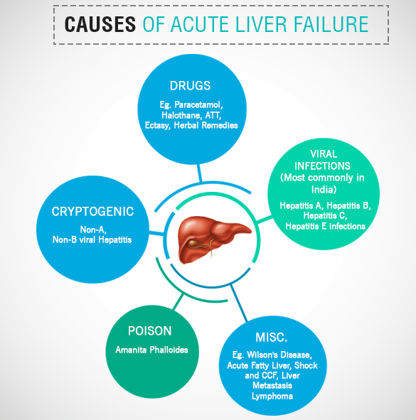
acute liver failure causes

1. Viral Hepatitis

Common Viral Causes

  • Hepatitis A
  • Hepatitis B
  • Hepatitis E
  • HSV (especially neonates)
  • EBV
  • CMV
  • Adenovirus
  • Enteroviruses

Important Point

In developing countries including Nepal and South Asia:

  • Hepatitis A and E remain major causes

2. Drug-Induced Liver Injury (DILI)

Common Drugs

  • Acetaminophen (Paracetamol)
  • Antitubercular drugs
  • Valproate
  • Antiepileptics
  • Herbal medications

Acetaminophen Toxicity

Most common cause in many developed countries.

Toxic metabolite:

  • NAPQI

Normally detoxified by glutathione.


3. Metabolic Disorders

Especially important in infants.

Major Causes

  • Galactosemia
  • Tyrosinemia
  • Mitochondrial disorders
  • Fatty acid oxidation defects
  • Wilson disease
  • Neonatal hemochromatosis

4. Autoimmune Hepatitis

Can present dramatically with:

  • Jaundice
  • Coagulopathy
  • Encephalopathy

Look for:

  • ANA
  • ASMA
  • Elevated IgG

5. Ischemic and Toxic Causes

  • Shock liver
  • Sepsis
  • Mushroom poisoning
  • Toxins

6. Indeterminate Causes

A substantial number of pediatric cases remain unexplained despite extensive workup.


Pathophysiology of Acute Hepatic Failure

Hepatocyte Injury

Massive hepatocyte necrosis leads to:

  • Failure of detoxification
  • Reduced clotting factor synthesis
  • Metabolic instability

Hyperammonemia

Ammonia accumulates due to impaired hepatic detoxification.

This causes:

  • Astrocyte swelling
  • Cerebral edema
  • Increased intracranial pressure

Coagulopathy

Liver cannot synthesize:

  • Factors II
  • V
  • VII
  • IX
  • X

Result:

  • Severe bleeding tendency

Immune Dysfunction

Patients become highly susceptible to:

  • Sepsis
  • Fungal infections

Clinical Features of Acute Hepatic Failure

Early Symptoms

  • Nausea
  • Vomiting
  • Malaise
  • Fever
  • Abdominal pain
  • Poor feeding
  • Irritability

Liver-Specific Findings

  • Jaundice
  • Hepatomegaly
  • Tender liver
  • Dark urine
  • Pale stools

Features of Hepatic Encephalopathy

Stage I

  • Irritability
  • Sleep disturbances
  • Behavioral changes

Stage II

  • Confusion
  • Drowsiness
  • Asterixis

Stage III

  • Stupor
  • Hyperreflexia

Stage IV

  • Coma

Signs of Cerebral Edema

  • Hypertension
  • Bradycardia
  • Unequal pupils
  • Abnormal posturing

This is a life-threatening emergency.


Diagnostic Evaluation of Acute Hepatic Failure

Initial Laboratory Workup

Liver Function Tests

  • AST/ALT
  • Bilirubin
  • Albumin
  • ALP
  • GGT

Synthetic Function

  • PT/INR
  • Fibrinogen

Metabolic Evaluation

  • Blood glucose
  • Lactate
  • Serum ammonia
  • ABG

Viral Studies

  • HAV IgM
  • HBsAg
  • Anti-HBc IgM
  • HEV serology
  • HSV PCR

Autoimmune Tests

  • ANA
  • ASMA
  • Anti-LKM
  • IgG

Metabolic Tests

  • Ceruloplasmin
  • Urine succinylacetone
  • Plasma amino acids
  • Urine organic acids

Imaging

Ultrasound Abdomen with Doppler

Useful for:

  • Liver size
  • Vascular patency
  • Ascites
  • Chronic liver disease exclusion

Important ICU Monitoring

Continuous monitoring of:

  • Mental status
  • Blood glucose
  • ICP signs
  • Urine output
  • Electrolytes
  • INR
  • Ammonia

Management of Acute Hepatic Failure

Core Principles

  • PICU admission
  • Aggressive supportive care
  • Prevent cerebral edema
  • Treat underlying cause
  • Early transplant referral

Stabilization

Airway

Intubate if:

  • Grade III/IV encephalopathy
  • Airway compromise

Circulation

Maintain:

  • Adequate perfusion
  • MAP
  • Renal function

Avoid fluid overload.


Management of Hypoglycemia

Frequent glucose monitoring is mandatory.

Treatment:

  • Dextrose bolus
  • Continuous glucose infusion

Cerebral Edema Management

General Measures

  • Head elevation to 30°
  • Avoid neck compression
  • Minimize stimulation

Osmotherapy

Mannitol

  • 0.5–1 g/kg IV

OR

Hypertonic Saline

Target serum sodium:

  • 145–150 mEq/L

Ammonia Reduction

Lactulose

Reduces ammonia absorption from gut.

Renal Replacement Therapy

Indicated for:

  • Severe hyperammonemia
  • Renal failure

Coagulopathy Management

Important principle:

  • Do NOT correct INR routinely unless bleeding or procedure planned.

Options

  • Vitamin K
  • FFP
  • Cryoprecipitate
  • Platelets

Infection Control

High suspicion for:

  • Bacterial infections
  • Fungal sepsis

Empiric antibiotics are often used in critically ill patients.


Etiology-Specific Treatment

Acetaminophen Toxicity

N-acetylcysteine (NAC)

Acts by:

  • Replenishing glutathione
  • Improving hepatic perfusion

N-acetylcysteine restores glutathione and reduces NAPQI toxicity\text{N-acetylcysteine restores glutathione and reduces } NAPQI \text{ toxicity}N-acetylcysteine restores glutathione and reduces NAPQI toxicity


HSV Hepatitis

  • IV acyclovir

Autoimmune Hepatitis

  • Corticosteroids

Wilson Disease

Usually requires urgent liver transplantation.


Nutrition in Acute Hepatic Failure

Key Principles

  • Early enteral nutrition preferred
  • Avoid prolonged fasting
  • Adequate calories essential

Protein

Previously restricted heavily, but modern pediatric guidelines recommend:

  • Avoid excessive restriction
  • Individualize according to encephalopathy severity

Liver Transplantation in Acute Hepatic Failure

Indications

  • Progressive encephalopathy
  • Refractory coagulopathy
  • Severe acidosis
  • Persistent hyperammonemia
  • Multi-organ failure

Poor Prognostic Factors

  • INR worsening
  • Severe encephalopathy
  • Cerebral edema
  • Renal failure
  • Rising bilirubin
  • Persistent lactic acidosis

Complications of Acute Hepatic Failure

Neurologic

  • Cerebral edema
  • Seizures
  • Herniation

Hematologic

  • Bleeding
  • DIC

Renal

Infectious

  • Sepsis
  • Fungal infections

Prognosis

Outcome depends on:

  • Etiology
  • Speed of recognition
  • Availability of transplant
  • Degree of encephalopathy

Better Prognosis

  • Hepatitis A
  • Acetaminophen toxicity (early NAC)

Worse Prognosis

  • Wilson disease
  • Indeterminate PALF
  • Severe cerebral edema

High-Yield Exam Points on Acute Hepatic Failure

Most Important Diagnostic Marker

  • Elevated INR

Most Common Cause in Developed Countries

  • Acetaminophen toxicity

Major Cause of Death

  • Cerebral edema and sepsis

Drug of Choice in Acetaminophen Toxicity

  • N-acetylcysteine

Key Emergency

  • Raised intracranial pressure

Acute Hepatic Failure Flowchart

Acute Liver Injury

Coagulopathy (INR ↑)

Evaluate Etiology

PICU Supportive Care

Prevent Cerebral Edema

Treat Specific Cause

Assess for Liver Transplant

Frequently Asked Questions (FAQs)

Is acute hepatic failure reversible?

Yes. Some causes recover completely with early management, while others require liver transplantation.

What is the most dangerous complication?

Cerebral edema leading to brain herniation.

Why is ammonia elevated?

The failing liver cannot convert ammonia into urea effectively.

Can children survive without liver transplant?

Yes, depending on etiology and severity. Hepatitis A-related PALF often recovers spontaneously.


Key Takeaway

Acute hepatic failure in children is a rapidly progressive and potentially fatal condition requiring:

  • Early diagnosis
  • Intensive monitoring
  • Prevention of cerebral edema
  • Etiology-directed therapy
  • Timely liver transplantation evaluation

Rapid recognition and evidence-based critical care significantly improve survival outcomes.

5 Critical Mistakes Pediatricians Make During Neonatal Resuscitation (And How to Avoid Them)


Introduction

Neonatal resuscitation is one of the most time-sensitive and high-stakes procedures in pediatrics. Despite structured guidelines like the American Academy of Pediatrics and the Neonatal Resuscitation Program, errors still occur—even among experienced clinicians.

These mistakes can lead to preventable morbidity and mortality. Understanding them is essential not only for pediatricians but also for residents, nurses, and delivery room staff.

In this article, we’ll break down 5 common mistakes pediatricians make during neonatal resuscitation and how to correct them using evidence-based practice.


1. Delayed Initiation of Positive Pressure Ventilation (PPV)

❌ The Mistake

Failure to initiate Positive Pressure Ventilation (PPV) within the “Golden Minute” when the neonate is apneic, gasping, or has a heart rate <100 bpm.

⚠️ Why It Matters

  • Ventilation—not chest compressions—is the most critical intervention
  • Delays increase risk of:
    • Hypoxic-ischemic injury
    • Bradycardia progression
    • Poor neurological outcomes

✅ Best Practice

  • Assess breathing and heart rate immediately after birth
  • Start PPV within 60 seconds (“Golden Minute”)
  • Use pulse oximetry early

2. Inadequate Mask Seal and Poor Ventilation Technique

❌ The Mistake

  • Improper mask size or seal
  • Incorrect head positioning
  • Ineffective ventilation despite apparent effort

⚠️ Why It Matters

  • Most failed resuscitations are due to ineffective ventilation
  • Leads to persistent hypoxia and bradycardia

✅ Best Practice (MR SOPA Approach)

Follow the MR SOPA corrective steps:

  • Mask adjustment
  • Reposition airway
  • Suction mouth and nose
  • Open mouth
  • Pressure increase
  • Airway alternative (ET tube or LMA)

3. Premature Initiation of Chest Compressions

❌ The Mistake

Starting chest compressions before ensuring adequate ventilation

⚠️ Why It Matters

  • Neonatal cardiac arrest is usually respiratory in origin
  • Without proper ventilation:
    • Compressions are ineffective
    • Oxygen delivery remains inadequate

✅ Best Practice

  • Ensure effective ventilation for at least 30 seconds
  • Start compressions only if:
    • HR <60 bpm despite effective PPV

4. Incorrect Oxygen Use (Too Much or Too Little)

❌ The Mistake

  • Starting all neonates on 100% oxygen
  • Failure to titrate oxygen using pulse oximetry

⚠️ Why It Matters

  • Hyperoxia → oxidative stress, especially in preterms
  • Hypoxia → organ damage

✅ Best Practice

  • Term babies: start with 21% oxygen (room air)
  • Preterm babies: start with 21–30% oxygen
  • Adjust based on preductal SpO₂ targets

5. Poor Team Communication and Role Assignment

❌ The Mistake

  • Lack of clear leadership
  • Unassigned roles
  • Ineffective communication during resuscitation

⚠️ Why It Matters

  • Leads to:
    • Delayed interventions
    • Duplicate or missed actions
    • Increased stress and errors

✅ Best Practice

  • Assign roles before delivery:
    • Airway manager
    • Compressor
    • Medication nurse
    • Team leader
  • Use closed-loop communication
  • Conduct pre-resuscitation briefing

Pro Tips for Better Neonatal Resuscitation

  • Always prepare equipment before delivery
  • Anticipate high-risk deliveries
  • Use checklists
  • Practice simulation training regularly
  • Follow updates from American Heart Association

Conclusion

Even skilled pediatricians can make errors during neonatal resuscitation—but most are preventable. The key lies in:

  • Mastering ventilation techniques
  • Following structured protocols
  • Practicing teamwork and communication

By avoiding these common mistakes, clinicians can significantly improve neonatal outcomes and reduce mortality.

FAQs

What is the most common mistake in neonatal resuscitation?

The most common mistake is ineffective ventilation due to poor mask seal or technique.

When should chest compressions be started in neonates?

Only when the heart rate is below 60 bpm after 30 seconds of effective ventilation.

Why is oxygen titration important in newborn resuscitation?

Both hypoxia and hyperoxia are harmful, especially in preterm infants.

What is enesthitis and how is it treated?

Introduction

Enthesitis is the painful inflammation of an enthesis, the site where ligaments or tendons attach to bone. Common in spondyloarthritis (e.g., psoriatic arthritis), it typically causes pain, stiffness, and tenderness, often in the heels, elbows, or hips. Treatment includes NSAIDs, biologics, rest, and physical therapy to manage symptoms and prevent potential joint damage. 

Symptoms of Enthesitis

  • Pain: Often described as severe or burning, specifically at tendon insertion sites, such as the Achilles tendon or bottom of the foot.
  • Stiffness: Increased difficulty moving the affected joint.
  • Tenderness: Sensitivity to touch.
  • Swelling: While not always present, swelling can occur in the soft tissue surrounding the site.
  • Impact on Mobility: Chronic cases can limit mobility and cause damage to adjacent bone and joints.

Causes and Risk Factors

  • Inflammatory Arthritis: Most frequently associated with psoriatic arthritis (PsA), ankylosing spondylitis (AS), and other forms of spondyloarthritis (SpA).
  • Physical Stress/Overuse: Repeated physical activity causing strain at the attachment site.
  • Immune System Dysfunction: Pro-inflammatory cytokine activity (specifically IL-17 and TNF-) is a major contributor to this inflammatory cascade.

Treatment Approaches

  • Medication: Non-steroidal anti-inflammatory drugs (NSAIDs) are the first line of treatment. For more chronic cases, biologics targeting TNF or IL-17 are often used.
  • Physical Interventions: Rest, immobilization, and gentle stretching.
  • Injections: Local corticosteroid injections, though used with caution near tendons.
  • Lifestyle Changes: Maintaining a healthy weight to reduce pressure on joints. CreakyJoints

Common Sites

Enthesitis can occur throughout the body, with more than 100 potential locations. Common sites include:

  • Achilles tendon (heel)
  • Plantar fascia (bottom of the foot)
  • Elbows (epicondylitis)
  • Hips
  • Patellar tendon (knee)

JIA, Classifications, Type and Diagnostic Criteria

JIA = Juvenile Idiopathic Arthritis
A chronic inflammatory arthritis of unknown cause beginning before age 16 and lasting ≥ 6 weeks, after exclusion of other causes.


✅ Definition (Juvenile Idiopathic Arthritis)

  • Arthritis in ≥1 joint
  • Onset < 16 years
  • Duration ≥ 6 weeks
  • Other causes excluded (infection, malignancy, trauma, connective tissue diseases)

🧬 ILAR Classification (Most used worldwide)

1️⃣ Oligoarticular JIA

Most common type (~50%)

Criteria

  • ≤4 joints involved in first 6 months

Subtypes

  • Persistent: remains ≤4 joints
  • Extended: becomes >4 joints after 6 months

Features

  • Large joints (knee most common)
  • Asymmetric
  • ANA positive common
  • Risk of uveitis

2️⃣ Polyarticular JIA (RF negative)

Criteria

  • ≥5 joints in first 6 months
  • RF negative

Features

  • Small joints of hands/feet
  • Symmetrical
  • Chronic course

3️⃣ Polyarticular JIA (RF positive)

Criteria

  • ≥5 joints
  • RF positive (≥2 tests, 3 months apart)

Features

  • Similar to adult rheumatoid arthritis
  • Severe, erosive disease
  • Adolescents (girls common)

4️⃣ Systemic JIA (Still disease)

Criteria

  • Arthritis with or preceded by fever ≥2 weeks
  • Daily (quotidian) fever for ≥3 days
    PLUS ≥1:
  • Evanescent salmon-pink rash
  • Hepatosplenomegaly
  • Lymphadenopathy
  • Serositis

Features

  • High ferritin
  • Risk of MAS (macrophage activation syndrome)

Criteria
Arthritis + enthesitis
OR arthritis/enthesitis + ≥2:

  • Sacroiliac tenderness/inflammatory back pain
  • HLA-B27 positive
  • Male >6 years
  • Acute anterior uveitis
  • Family history of spondyloarthropathy

Features

  • Lower limb arthritis
  • May progress to ankylosing spondylitis

6️⃣ Psoriatic Arthritis

Criteria
Arthritis + psoriasis
OR arthritis + ≥2:

  • Dactylitis
  • Nail pitting/onycholysis
  • Psoriasis in first-degree relative

7️⃣ Undifferentiated JIA

  • Does not fit above categories
    OR fits more than one category

🧪 Diagnostic Criteria & Workup

🔹 Clinical Diagnosis (Primary)

No single confirmatory test.

🔹 Essential Features

✔ Chronic joint swelling or limitation
✔ Morning stiffness
✔ Pain improves with activity
✔ Reduced range of motion


🔬 Laboratory Findings (Supportive)

TestSignificance
CBCanemia, leukocytosis (systemic JIA)
ESR / CRPinflammation
ANAoligoarticular JIA, uveitis risk
RFpolyarticular RF+
Anti-CCPerosive disease predictor
Ferritinvery high in systemic JIA
HLA-B27ERA subtype

🩻 Imaging

Early

  • Ultrasound → synovitis, effusion

Later

  • X-ray → joint space narrowing, erosions, growth abnormalities
  • MRI → early synovitis & sacroiliitis

🚨 Important Diagnostic Exclusions

Before diagnosing JIA, rule out:

  • Septic arthritis
  • Tuberculosis
  • Leukemia
  • SLE
  • Rheumatic fever
  • Trauma
  • Hemophilia

⚠️ Red Flags suggesting other diagnosis

  • Severe bone pain at night → leukemia
  • High fever with toxicity → infection
  • Weight loss, pallor → malignancy
  • Acute monoarthritis → septic arthritis

🧠 Exam Pearls (High Yield)

✔ Oligoarticular = most common
✔ Systemic JIA = quotidian fever + rash
✔ ANA+ girls → high uveitis risk
✔ RF+ polyarticular → resembles adult RA
✔ ERA → HLA-B27 boys, lower limb arthritis
✔ Screen for uveitis regularly even if asymptomatic

Classification of Newborn Care: 3 Level Care for Future

Newborn care is broadly classified into three levels based on the complexity of care, monitoring, and interventions required.


1. Level I – Basic Newborn Care (Well Newborn Nursery)

Definition

Care provided to healthy term newborns and stable late preterm infants who require only routine monitoring.

Babies managed

  • Term newborns (≥37 weeks)
  • Birth weight ≥2500 g
  • Babies with no complications
  • Stable late preterm (≥35 weeks)

Services provided

  • Routine neonatal examination
  • Thermal care
  • Early breastfeeding support
  • Cord care
  • Monitoring:
    • Temperature
    • Feeding
    • Urination/stooling
  • Immunization (BCG, OPV, Hep B depending on country policy)
  • Screening tests
  • Parent education

Procedures allowed

  • Basic resuscitation
  • Oxygen by hood
  • IV fluids if needed briefly

Staffing

  • Pediatrician or trained medical officer
  • Nurses trained in newborn care

2. Level II – Special Care Newborn Unit (SCNU / Special Care Nursery)

Definition

Care for moderately ill newborns who require closer monitoring or short-term intensive support.

Babies managed

  • Preterm infants ≥32 weeks
  • Birth weight ≥1500 g
  • Neonates recovering from severe illness
  • Neonates needing short-term respiratory support

Indications

  • Respiratory distress
  • Neonatal jaundice requiring phototherapy
  • Sepsis (suspected)
  • Feeding difficulty
  • Hypoglycemia
  • Hypothermia
  • Apnea of prematurity

Services provided

  • Continuous monitoring
  • Phototherapy
  • IV fluids and medications
  • Tube feeding
  • Oxygen therapy
  • CPAP (in many Level II units)
  • Short-term mechanical ventilation (<24 hr)

Procedures

  • Umbilical catheterization
  • Lumbar puncture
  • Exchange transfusion (in some units)

Staffing

  • Pediatricians
  • Neonatal nurses
  • Access to laboratory and imaging

3. Level III – Neonatal Intensive Care Unit (NICU)

Definition

Provides comprehensive intensive care for very sick or extremely premature neonates.

Babies managed

  • Gestational age <32 weeks
  • Birth weight <1500 g (VLBW)
  • Extremely low birth weight (<1000 g)
  • Neonates requiring advanced respiratory support

Conditions managed

  • Severe respiratory distress syndrome
  • Birth asphyxia
  • Persistent pulmonary hypertension
  • Sepsis/septic shock
  • Major congenital anomalies
  • Surgical conditions
  • Severe prematurity complications

Services provided

  • Mechanical ventilation
  • High frequency ventilation
  • Surfactant therapy
  • Total parenteral nutrition (TPN)
  • Continuous cardiorespiratory monitoring
  • Central lines
  • Invasive procedures
  • Advanced imaging
  • Neonatal surgery (in Level III C)

Sub-classification (sometimes used)

  • Level III A – mechanical ventilation
  • Level III B – advanced ventilation + surgery
  • Level III C – ECMO capable units

Staffing

  • Neonatologist
  • Pediatric intensivists
  • Specialized neonatal nurses
  • Respiratory therapists
  • Full laboratory and imaging services

Simplified Summary Table

LevelUnitBabies caredKey features
Level IWell newborn nurseryHealthy term babiesRoutine care
Level IISpecial care nursery / SCNUModerately ill, ≥32 weeksMonitoring, CPAP, phototherapy
Level IIINICUVery sick, <32 weeksFull intensive care

Alternative Public Health Classification (Used in Many Countries)

  1. Home care
  2. Community level care
  3. Facility based newborn care
    • NBCC (Newborn Care Corner)
    • SNCU
    • NICU

Exam Pearls (Pediatrics / Neonatology)

  • Level I → routine care
  • Level II → moderate illness, ≥32 weeks
  • Level III → intensive care, <32 weeks
  • VLBW infants (<1500 g) should be managed in Level III NICU

Pulsus Paradoxus: Definition, common cause, Pathology and Clinical relevance

Definition

Pulsus paradoxus is an exaggerated fall in systolic blood pressure (>10 mmHg) during inspiration.

Normally, systolic BP falls slightly during inspiration (<10 mmHg).
When the drop is >10 mmHg, it is called pulsus paradoxus.

Despite the name “paradoxus”, the pulse does not actually disappear; it only becomes weaker or unpalpable during inspiration.


Normal Physiology

During inspiration:

  • Intrathoracic pressure decreases
  • Venous return to the right ventricle increases
  • RV expands
  • Interventricular septum shifts slightly toward the left ventricle
  • LV filling decreases slightly
  • Systolic BP falls by <10 mmHg

Mechanism of Pulsus Paradoxus

In certain conditions:

  1. Excess RV filling or external compression
  2. RV expansion pushes interventricular septum toward LV
  3. LV filling markedly decreases
  4. LV stroke volume falls
  5. Systolic BP drops >10 mmHg during inspiration

Causes of Pulsus Paradoxus

Cardiac Causes

  • Cardiac Tamponade (classic cause)
  • Constrictive Pericarditis
  • Large pericardial effusion

Respiratory Causes

  • Severe Asthma
  • Chronic Obstructive Pulmonary Disease exacerbation

Other Causes

  • Tension Pneumothorax
  • Massive pulmonary embolism
  • Severe hypovolemia
  • Upper airway obstruction

How to Measure Pulsus Paradoxus

Using a sphygmomanometer:

  1. Inflate cuff above systolic pressure.
  2. Slowly deflate.
  3. Note two pressures:
  • First pressure: Korotkoff sounds heard only during expiration
  • Second pressure: Sounds heard throughout inspiration and expiration

Difference between the two = Pulsus paradoxus

Example:

  • Expiration only = 120 mmHg
  • Throughout respiration = 105 mmHg

Pulsus paradoxus = 15 mmHg


Clinical Significance

A pulsus paradoxus >10 mmHg suggests serious cardiopulmonary disease, especially:

  • Cardiac tamponade
  • Severe asthma attack

Reverse Pulsus Paradoxus

Reverse pulsus paradoxus is a rare cardiovascular finding in which systolic blood pressure increases during inspiration, opposite to the normal drop seen in typical pulsus paradoxus. It is most commonly associated with conditions that alter intrathoracic pressure dynamics or ventricular interaction, such as positive pressure ventilation, hypertrophic obstructive cardiomyopathy (HOCM), isovolumetric ventricular pacing, and sometimes aortic regurgitation.

The mechanism usually involves enhanced left ventricular filling or reduced afterload during inspiration, leading to a paradoxical rise in systolic pressure. Clinically, it is less frequently encountered than classic pulsus paradoxus and is often identified in intensive care settings where patients are mechanically ventilated. Recognition of reverse pulsus paradoxus is important because it can provide clues about underlying cardiac physiology and ventilatory influences rather than indicating conditions like cardiac tamponade, which are linked to the traditional form.


Quick Exam Definition

Pulsus paradoxus is an inspiratory fall in systolic blood pressure greater than 10 mmHg.

6 Steps of How to Demonstrate / Measure Pulsus Paradoxus (Bedside Method)

Pulsus paradoxus is demonstrated using a blood pressure cuff and stethoscope.


Step-by-Step Procedure

1. Position the Patient

  • Patient should be lying supine or semi-recumbent
  • Ask the patient to breathe normally

2. Inflate the BP Cuff

  • Inflate the cuff 20–30 mmHg above systolic pressure

3. Slowly Deflate the Cuff

Deflate slowly at 2–3 mmHg per second while auscultating the brachial artery.


4. Identify the First Pressure

At first you will hear Korotkoff sounds only during expiration and they disappear during inspiration.

Record this pressure.

Example: 120 mmHg

Reason: During inspiration the systolic pressure falls below cuff pressure.


5. Continue Deflating

As the cuff pressure falls further, Korotkoff sounds will be heard throughout the respiratory cycle (during both inspiration and expiration).

Record this second pressure.

Example: 105 mmHg


6. Calculate Pulsus Paradoxus

Pulsus paradoxus=First pressure−Second pressure\text{Pulsus paradoxus} = \text{First pressure} – \text{Second pressure}Pulsus paradoxus=First pressure−Second pressure

Example:120−105=15 mmHg120 – 105 = 15\ mmHg120−105=15 mmHg

If >10 mmHg → Pulsus paradoxus present


Clinical Example

If:

  • Sounds heard only in expiration at 118 mmHg
  • Sounds heard throughout breathing at 102 mmHg

Then:118−102=16 mmHg118 – 102 = 16\ mmHg118−102=16 mmHg

Positive pulsus paradoxus


How to Demonstrate in Clinical Exam (Short Viva Answer)

You can say:

Pulsus paradoxus is demonstrated using a sphygmomanometer. The cuff is inflated above systolic pressure and slowly deflated. The first pressure at which Korotkoff sounds are heard only during expiration is noted. The pressure at which sounds are heard during both inspiration and expiration is then noted. The difference between these pressures is the pulsus paradoxus. A difference greater than 10 mmHg indicates pulsus paradoxus.


Important Clinical Associations

  • Cardiac Tamponade (classic)
  • Severe Asthma
  • Tension Pneumothorax
  • Constrictive Pericarditis

Exam Tip:
If a patient has hypotension + raised JVP + pulsus paradoxus, suspect cardiac tamponade (Beck’s triad).

Heart Failure: Classes and Stages


Introduction

Heart failure (HF) severity and progression are described using two complementary systems:

  1. NYHA Functional Classification → based on symptoms and activity limitation
  2. ACC/AHA Stages of Heart Failure → based on disease progression and structural changes

1. NYHA Functional Classification

Used in patients with established heart disease to assess symptom severity and functional limitation.

Class I

  • No limitation of physical activity
  • Ordinary physical activity does not cause symptoms
  • Comfortable at rest

Examples

  • Walking
  • Climbing stairs normally
  • Daily activities without dyspnea

Class II

  • Slight limitation of physical activity
  • Comfortable at rest
  • Ordinary activity causes symptoms

Symptoms may include:

  • Fatigue
  • Palpitations
  • Shortness of breath

Example

  • Dyspnea when climbing several flights of stairs

Class III

  • Marked limitation of physical activity
  • Comfortable at rest
  • Less than ordinary activity causes symptoms

Symptoms:

  • Dyspnea
  • Fatigue
  • Palpitations with mild activity

Example

  • Breathlessness when walking short distances

Class IV

  • Severe limitation
  • Symptoms even at rest
  • Unable to perform any physical activity without discomfort

Examples

  • Dyspnea at rest
  • Orthopnea
  • Severe fatigue

2. ACC/AHA Stages of Heart Failure

Describes development and progression of HF and emphasizes prevention.

Unlike NYHA classes, stages are progressive and irreversible.


Stage A – At Risk for Heart Failure

Patients without structural heart disease or symptoms but with risk factors.

Common risk factors:

  • Hypertension
  • Coronary artery disease
  • Diabetes mellitus
  • Metabolic syndrome
  • Obesity
  • Exposure to cardiotoxic drugs
  • Family history of cardiomyopathy

Goal:

  • Prevent development of structural heart disease

Stage B – Pre-Heart Failure

Patients without symptoms but with structural heart disease.

Examples:

  • Left ventricular hypertrophy
  • Previous myocardial infarction
  • Reduced ejection fraction
  • Valvular heart disease

Features:

  • No symptoms yet
  • Evidence of cardiac structural abnormalities

Goal:

  • Prevent symptomatic HF

Stage C – Symptomatic Heart Failure

Structural heart disease with current or previous symptoms.

Common symptoms:

  • Dyspnea
  • Fatigue
  • Reduced exercise tolerance
  • Fluid retention (edema)

Patients often correspond to NYHA Class II–III.

Goal:

  • Symptom control and prevention of hospitalization

Stage D – Advanced Heart Failure

  • Severe symptoms despite optimal medical therapy
  • Recurrent hospitalizations
  • Marked limitation of daily activities

Management may include:

  • Advanced therapies
  • Mechanical circulatory support
  • Heart transplant
  • Palliative care

Patients usually correspond to NYHA Class IV.


Key Differences Between NYHA and ACC/AHA

FeatureNYHA ClassificationACC/AHA Staging
BasisSymptoms and activity limitationDisease progression
ReversibilityCan improve or worsenUsually progressive
UseFunctional assessmentPreventive and therapeutic planning
RangeClass I–IVStage A–D

Clinical Correlation

Typical relationship:

ACC/AHA StageNYHA Class
Stage ANo class
Stage BClass I
Stage CClass II–III
Stage DClass IV

Exam Pearl

  • ACC/AHA = structural disease progression
  • NYHA = symptom severity

Postcoital Vaginal Bleeding in Woman: top 5 Causes, Evaluation, and When to Worry (is it dangerous?)

Postcoital bleeding (bleeding after sexual intercourse / vaginal bellding after sex) can be alarming, especially in young women. If a woman notices spotting only on the same day after sex, the cause is usually benign — but it should never be ignored.

This article explains the common causes, red flags, and when medical evaluation is necessary.


What Is Postcoital Bleeding?

Postcoital bleeding refers to vaginal bleeding that occurs immediately after sexual intercourse and is not related to menstruation.

It may present as:

  • Light spotting
  • Pink or brown discharge
  • Fresh red bleeding
  • Bleeding that stops within a few hours

Common Causes in a young Woman

In this age group, most causes are non-cancerous.


1. Cervical Ectropion (Most Common Cause)

Cervical ectropion occurs when the delicate inner cervical cells are exposed on the outer surface of the cervix.

These cells are fragile and bleed easily when touched during intercourse.

It is common in:

  • Young women
  • Women taking oral contraceptive pills
  • Pregnancy (due to high estrogen levels)

Typically:

  • Bleeding is mild
  • Occurs only after sex
  • No severe pain

2. Cervicitis (Cervical Infection)

Inflammation of the cervix can cause contact bleeding.

Common sexually transmitted infections include:

  • Chlamydia trachomatis
  • Neisseria gonorrhoeae
  • Trichomonas vaginalis

Associated symptoms:

  • Abnormal vaginal discharge
  • Foul smell
  • Pain during intercourse
  • Burning urination

Young sexually active women are at higher risk.


3. Vaginal Infections (Vaginitis)

Infections make vaginal tissue inflamed and fragile.

Common causes:

  • Candida albicans
  • Trichomonas vaginalis

Symptoms:

  • Itching
  • Thick or foul discharge
  • Pain during sex

4. Mechanical Trauma

Minor tears may occur due to:

  • Rough intercourse
  • Inadequate lubrication
  • First sexual intercourse
  • Vaginal dryness

Bleeding is usually mild and short-lived.


Even light bleeding after sex can occur in early pregnancy due to a fragile cervix.

Important causes:

  • Implantation bleeding
  • Threatened miscarriage
  • Ectopic pregnancy

A urine pregnancy test should be done if periods are delayed.


Less Common but Important Causes

Although rare at 23 years of age:

Cervical Dysplasia

Often associated with:

  • Human papillomavirus

May present with recurrent postcoital bleeding.

Cervical Cancer

Rare in young women but should be considered if:

  • Bleeding is persistent
  • There is unexplained weight loss
  • There is pelvic pain
  • Abnormal Pap smear

When Is It Likely Benign?

Postcoital bleeding is usually harmless if:

  • It occurs only on the day of intercourse
  • Bleeding is light
  • It stops within hours
  • There is no pain or foul discharge
  • It does not happen frequently

When to See a Doctor

Seek medical evaluation if:

  • Bleeding happens repeatedly
  • Bleeding becomes heavy
  • There is pelvic pain
  • There is abnormal discharge
  • Periods are missed
  • Bleeding occurs unrelated to intercourse

How Is It Evaluated?

A doctor may perform:

  • Detailed history
  • Speculum examination
  • Pregnancy test
  • STI testing (NAAT)
  • Vaginal swab
  • Pap smear (if due)
  • Colposcopy if cervix looks abnormal

Key Takeaway

In woman, postcoital bleeding occurring only on the same day after intercourse is most commonly due to cervical ectropion, mild infection, or minor trauma.

However, repeated episodes should always be medically evaluated to rule out infection and cervical pathology.

Early evaluation ensures reassurance in benign cases and timely treatment if needed.

Exit mobile version