Mechanical Ventilation: Definition, Settings, benefits of invasive mechanical ventilation

Introduction to Mechanical Ventilation

The mechanical ventilation is basically a replaement of assistance to the spontaneous ventilation/ breathing. 
The concept of mechanical ventilation was first introduced by a scientist “Andreas Vesalius in 1555
He said that : “an opening must be attempted in the trunk of the trachea, into which a tube of reed
or cane should be put; you will then blow into this, so that the lung may rise again . . .
and the heart becomes strong”
This was the fundamental of development of mechanical ventilation and intubation but it took 400 years to apply this and actually use it in medical field. 
Now a days mechanical ventilation has been the very vital part of medical sciece and has been used for many purposed in daily basis for saving millions of the lives worldwide.
Mecahnical ventilation and mechanicala ventilators have been used to save lives in emergency situations as well as in elective and emergency surgeries. They are the majr part of modern day ICU care.
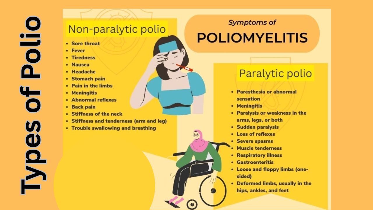
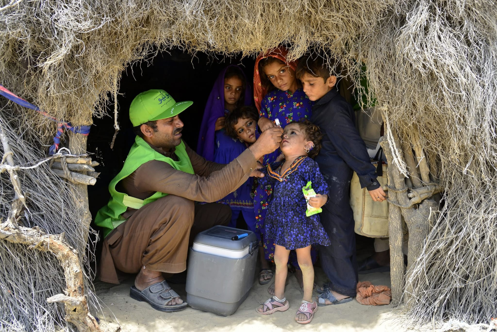
The first use of assisted ventilation (nt mechanical ventilations) was done in Europe during Polio outbreak (Read more about polio here). 
Back then even human were used to a rtificailly ventilate the lung of sick people making them the human ventilators, and they even worked upto 8 hours a day and the medical schools were shut down for same purpose. 
Later a company named “Ëmerson company” made a prototype positive pressure ventilator which was used in the Massachusetts General Hospital and it became instant succcess and started the era of Modern day intennsive care medicine. 

What is Mechanical Ventilation? What happens when youre on a ventilator?

The basic concept of mechanical ventilation or positive pressure ventilation is to create a positive pressure the moves air/oxygen into the lungs. DUring breathing the pressure in our alveoli keeps on changing and during inspiration it becomes subatmospheric which drags air into the lungs and aveoli.
But if a person is not able to breathe by themselves in that condition positive pressure more than that of alveoli and lungs needs to be created by using artificial means and air or oxygen is pushed into the lungs.
A ventilator machine labelled different parts

Mechanical Ventilation Definition

By undertanding above concept, mechanical ventilation  can be difined as a process in which the lungs of a person who is not able to breath by oneself are inflated using external force, person or machine in order to push air into them and pull it back, in order to complete gaseous exchange, or for purpose of delivering medicine (in case of anesthesia or other critical care). 

Understanding Mechanical Ventilation Settings

a bag-valve-mask

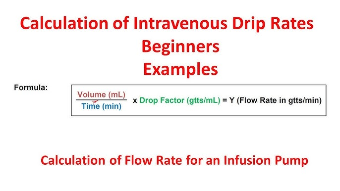
In general there are two methods of positive pressure ventilation:

Volume controlled ventilation VC mode:

In this mode the initially the volume of air that is needed to be pushed into the ungs  is selectd and ventilator selects the pressure needed to push that much volume into lungs by itself. Depending on that the rate of lung inflation, that if respiratory rate can be kept constant ot adjusted. 

Pressure control ventilation PC mode:

In this mode the pressure at which the air/oxygen is pushed into lungs is preselected and duration and rate of lung inflation can be adjusted on the basis of need by the operator or doctor. by this desired tidal volume and sufficient oxygenation can be achieved.
The rate of lung inflation is initially high then is reduced so that presssure is same throughout(constant).
In other classification it can be classified as invasive and noninvasive ventilation:

Invasive ventilation

Invasive ventilation means ventilation with intubation in which a person a person has Endotrachea tube (ET Tube) placed in his trachea and through this air is supplied or his lungs are ventilated.
Endotracheal tube

Non-inasive ventilation

Non invasive ventilation consists of CPAP and BiPAP. CPAP standa for continuous positive pressure ventilation and BiPAP mean bilevel positive pressure ventilation.
These both are achieved through a fitting mask kept covering nose and mouth of the patient.
Though these types of ventilation can also be achieved in invasive ventilation as well, unless not necessary a person is not intubated but CPAP / BiPAP mas is used for this purpose.
Volume control Vs Pressure control technique in mechanical ventilation

Invasive Mechanical Ventilation: Benefits and Risks

Indication of mechanical ventilation

  • Respiratory failure
    • Hypoxemic respiratory failure : low oxygen saturation (sa02 or sp02)
    • Hypercarbic respiratory failure: High carbondioxide content in blood (paCO2)
    • Acute respiratory failure
  • Ventilatory failure
  • To reduce cerebral blood flow in case of raise ICP
  • Prevent aspiration of gastric content or other foreign body
  • Protect airway in seerely ill patient
  • In intoxicated, poisoned patients
  • To delever medicine and artificially breathe in case of general anesthesia
  • Hemodynamic instability
  • Heart attack
  • Head, face and neck surgery
  • Unstable or risky airway
  • Cardiac arrest
  • Encephalopathy
  • Coma or deterirating GCS
Ventilated patient depiction drawing

Befefits of mechanical ventilation:

  1. Proper and adequate oxygenation
  2. Accurate measurement of pressure and volumes
  3. Decreases work of breathing
  4. Improves gas exhange
  5. Reduces mortality
  6. Low failure rate than noninvasive ventilation
  7. Improves general pulmonary function
  8. Multiple modes available and settings can be changed according to need
  9. Stabilize and protects the airwat from collapse or obstruction
  10. Prevents aspiration
  11. Prevents atelectasis
  12. No airleaks

Risks associated with mechcanical ventilation:

  1. High pressure related lung injury : Barotrauma
  2. Volume trauma
  3. Oxygen toxicity
  4. Ventilator dependence
  5. Infection of airway
  6. Pneumonia, also called VAP : Ventilator associated pneumonia
  7. Mucus plug and lung collapse
  8. Airway trauma, mediastinal perforation
  9. Cuff presure injury to trachea and fistula formation, tracheal necrosis
  10. Hemodynamic collapse
  11. Tube malposition
  12. Electrolyte and aid base imbalance
  13. Mouth, teeth and lip trauma
  14. Infection of sinuses
  15. Muscle weakness and wasting and difficulty breathing after intubation later
  16. Position related complication
  17. Deep venous thrombosis and Pulmonary embolism (PE)
  18. Presure sores, ulcers
  19. Psychaitric problems
  20. Vocal cord injury and difficulty prducing speech

Mechanical Ventilation Systems: Types and Components

  1. Invasive ventilator with modern mamchine
  2. Bag-valve-mask ventilator (manual)
                Though bag valve mask can also be connected to endotracheal tube.
ventilator


Components of mechanical ventilator:

  • Connector tube
  • circuits
  • t-piece
  • HME filter
  • monitor
  • air and oxygen ports and supply
  • ventilator machine
  • power supply
  • control knobs and butttons
ventilator parts

Instruments needed for mechanical ventilation

  1. Ventilator machine
  2. Connecting tubes for oxygen deivery
  3. Endotracheal ET tube
  4. Syringes
  5. Stethoscope
  6. Direct laryngoscope
  7. Bag-Valve-Mask
  8. Suction machine
  9. Suction tube
  10. Source of oxygen
  11. Fixating tapes/dresings
  12. Medications for anesthesia and muscle relaxation
  13. Monitor for vitals monitering
  14. Trained manpower

Process of putting one into mechanical ventilator or starting mechnical ventilation

  • The patient is decided for mechanical ventilation on basis of above mentioned criteria
  • Patient is preoxygenatied
  • Rapid sequence intubation is done using anesthetic agent and muscle relaxants
  • Once intubation is done then patient is connected to ventilator machine with appropriate setttings
  • The  settings of the ventilator can be changed as need and patiend is intensively monitored
  • Regularly check ABG to maintain arterial blood ga and electrolyte in limits
  • Keep arterial blood pH 7.35-7.45
  • Reularly check patient efforts, improvements, and treat the condition and cause of patiet’s need of intubation
  • Follow strict precaution for infection prevention
  • Prevent other complication
    How does a ventilator work

Method of ventilation-Lung Protective ventilatory strategy

  • Set tidal volume to 6ml/kg
    • calculate ideal body weight
    • use VC and set initial tidal volume Vt to 8 ml/kg
    • set RR to match baseline minute ventilation but not >35 bpm
    • set PEEP to 5 cm of H2O
    • reduce Vt by 1 ml/kg every 1-2 hr till its 6ml/kg
    • adjust PEEP and FiO2 to maintain SpO2 88-95% 
  • Prevent plateau pressure exceedig 30 cm of water
    • if plateau pressure .30 cm of water and Vt 6 ml/kg decrease vt 1 ml/kg until plateau pressure falls below 30 cm of water or Vt reached minimum of 4ml/kg
  • use least possible concentreation of oxygent (fraction of inspired oxygen or FiO2) to maintain saturation more than 90%
  • Adjust PEEP to maintain alveolar potency while preventing overdistention and closure reopening
  • Ph goal to 7.30 to 7.45
    • if pH 7.15-7.30 increase RR until ph >7.3, paco2 <25 or RR = 35
    • if pH <7.15, increase RR=35. if still remains <7.15, increase Vt in 1ml/kg until pH>7.15
    • if pH>7.45 decrease RR if possible
Read Normal ABG finding of a health person here: 

Care during mechanical ventilation:

  • Sedation and muscle relaxants or paralysis
  • Analgesia
  • Intensive monitoring
  • Chest physiotherapy
  • Suctioning and secretion clearing
  • Nutrition
  • Humidification
  • Prevention of infection: common site of infections include lungs, urine, oral cavity, skin, and blood 
  • Mobilization
  • Pressure prevention
  • Thromboembolic prophylaxis
  • Skincare
  • Oralcare
  • Eye care
  • Other general care
ventilator in action

Indication for discntinuation mechanical ventilation or indication of extubation

  • Patients recovery
  • Weaning
  • Completion of indication
  • Death

Technique of weaning:

    SIMV wean
    PS wean
    T-piece trial

Some terminlogies related to mechanical ventilation:

Respiratory rate RR

Number of breathes  (insiration and expiration = one breathe) per minute

Tidal volume Vt

The volume of air inhaled or exhaled in each breath / respiration by the person

Minute ventilation MV

Total volume inhald or exhaled by the person in a minute

FiO2

Fraction of the inhaled oxygen i.e percentage of oxygen in inhaled air

Peak pressure

The maximum pressure during inspiration generated by machine to push air into the lungs.

PEEP

Positive end expiratory pressure. It is the pressure at aveoli at the end of the expiration cycle before beginning of inspiration.

ACMV

Assist controlled mechanical ventilation

IMV

Intermittent mandatory ventilation

SIMV

Synchronized intermittent mandatory ventialtion

PSV

Pressure Support Ventilation

A/C, PCV

Assisted/Controlled, Pressure Controlled Ventilation (time cycled)

A/C, VCV

Assisted/Controlled, Volume Cycled Ventilation.

IRV

Inverse ratio ventilation

TCV

TIme controlled ventilation

ZEEP

Zero end expiratory pressure

NEEP

Negative end expiratry pressure

I:E ratio

Ratio of inspiratory time to expiratory time

SIMV + pressure support

Synchronized Intermittent Mandatory Ventilation with PEEP

CPAP

Continuous positive airway presssure

BiPAP

Bilevel positive aiway pressure

If you want to read more about above termnologie write us at mail@chaitanya.com.np

Some non-convetional ventilation strategies

  • HFOV- high frequency oscillatory ventillation
  • APRV- airway pressure release ventilation
  • PLV- partial liquid ventilation
  • ECMO- extracorporeal membrane oxygenation
  • PAV- proportional assist ventilation
  • NAV- neurally adjusted ventillatory-assist ventilation
  • PAV-NAV : proportional assist ventilation-neurally adjusted ventillatory-assist ventilation

Read my friend’s blog at doctorhealthrx.com

Conclusion: Importance of Mechanical Ventilation

Mechanical ventilation is a medical technique that replaces or assists spontaneous breathing. The concept was first introduced in 1555 by Andreas Vesalius, but it took 400 years to develop mechanical ventilation into a medical practice.
Today, mechanical ventilation and ventilators are widely used for various purposes in medical care, including elective and emergency surgeries and emergency situations. There are two main methods of positive pressure ventilation, volume-controlled and pressure-controlled ventilation, which can be invasive or noninvasive.
Mechanical ventilation has many benefits, such as improving gas exchange, reducing the work of breathing, and improving general pulmonary function, but there are also risks, such as high pressure or lung damage. Overall, mechanical ventilation has been a crucial part of modern-day intensive care medicine and has saved millions of lives worldwide.

Understanding Polio: Causes, Symptoms, Prevention, and Treatment

Table of Contents(toc)

What is Polio?

Poliomyelitis, commonly known as polio, is a highly contagious viral disease caused by the poliovirus. It primarily affects the nervous system and can lead to paralysis, permanent disability, or even death in severe cases. Polio mostly affects children under the age of five, but it can also infect unvaccinated adults.

Types of Poliomyelitis

1. Non-paralytic polio: This form causes flu-like symptoms but does not lead to paralysis.
2. Paralytic polio: The most severe form, leading to muscle weakness, loss of reflexes, and paralysis. Paralysis can be permanent.
3. Post-polio syndrome (PPS): This can occur years after recovery, causing muscle weakness, fatigue, and pain.

Transmission

  • Polio spreads through:
  • Fecal-oral route: Contaminated food or water.
  • Person-to-person contact: Through saliva or respiratory droplets.

Prevention

Polio has no cure, but it can be prevented with vaccines:
  • Oral Polio Vaccine (OPV).
  • Inactivated Polio Vaccine (IPV).
Global vaccination programs have significantly reduced polio cases, bringing the disease close to eradication.

Causes of Polio

Polio is caused by the poliovirus, a highly infectious virus that belongs to the Picornaviridae family. It primarily spreads through the fecal-oral route, often in areas with poor sanitation, where contaminated food or water can transmit the virus. It can also spread through direct contact with an infected person’s saliva or respiratory droplets. Once inside the body, the virus multiplies in the throat and intestines before entering the bloodstream and, in some cases, attacking the central nervous system. This attack can lead to inflammation and destruction of motor neurons, resulting in paralysis. Poor hygiene, lack of vaccination, and overcrowding increase the risk of transmission.

Symptoms of Polio

  • Mild cases may show no symptoms. Severe cases can present:
  • Fever, sore throat, and fatigue.
  • Muscle stiffness and pain.
  • Sudden paralysis in severe cases.

How is Polio Diagnosed?

Polio is diagnosed through a combination of clinical evaluation and laboratory tests. A healthcare provider may suspect polio based on symptoms such as acute flaccid paralysis, fever, muscle weakness, and reduced reflexes. 
To confirm the diagnosis, laboratory tests are conducted on samples of throat secretions, stool, or cerebrospinal fluid (CSF) to detect the presence of the poliovirus. 
Stool samples are the most reliable for isolating the virus. In some cases, molecular techniques like polymerase chain reaction (PCR) are used to identify the virus’s genetic material. 
Early and accurate diagnosis is essential for managing the disease and preventing its spread.

Prevention and Treatment of Polio

Polio prevention is primarily achieved through vaccination. The Oral Polio Vaccine (OPV) and the Inactivated Polio Vaccine (IPV) are highly effective in protecting against your disease. OPV is widely used in mass immunization campaigns, particularly in endemic areas, while IPV is administered via injection and is part of routine immunization schedules in many countries. Maintaining good hygiene, proper sanitation, and access to clean drinking water also help reduce the risk of transmission.
Treatment for polio focuses on supportive care, as there is no cure for the disease. This includes managing symptoms, such as using pain relievers, physical therapy to reduce muscle stiffness, and mobility aids like braces or wheelchairs for those with paralysis. In severe cases, mechanical ventilation may be necessary if breathing muscles are affected. Rehabilitation programs can help improve quality of life for individuals recovering from polio or managing post-polio syndrome.

Post-Polio Syndrome: Understanding the Long-Term Effects

The Global Efforts to Eradicate Polio

Global efforts to eradicate polio have been spearheaded by initiatives like the Global Polio Eradication Initiative (GPEI), launched in 1988. This collaborative effort involves organizations such as the World Health Organization (WHO), UNICEF, Rotary International, and the Centers for Disease Control and Prevention (CDC), among others. These programs focus on widespread vaccination campaigns, surveillance to detect and respond to outbreaks, and improving access to clean water and sanitation in high-risk regions. As a result, polio cases have declined by over 99%, with wild poliovirus now endemic in only a few countries, such as Afghanistan and Pakistan. Continued global collaboration and funding are crucial to overcome challenges like vaccine resistance, conflict, and gaps in healthcare systems to achieve a polio-free world.

Polio vaccine 

The polio vaccine is a crucial tool in preventing poliomyelitis and achieving global eradication of the disease. There are two primary types of polio vaccines: the Oral Polio Vaccine (OPV) and the Inactivated Polio Vaccine (IPV). OPV is administered orally and contains a weakened form of the virus, which stimulates strong immunity and can also reduce community transmission through shedding of the weakened virus. IPV, given by injection, uses an inactivated (killed) virus to provide individual protection without the risk of vaccine-derived poliovirus. 
Both vaccines are safe and effective, with IPV being the preferred choice in countries with robust healthcare systems. Mass immunization campaigns using OPV have been instrumental in significantly reducing global polio cases, while routine immunization programs with IPV help maintain long-term immunity in polio-free regions.

Polio vaccine storage

Proper storage of the polio vaccine is critical to maintaining its effectiveness. Both the Oral Polio Vaccine (OPV) and the Inactivated Polio Vaccine (IPV) are sensitive to temperature changes and must be stored under strict conditions. OPV requires storage at temperatures between -20°C to -25°C, typically in freezers, to preserve the live attenuated virus. IPV, on the other hand, should be stored at temperatures between 2°C to 8°C in a refrigerator. 
Vaccines should be kept in insulated containers during transportation to maintain the cold chain and prevent spoilage. Any exposure to higher temperatures or freezing (in the case of IPV) can degrade the vaccine’s potency. Adhering to proper storage protocols ensures the vaccines remain effective, especially in areas with limited infrastructure or during mass immunization campaigns.

Some frequently asked question regarding polio vaccine in Nepali

एफ आइ पि भि ले कुन रोग बाट जोगाउछ?

पोलियो थोपा जस्तै यसले पनि पोलियो रोग लाग्न बाट जोगाउछ।

एफ आइ पि भि पहिलेको तरिका:

  • एक बर्षमुनि: ६ र १४ हफ्ता दुइ डोज
  • छुट भएमा: पांच बर्ष सम्म ६ हफ्ताको फरकमा दुइ डोज

एफ आइ पि भि नया तरिका: FIPV new technique

  • एक बर्षमुनि: १४ हफ्ता र ९ महिनामा दुइ डोज
  • छुट भएमा: पांच बर्ष सम्म ४ महिना अर्थात १६ हफ्ताको फरकमा दुइ डोज

खोप लगाउने स्थान साइट: FIPV vaccine injection site:

  • १४ हफ्ता र ९ महिना मा दाया पाखुरामा छालामा (intradermal) दिने। 
  • बि सि जि संगै परे सो बेला बाया पखुरा मा छालामा (intradermal) दिने।
  • अन्यबेला दिन परे या दिएको भए १६ हफ्ता अर्थात ४ महिना फरकमा दिने।

पोलियो रोग बारे पढ्न यहा क्लिक गर्नुहोस

जान्नुहोस पोलियो थोपा किन खुवाउने?

Amoebic liver abscess, causes and treatment, clinical features

Amoebic liver abscess, causes and treatment, clinical features

Table of contents(toc)

Introduction

Liver abscess is a rare but severe infectious disaese. 
It is commonly caused by entamoeba histolytics and some other species as well. 
Threr are total 4 different species of Entamoeba includes:
  1. E. histolytica
  2. E. 
  3. E. 
  4. E.
Of these only E, histolytica and, are capable of causing human infections. 
Investigations to diagnose and manage liver abscess
  1. LFT
  2. Ultrasonography
  3. CT scan
  4. Laparoscopic or Open diagnostic surgery

Powerpoint on Amobeic liver abscess:

Thank you for reading it. 
Please share. 

Hepatitis Full lecture PPT

Hepatitis introduction, causes, viral hepatitis, treatment of hepatitis and liver function test interpretation

Table of Contents(toc)

Introduction

Hepatitis means inflammation of liver. 
It is causes by variety of reasons including following. 
  1. Viral
  2. Toxin including alcohol
  3. Bacterial
  4. Autimmune
  5. Traumatic
Diagnosis of Hepatitis:
Hepatitis is diagnosed by checkling liver function tests (LFTs). They are serological markers of liver cell damage. 

Hepatitis PPT here:

Thank you for reading.

Swasthya sewa niyamawali : A summary for entrance and loksewa preparation

Swasthya sewa niyamawali download free 2024

Swasthya Sewa Niyamawali 2053 is a set of health service regulations implemented in Nepal. It outlines guidelines and standards for the provision of healthcare services in the country. The regulations cover various aspects such as health facility management, service delivery, infrastructure requirements, human resources, and quality assurance. The goal of the Niyamawali is to ensure the availability of quality healthcare services and protect the rights of patients. It plays a crucial role in regulating the healthcare sector and improving the overall health system in Nepal.

Summary of Swasthya sewa niyamawali 2055

Here are some additional facts about Swasthya Sewa Niyamawali in Nepal:
  • It was made in BS 2055.
  • Last edition was on BS 2075
  • It has been edited four times since then.

Impotant points regarding swastya sewa niyamawali:

  1. Development and Implementation: Swasthya Sewa Niyamawali 2055 was developed by the Government of Nepal in collaboration with health experts and stakeholders. It was implemented in 1996 and has undergone subsequent revisions to address evolving healthcare needs.
  2. Coverage: The Niyamawali applies to a wide range of health facilities, including hospitals, clinics, nursing homes, dispensaries, and health posts. It ensures that both public and private health institutions adhere to the prescribed standards.
  3. Service Delivery: The regulations define the responsibilities and obligations of healthcare providers in delivering services. It covers areas such as patient care, emergency services, referral mechanisms, pharmaceutical services, and preventive measures.
  4. Infrastructure Requirements: The Niyamawali sets forth standards for infrastructure and physical facilities. It includes provisions for hygiene, sanitation, waste management, medical equipment, and adequate space for patient care.
  5. Human Resources: The regulations address the requirements for healthcare professionals, including qualifications, licensing, and registration. It emphasizes the need for qualified medical personnel and skilled staff in health facilities.
  6. Quality Assurance: Swasthya Sewa Niyamawali emphasizes the importance of quality assurance in healthcare. It establishes mechanisms for monitoring, evaluation, and accreditation of health services to ensure compliance with defined standards.
  7. Patient Rights and Safety: The Niyamawali places a strong emphasis on protecting the rights and safety of patients. It outlines measures to ensure confidentiality, informed consent, patient education, and handling of complaints and grievances.
  8. Capacity Building and Training: The regulations promote continuous capacity building and professional development of healthcare providers. It encourages training programs, workshops, and research to enhance the skills and knowledge of medical personnel.
  9. Enforcement and Penalties: Swasthya Sewa Niyamawali empowers regulatory bodies to enforce compliance with the regulations. It outlines penalties and legal consequences for non-compliance, including fines, suspension, or revocation of licenses.
  10. Ongoing Revisions: The Niyamawali is periodically reviewed and revised to align with emerging healthcare needs and advancements. This ensures that the regulations remain relevant and effective in addressing the evolving healthcare landscape in Nepal.

swasthya sewa niyamabali 2055 download below:

Summary of swasthya sewa niyamawali: 

Swasthya Sewa Niyamawali 2055 is a set of health service regulations implemented in Nepal. It outlines guidelines and standards for the provision of healthcare services in the country. The regulations cover various aspects such as health facility management, service delivery, infrastructure requirements, human resources, and quality assurance. 

The goal of the Niyamawali is to ensure the availability of quality healthcare services and protect the rights of patients. It plays a crucial role in regulating the healthcare sector and improving the overall health system in Nepal.

Thank you.

Gastritis for medical students: GERD, gastritis, esophagitis, duodenitis, gastroduodenitis, Peptic ulcer disease and more

Introdiction:

APD (acid peptic disease/disorders) consists  of different spectrum of disease which includes GERD gastritis, esophagitis, Gastroduodenitis, peptic ulcer disease (gastric and duodental ulcers).

Symptoms of Gastritis and Peptic ulcer disease:

The common symptoms of it innclude heartburn, acid reflux, belching and abdominal pain. These symptoms are usually aggravated by empty stomach and occasionally the symptoms are also precipitated by eating food depending upon where  the pathology is. 

Pathophysiology:

Acid and pepsin levels that are too high can lead to acid peptic diseases. The gastric and duodenal ulcer are the two main varieties of acid peptic diseases. The acid corrodes and injures the unprotected gastric mucsa and pepsin also plays vital role in it.

But they can also happen at Meckel’s diverticulum, the lower end of the oesophagus, and the GJ Stoma. Acute or chronic symptoms can be present for a long time or be more complicated.

Role of Helicobacter pylori:

A bacterium names Helicobacter pylori is also associated with pathology of this disease. This bacteria can bear strong acidic  environment hence it lives in human stomach and helps frmation of the ulcers in the esophagus, stomach and the duodenum.

Complications of gastritis and pepic ulcer disease PUD:

The complications of untreated APD include but no limited to ulceration and bleeding, pain, weight loss, malnutrition, perforation, peritonitis, hospital stay and need for  surgery. in some cases the patient may also develop carcinoma (cancer) as well.

The gastric ulcer commonly changes into carcinoma while the duodenal ucer mostly perforates. 

APD/ GERD treatment

The treatment of  this disaese includes acid supressants i e PPIs, H2 antihistamines, Prostaglandin analogues, antacid tablets, and sometimes the patient may need antibiotcs as well for eradication of H pylori.

Occasionally ulcer protective  agents are also used for helping the ulcers heal, like sucralfate and the bismuth.

The complications like perforations are managed surgically while if any ulcer present should be checked for malignancy.

Here is the powerpoint presentation ppt for my studdents to go htorugh and revise while preparing for the competitive exams. 

Video on Peptic ulcer disease

Tags; ( please ignore )Health exams, Exam preparation, Medical exams, Health test, Study tips, Health education, Health knowledge, Exam stress, Health science, Health information, Exam strategies, Health conditions, Test anxiety, Health promotion, Exam resources, Health resources, Health research, Exam techniques, Health coaching, Exam success, PSC exam preparation, Lok Sewa exam tips, License exam study material, Exam syllabus, Exam pattern, Previous year question papers, Mock exams, General knowledge for exams, Exam eligibility criteria, Exam registration, Exam dates, Exam centers, Exam results, Exam analysis, Exam cutoff marks, Exam books and guides, Exam coaching, Exam time management, Exam revision techniques, Exam success stories, Public service commission exam, Civil service exam preparation, PSC exam syllabus, PSC exam pattern, PSC exam eligibility, PSC exam registration, PSC exam dates, PSC exam centers, PSC exam results, PSC exam preparation tips, Lok Sewa Aayog exam preparation, Lok Sewa Aayog exam syllabus, Lok Sewa Aayog exam pattern, Lok Sewa Aayog exam eligibility, Lok Sewa Aayog exam registration, Lok Sewa Aayog exam dates, Lok Sewa Aayog exam centers, Lok Sewa Aayog exam results, License exam preparation, License exam syllabus, License exam pattern, License exam eligibility, License exam registration, License exam dates, License exam centers, License exam results, License exam preparation tips, General knowledge for PSC, Lok Sewa, and license exams, Current affairs for exams, Mental ability for exams.

How to put a cannula: the easiest way in 2026?

Introduction

Intravenous (IV) cannulation is the process of inserting a small, hollow tube called a cannula into a vein to administer fluids, medications, or blood products directly into the bloodstream. This procedure has become an essential part of modern medicine and is commonly used in hospitals, clinics, and emergency settings.

History of IV Cannulation

The concept of intravenous administration of fluids dates back to the 17th century when Sir Christopher Wren, an English physician, used a goose quill to introduce a mixture of wine and opium into the bloodstream of dogs. In the 19th century, the use of glass syringes and metal needles made intravenous injection more reliable and efficient. However, it was not until the 20th century that the first cannula was developed by Dr. Werner Forssmann in 1929. Dr. Forssmann inserted a catheter into his own arm and then walked upstairs to the X-ray department, where he successfully took an X-ray of his own heart. This groundbreaking procedure earned Dr. Forssmann the Nobel Prize in Medicine in 1956.

Indications for IV Cannulation

IV cannulation is used in a wide range of medical situations where intravenous access is required. Some of the most common indications for IV cannulation include:

  1. Dehydration: IV fluids can be administered to replace fluids lost through vomiting, diarrhea, or excessive sweating.
  2. Medication administration: Many medications, including antibiotics, pain relievers, and chemotherapy drugs, are administered through IV cannulation.
  3. Blood transfusion: IV cannulation is used to administer blood products in cases of severe anemia or blood loss.
  4. Emergency situations: In emergency situations, such as cardiac arrest or severe trauma, IV cannulation may be required to administer life-saving medications or fluids.
  5. Monitoring purposes: In some cases, IV cannulation may be used to monitor blood pressure, administer contrast dye for imaging studies, or measure blood gases.

Procedure for IV Cannulation

The procedure for IV cannulation involves several steps:

  1. Patient preparation: The patient is typically placed in a comfortable position, and the site for cannulation is selected. The healthcare provider will clean the skin with an antiseptic solution to reduce the risk of infection.
  2. Vein location: The healthcare provider will use their fingers to feel for a suitable vein, which is typically a superficial vein located on the back of the hand, wrist, or antecubital fossa (the bend of the elbow).
  3. Cannulation: The healthcare provider will insert a small needle through the skin and into the vein. Once the needle is in place, a small plastic tube called a cannula is threaded over the needle and into the vein. The needle is then removed, leaving the cannula in place.
  4. Securement: The cannula is secured in place using adhesive tape or a dressing.
  5. Flush and test: The healthcare provider will flush the cannula with saline to ensure that it is working correctly. They may also attach tubing and a bag of fluids or medication to the cannula.
IV cannula insertion technique

Complications of IV Cannulation

Although IV cannulation is a relatively safe procedure, there are some potential complications. These may include:

  1. Infection: The risk of infection can be minimized by using sterile technique during the procedure and monitoring the site for signs of infection, such as redness, swelling, or pain.
  2. Bleeding: In rare cases, the cannula may puncture the vein or an artery, causing bleeding or bruising. This can be minimized by selecting an appropriate vein and monitoring the site after the procedure.
  3. Phlebitis: Phlebitis is the inflammation of the vein and can occur as a result of irritation from the cannula or from the medication or fluid being administered. Symptoms of phlebitis include pain, redness, and swelling around the cannula site. The risk of phlebitis can be minimized by selecting an appropriate vein and changing the cannula every 72 to 96 hours.
  4. Extravasation: Extravasation occurs when fluid or medication leaks out of the vein and into the surrounding tissue. This can cause tissue damage and pain. The risk of extravasation can be minimized by monitoring the site during administration and stopping the infusion if any signs of extravasation occur.
  5. Air embolism: An air embolism occurs when air enters the bloodstream through the cannula. This can be a life-threatening complication and can cause symptoms such as shortness of breath, chest pain, and confusion. The risk of air embolism can be minimized by ensuring that all air is removed from the tubing before administration and by securing the cannula in place.
  6. Occlusion: Occlusion occurs when the cannula becomes blocked, preventing the administration of fluids or medication. This can be caused by a blood clot, kinked tubing, or a dislodged cannula. The risk of occlusion can be minimized by flushing the cannula regularly and monitoring the site for any signs of blockage.

IV cannulation is an essential procedure in modern medicine, used in a wide range of medical situations where intravenous access is required. The procedure involves inserting a small, hollow tube called a cannula into a vein to administer fluids, medications, or blood products directly into the bloodstream. Although IV cannulation is a relatively safe procedure, there are potential complications that healthcare providers must be aware of, including infection, bleeding, phlebitis, extravasation, air embolism, and occlusion. By using sterile technique, selecting appropriate veins, and monitoring the site for signs of complications, healthcare providers can minimize the risk of complications and ensure that IV cannulation is a safe and effective procedure for their patients.

 IV Cannulation Procedure made simple why to put it and what is it? Guide Health exams preparation study tips health science Coaching syllabus wise MCQ test loksewa psc license free institute analog 

Exit mobile version