How to fill HA online registration form: NHPC? अनलाइन रजिस्ट्रेसन

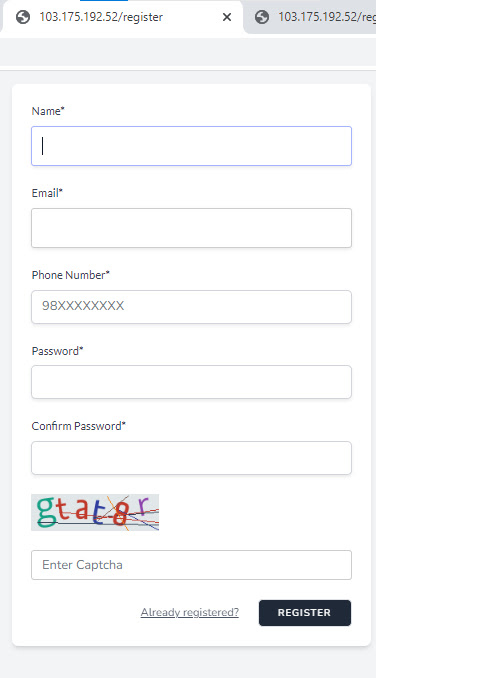
How to fill HA online registration form NHPC

HA अनलाइन रजिस्ट्रेसन फर्म भर्ने तरिका

  1. First visit NHPC official website at nhpc.gov.np सुरुमा कुनै ब्राउजर (eg chrome, firefox) खोलेर NHPC को वेवसाइट खोल्नुहोस। 
    new regiatration button

  2. Then click on new registration button on top right दायापट्टि टुप्पोमा रहेको “new registration” बटनमा क्लिक गर्नुहोस।
    New registration window

  3. Then a new tab will open नया ट्याब खुल्नेछ। 
  4. Fill the requred information and proceed. मागेको जानकारि र कागजात अपलोड गर्नुहोस।

रजिस्ट्रेसनको लागि चाहिने कागजात

विशिष्ट तहको लागि

1. नेपाली नागरिकताको प्रमाण पत्र
2. साधारण योग्यता (टेष्ट÷एस.एल.सी.) को लब्धाङ्क पत्र र चारित्रिक प्रमाण पत्र
3. प्रमाण पत्र तह वा सो सरह शैक्षिक योग्यताको लब्धाङ्क पत्र र चारित्रिक प्रमाण पत्र
4. व्यवसायिक÷ प्राविधिक शिक्षाको स्नातक वा सो सरह (स्वास्थ्य व्यवसाय) संग सम्बन्धीत उतिर्ण गरेको लब्धाङ्क पत्र र चारित्रिक प्रमाण पत्र
5. संबन्धित विश्वविद्यालय÷परिषद÷बोर्ड बाट प्रदान गरीएको ओरिजिनल÷प्रोभिजनल प्रमाण पत्र
6. सिप अध्यापन गराउन सक्षम योग्य जनशक्ति भएको स्तरीय स्वास्थ्य संस्था बाट जारी भएको OJT / INTERNSHIP गरेको प्रमाण पत्र
7. विदेशी शिक्षण संस्थाबाट उतिर्ण गरेको भए समकक्षताको प्रमाण पत्र र पासपोर्ट चाहिने मुलुक भए पासपोर्ट र भिसाको प्रतिलिपी
8. विदेशी शिक्षण संस्थाबाट उतिर्ण गरेको भए शैक्षिक योग्यताको CURRICULUM, शिक्षण संस्थाको PROSPECTUS समेतको सक्कल तथा प्रतिलिपी परिषद कार्यालयमा बुझाउनु पर्नेछ (UPLOAD गर्नु नपर्ने) ।
9. परीक्षा दस्तुर रु. ३०००। नेपाल एस.वि.आई बैंक खाता नं. २०४३५२४०१००००८, वा राष्ट्रिय बाणिज्य बै.क खाता नं. 115000213301 वा हिमालयन बैंक खाता नं. ००२००५७४६६००१६ मा दाखिला गरेको भौचर ।
10. परीक्षामा उतिर्ण भए पश्चात नाम दर्ता दरखास्त दस्तुर रु. २०५०।– नेपाल एस.वि.आई बैंक खाता नं. २०४३५२४०१००००८, वा राष्ट्रिय बाणिज्य बै.क खाता नं. 115000213301 वा हिमालयन बैंक खाता नं. ००२००५७४६६००१६ मा दाखिला गरेको भौचर (विदेशि शिक्षण संस्थाबाट अध्ययन गरेकाहरुको हकमा दस्तुर रु. ४०५०।– वैंक दाखिला गरेकोे भौचर)
11. दफा २ अनुसारका SCAN गरी UPLOAD गरेका प्रमाण पत्र, कागजातहरुको सत्यापन पक्ष (ORIGINALITY) को जिम्मेवारी आवेदक स्वयं हुनेछ र UPLOAD गरेका सबै कागजातहरुको सक्कलै कपी प्रमाणपत्र लिन आउँदा लिएर आउनु पर्नेछ ।
12. कृपया सक्कल कागजातको स्कयान मात्र Upload गर्नु होला ।  (Please upload the scan of Original Document Only)
Nepal SBI Bank= 20435240100008
   Himalayan Bank= 00200574660016
   Rastriya Banijya Bank= 115000213301

प्रथम तहको लागि

1. नेपाली नागरिकताको प्रमाण पत्र
2. साधारण योग्यता (एस.एल.सी.) को लब्धाङ्क पत्र र चारित्रिक प्रमाण पत्र
3. प्रमाण पत्र तह वा सो सरह शैक्षिक योग्यताको लब्धाङ्क पत्र र चारित्रिक प्रमाण पत्र
4. व्यसायिक÷ प्राविधिक शिक्षाको स्नातक वा सो सरह (स्वास्थ्य व्यवसाय) संग सम्बन्धीत उतिर्ण गरेको लब्धाङ्क पत्र र चारित्रिक प्रमाण पत्र
5. संबन्धित विश्वविद्यालय÷परिषद÷बोर्ड बाट प्रदान गरीएको ओरिजिनल÷प्रोभिजनल प्रमाण पत्र
6. सिप अध्यापन गराउन सक्षम योग्य जनशक्ति भएको स्तरीय स्वास्थ्य संस्था बाट जारी भएको OJT / INTERNSHIP गरेको प्रमाण पत्र (शिक्षण अस्पतालमा अध्ययन गरेकाहरुको हकमा र जनस्वास्थ्य विषय अध्ययन गरेकाहरुको हकमा आवश्यक नपर्ने
7. विदेशी शिक्षण संस्थाबाट उतिर्ण गरेको भए समकक्षताको प्रमाण पत्र र पासपोर्ट चाहिने मुलुक भए पासपोर्ट र भिसाको प्रतिलिपी
8. विदेशी शिक्षण संस्थाबाट उतिर्ण गरेको भए शैक्षिक योग्यताको CURRICULUM, शिक्षण संस्थाको PROSPECTUS समेतको सक्कल तथा प्रतिलिपी परिषद कार्यालयमा बुझाउनु पर्नेछ (UPLOAD गर्नु नपर्ने)।
9. परीक्षा दस्तुर रु. ३०००। नेपाल एस.वि.आई बैंक खाता नं. २०४३५२४०१००००८ वा हिमालयन बैंक खाता नं. ००२००५७४६६००१६ मा दाखिला गरेको भौचर।
10. परीक्षामा उतिर्ण भए पश्चात नाम दर्ता दरखास्त दस्तुर रु. १५५०।– नेपाल एस.वि.आई बैंक खाता नं. २०४३५२४०१००००८, हिमालयन बैंक खाता नं. ००२००५७४६६००१६ वा वा राष्ट्रिय बाणिज्य बै.क खाता नं. 115000213301 मा दाखिला गरेको भौचर (विदेशि शिक्षण संस्थाबाट अध्ययन गरेकाहरुको हकमा दस्तुर रु. ३०५०।– वैंक दाखिला गरेकोे भौचर)
11. दफा २ अनुसारका SCAN गरी UPLOAD गरेका प्रमाण पत्र, कागजातहरुको सत्यापन पक्ष (ORIGINALITY) को जिम्मेवारी आवेदक स्वयं हुनेछ र UPLOAD गरेका सबै कागजातहरुको सक्कलै कपी प्रमाणपत्र लिन आउँदा लिएर आउनु पर्नेछ ।
12. कृपया सक्कल कागजातको स्कयान मात्र Upload गर्नु होला ।  (Please upload the scan of Original Document Only)
13. Nepal SBI Bank= 20435240100008
          Himalayan Bank= 00200574660016
          Rastriya Banijya Bank= 115000213301

द्वितिय तहको लागि

1. नेपाली नागरिकताको प्रमाण पत्र
2. साधारण योग्यता (एस.एल.सी.) को लब्धाङ्क पत्र र चारित्रिक प्रमाण पत्र
3. व्यवसायिक शिक्षा (स्वास्थ्य व्यवसाय संग सम्बन्धीत) प्रमाण पत्र तह वा सो सरह उतिर्ण गरेको योग्यताको लब्धाङ्क पत्र र चारित्रिक प्रमाण पत्र
4. सम्बन्धित विश्वविद्यालय÷परिषद÷बोर्ड बाट प्रदान गरीएको ओरिजिनल÷प्रोभिजनल प्रमाण पत्र
5. व्यवसायिक÷प्राविधिक शिक्षाको प्रमाण पत्र तह वा सो सरहको शैक्षिक योग्यताको लागि भर्ना योग्यता प्रमाण पत्र वा सो सरह (विज्ञान) भएकाहरुको हकमा सो योग्यताको लब्धाङ्क पत्र र चारित्रिक प्रमाण पत्र
6. सिप अध्यापन गराउन सक्षम योग्य जनशक्ति भएको स्तरीय स्वास्थ्य संस्थाबाट जारी भएको OJT / INTERNSHIP गरेको प्रमाण पत्र
7. विदेशी शिक्षण संस्थाबाट उतिर्ण गरेको भए समकक्षताको प्रमाण पत्र
8. विदेशी शिक्षण संस्थाबाट उतिर्ण गरेको भए उक्त योग्यताको CURRICULUM र शिक्षण संस्थाको Prospectus समेतको सक्कल तथा प्रतिलिपी परिषद कार्यालयमा बुझाउनु पर्नेछ (Upload गर्नु नपर्ने)
9.परीक्षा दस्तुर रु. ३०००। नेपाल एस.वि.आई बैंक खाता नं. २०४३५२४०१००००८ वा हिमालयन बैंक खाता नं. ००२००५७४६६००१६ मा दाखिला गरेको भौचर।
10. परीक्षामा उतिर्ण भए पश्चात नाम दर्ता दरखास्त दस्तुर रु. १०५०।– नेपाल एस.वि.आई बैंक खाता नं. २०४३५२४०१००००८, हिमालयन बैंक खाता नं. ००२००५७४६६००१६ वा वा राष्ट्रिय बाणिज्य बै.क खाता नं. 115000213301 मा दाखिला गरेको भौचर (विदेशि शिक्षण संस्थाबाट अध्ययन गरेकाहरुको हकमा दस्तुर रु. २०५०।– वैंक दाखिला गरेकोे भौचर)
11. LOG BOOK बुझाउनु पर्ने व्यवसायीहरुको हकमा ती व्यवसायीहरुले आफ्नो शिक्षण संस्थामा सक्कलै LOG BOOK बुझाउने र अध्ययन गरेको शिक्षण संस्थाले आफ्ना विद्यार्थीहरुको सक्कलै LOG BOOK संकलन गरी १५ दिन भित्र परिषद समक्ष बुझाउनु पर्ने ।
12. दफा २ अनुसारका SCAN गरी UPLOAD गरेका प्रमाण पत्र, कागजातहरुको सत्यापन पक्ष (ORIGINALITY) को जिम्मेवारी आवेदक स्वयं हुनेछ र UPLOAD गरेका सबै कागजातहरुको सक्कलै कपी प्रमाणपत्र लिन आउँदा लिएर आउनु पर्नेछ ।
13. कृपया सक्कल कागजातको स्कयान मात्र Upload गर्नु होला ।  (Please upload the scan of Original Document Only)
14. Nepal SBI Bank= 20435240100008     
           Himalayan Bank= 00200574660016
           Rastriya Banijya Bank= 115000213301

तृतीय तहको लागि

1. नेपाली नागरिकताको प्रमाण पत्र
2. साधारण योग्यता (टेष्ट÷एस.एल.सी.) को लब्धाङ्क पत्र र चारित्रिक प्रमाण पत्र
3. व्यवसायिक योग्यता (T-SLC) (कम्तीमा एक शैक्षिक वर्षको तालीम) को लब्धाङ्क पत्र र चारित्रिक प्रमाण पत्र (पूनः परीक्षा दिइ उतिर्ण गरेको भए प्रत्येक सालको लब्धाङ्क पत्र)।
4. सम्बन्धीत परिषद बोर्डले प्रदान गरेको ओरिजिनल ÷प्रोभिजनल प्रमाण पत्र
5. सिप अध्यापन गराउन सक्षम योग्य जनशक्ति भएको स्तरीय स्वास्थ्य संस्था बाट जारी भएको OJT गरेको प्रमाण पत्र
6. विदेशी शिक्षण संस्थाबाट उतिर्ण गरेको भए समकक्षताको प्रमाण पत्र
7. नाम दर्ता दरखास्त दस्तुर रु. ७५०।– नेपाल एस.वि.आई बैंक खाता नं. २०४३५२४०१००००८, हिमालयन बै.क खाता नं. ००२००५७४६६००१६ वा राष्ट्रिय बाणिज्य बै.क खाता नं. 115000213301 मा दाखिला गरेकोे भौचर (विदेशि शिक्षण संस्थाबाट अध्ययन गरेकाहरुको हकमा दस्तुर रु. १५५०।– वैंक दाखिला गरेकोे भौचर)
8. LOG BOOK बुझाउनु पर्ने व्यवसायीहरुको हकमा ती व्यवसायीहरुले आफ्नो शिक्षण संस्थामा सक्कलै LOG BOOK बुझाउने र अध्ययन गरेको शिक्षण संस्थाले आफ्ना विद्यार्थीहरुको सक्कलै LOG BOOK संकलन गरी १५ दिन भित्र परिषद समक्ष बुझाउनु पर्ने ।
9. दफा २ अनुसारका SCAN गरी UPLOAD गरेका प्रमाण पत्र, कागजातहरुको सत्यापन पक्ष (ORIGINALITY) को जिम्मेवारी आवेदक स्वयं हुनेछ र UPLOAD गरेका सबै कागजातहरुको सक्कलै कपी प्रमाणपत्र लिन आउँदा लिएर आउनु पर्नेछ ।
10. कृपया सक्कल कागजातको स्कयान मात्र Upload गर्नु होला । (Please upload the scan of Original Document Only)
11. Nepal SBI Bank= 20435240100008
          Himalayan Bank= 00200574660016
          Rastriya Banijya Bank= 115000213301

बैंक अकाउन्ट NHPC   वेवसाइटबाट कन्फर्म गर्नुहोला! 

(NOTE: Taken from NHPC site for education purpose only) mail at mail@chaitanya.com.np for removal if copyright applies)

How does a stethoscope work? A wikihow to stethoscope

How does a stehoscope work: A complete guide on how a stethoscope works

A stethoscope works by transmitting sound waves from the patient’s body to the listener’s ears through a chestpiece, tubing, and earpieces. The vibrations caused by bodily functions are converted into audible sound waves, enabling healthcare professionals to assess and diagnose various conditions.
stethoscope 

The Science Behind Stethoscopes: How Do They Actually Work?

For centuries, the stethoscope has been an essential tool in the medical field, enabling physicians to listen to the sounds of the body and diagnose a variety of conditions. But have you ever wondered how exactly this simple device works? The science behind stethoscopes is fascinating and complex, involving principles of acoustics, physics, and human anatomy. From the diaphragm to the earpieces, every component of the stethoscope plays a crucial role in transmitting and amplifying the sounds of the body. Understanding how stethoscopes work can help us appreciate the remarkable precision and accuracy of modern medical technology. In this article, we will dive deeper into the science behind stethoscopes, exploring the mechanics of sound transmission, the anatomy of the human body, and the history of this iconic medical instrument. So, put on your lab coat and let’s explore the science of stethoscopes together!

The History of Stethoscopes

The stethoscope has a long and fascinating history, dating back to the early 19th century when it was first invented by a French physician named René Laennec. In those days, doctors would listen to the sounds of their patients’ bodies by placing their ear directly against the patient’s chest or back, which was not only uncomfortable but also unhygienic. Laennec was inspired to invent the stethoscope after observing two children playing with a long wooden stick, one end of which was placed against the ground while the other end was placed against the ear. He realized that this simple principle could be applied to listening to the sounds of the body, and thus the stethoscope was born.
The first stethoscopes were made of wood and were shaped like a tube with a bell-shaped end that was placed against the patient’s chest. The other end of the tube was placed against the doctor’s ear, allowing them to hear the sounds of the body more clearly. Over time, stethoscopes evolved to include a diaphragm that could be used to listen to higher-frequency sounds, such as heart murmurs, and a bell that could be used to listen to lower-frequency sounds, such as lung sounds.
Today, stethoscopes come in a variety of shapes and sizes and are made from a range of materials, including plastic, metal, and rubber. Despite these changes, the basic principle of the stethoscope remains the same: to amplify the sounds of the body and enable doctors to diagnose a variety of conditions.

How Do Stethoscopes Work?

At its most basic level, a stethoscope works by transmitting sound waves from the patient’s body to the doctor’s ear. Sound waves are created by the movement of air molecules, which vibrate as they travel through the body. These vibrations are then transmitted through the stethoscope to the doctor’s ear, where they are amplified and interpreted.
The key component of the stethoscope that enables this process to occur is the diaphragm. The diaphragm is a thin, flexible membrane that is placed against the patient’s skin. When sound waves from the body hit the diaphragm, they cause it to vibrate, creating a mechanical signal that is transmitted through the stethoscope tubing to the doctor’s ear.
The tubing of the stethoscope also plays a crucial role in sound transmission. The tubing is designed to be narrow and flexible, which helps to filter out unwanted background noise and amplify the sounds of the body. The length of the tubing can also affect the quality of sound transmission, with longer tubing generally producing a clearer sound.
The earpieces of the stethoscope are also important for sound transmission. The earpieces are designed to fit snugly in the doctor’s ears, creating a seal that helps to block out unwanted noise and ensure that the sounds of the body are heard clearly. Some stethoscopes also come with noise-cancelling features, which can help to further filter out background noise and improve sound quality.

The Anatomy of a Stethoscope

To understand how stethoscopes work, it’s important to first understand the anatomy of the device. A stethoscope is made up of several key components, each of which plays a crucial role in sound transmission.
The chestpiece is the part of the stethoscope that is placed against the patient’s skin. The chestpiece can be either a diaphragm or a bell, depending on the type of sound that needs to be heard. The diaphragm is a thin, flat piece of plastic or metal that vibrates when sound waves hit it. The bell, on the other hand, is a larger, concave piece of metal that is used to listen to lower-frequency sounds.
The tubing of the stethoscope connects the chestpiece to the earpieces. The tubing is typically made of rubber or plastic and is designed to be narrow and flexible, allowing sound waves to travel through it with minimal distortion.
The earpieces of the stethoscope are the part that is inserted into the doctor’s ears. The earpieces are usually made of soft rubber or plastic and are designed to fit comfortably in the ear canal. Some stethoscopes also come with adjustable earpieces, which can be moved up or down to accommodate different ear sizes.

Types of Stethoscopes and Their Uses

There are several different types of stethoscopes, each of which is designed for a specific purpose. The most common types of stethoscopes include:

  • – Acoustic stethoscopes: These are the most basic type of stethoscope and rely solely on sound transmission through the tubing and earpieces.
  • – Electronic stethoscopes: These stethoscopes use electronic amplification to enhance sound quality and filter out unwanted noise.
  • – Fetal stethoscopes: These stethoscopes are designed specifically for listening to the heartbeat of a fetus.
  • – Pediatric stethoscopes: These stethoscopes are designed for use on infants and young children and have smaller chestpieces and earpieces.
  • – Cardiology stethoscopes: These stethoscopes are designed for use in cardiology and have a larger diaphragm and a bell for listening to both high- and low-frequency sounds.
  • – Dual-head stethoscopes: These stethoscopes have both a diaphragm and a bell, allowing doctors to listen to a wider range of sounds.

Stethoscope Materials and Construction

  • Stethoscopes are made from a variety of materials, each of which has its own unique properties and benefits. The most common materials used in stethoscope construction include:
  • – Plastic: Plastic stethoscopes are the most basic type of stethoscope and are typically the least expensive. They are lightweight and easy to clean but may not provide the highest quality sound transmission.
  • – Metal: Metal stethoscopes are more durable and provide better sound transmission than plastic stethoscopes. They are typically more expensive but are also more reliable and long-lasting.
  • – Rubber: Rubber stethoscopes are soft and flexible, making them comfortable to use for extended periods of time. They are also easy to clean and maintain.

The construction of a stethoscope can also affect its sound quality and durability. High-quality stethoscopes are typically made with precision components and feature tight seals and well-constructed tubing to ensure clear sound transmission.

The Importance of Proper Stethoscope Maintenance

Proper maintenance of a stethoscope is essential for ensuring that it continues to function properly and provide accurate sound transmission. Some key tips for stethoscope maintenance include:

  • – Clean your stethoscope regularly with a mild soap and water solution.
  • – Store your stethoscope in a clean, dry place when not in use.
  • – Avoid exposing your stethoscope to extreme temperatures or sunlight.
  • – Replace worn or damaged parts of your stethoscope as needed.

By taking care of your stethoscope, you can ensure that it continues to provide reliable and accurate sound transmission for years to come.

Innovations in Stethoscope Technology

In recent years, there have been several exciting innovations in stethoscope technology that have helped to improve sound quality and diagnostic accuracy. One of the most notable of these is the electronic stethoscope, which uses advanced amplification and filtering technology to provide clearer sound transmission.
Other innovations include the development of stethoscopes with Bluetooth connectivity, which can allow doctors to transmit heart and lung sounds directly to a computer or mobile device for analysis. Some stethoscopes also come with built-in recording features, allowing doctors to record and review sounds at a later time.

Common Stethoscope Myths Debunked

There are several common myths and misconceptions surrounding stethoscopes. One of the most persistent of these is the idea that you can hear a patient’s thoughts or emotions through a stethoscope. In reality, stethoscopes are designed solely for the purpose of listening to the sounds of the body and cannot pick up on thoughts or emotions.
Another common myth is that stethoscopes are only used for listening to the heart. While the heart is certainly one of the most important organs that can be listened to with a stethoscope, doctors also use stethoscopes to listen to the lungs, stomach, and other parts of the body.

Conclusion

The stethoscope is a remarkable tool that has played a crucial role in the history of medicine. From its humble beginnings as a simple wooden tube to the high-tech electronic devices of today, the stethoscope has continued to evolve and improve over time. By understanding the science behind stethoscopes, we can appreciate the remarkable precision and accuracy of modern medical technology and the incredible insights it can provide into the workings of the human body. Whether you’re a physician, nurse, or simply someone with an interest in science and medicine, the stethoscope is a fascinating subject that is sure to captivate and inspire.

Council For Technical Education and Vocational Training CTEVT

Council For Technical Education and Vocational Training CTEVT

CTEVT Logo


Introduction to CTEVT

The Council for Technical Education and Vocational Training (CTEVT) constituted in 1989 (2045 BS) is a national autonomous apex body of Technical and Vocational Education and Training (TVET) sector committed for the production of technical and skillful human resources required to the nation. It mainly involves in policy formulation, quality control, preparation of competency based curriculum, developing skill standards of various occupations and testing the skills of the people, conduct various research studies and training needs assessment etc.

It has an assembly consisting of 24 members and a governing board known as Council comprising nine members. Minister of Education chairs both the Assembly and the Council. The Council has a full time Vice-Chairperson and a Member-Secretary.

Vision of CTEVT

Skilling Nepal for People’s Prosperity

Mission of CTEVT

CTEVT is a vibrant organization promoting TVET system to develop a competent workforce for national and international market needs

Goals of CTEVT

In order to achieve and attain the mission and vision, CTEVT has set the following six goals:
  1. Expand TVET programs for ensuring access and equity
  2. Ensure quality, relevant and efficient TVET system
  3. Enhance effectiveness and efficiency of CTEVT management
  4. Establish NVQF to ensure its compatibility with education framework
  5. Extend technical input to establish TVET Fund
  6. Establish effective coordination with and among TVET Stakeholders

Major Responsibilities of CTEVT

CTEVT mainly has the following responsibilities:

  1. Provide advice to the Government of Nepal regarding TEVT policy and programs.
  2. Determine scope and standards of TEVT programs.
  3. Arrange for and conduct TEVT Programs from basic level to higher education.
  4. Liaison and maintain coordination with national and international TEVT agencies for quality education and training.
  5. Extend technical input to establish TVET Fund
  6. Establish effective coordination with and among TVET Stakeholders

CTEVT contacts: 

CTEVT Location: Sanothimi Bhaktapur, Nepal

CTEVT Phone: +977 – 1 – 6631458, 6631459, 6630769

CTEVT E-mail: info@ctevt.org.np, research@ctevt.org.np

CTEVT E-mail: info@ctevtexam.org.np (For examination purpose)

CTEVT Website: http://www.ctevt.org.np

Check CTEVT result at

CTEVT P.O. Box: 3546, Kathmandu, Nepal

A trip to Dakshinkali mata darshan, Manakamana Darshan and Pathibhara mata darhan in Kathmandu and Lalitpur

A trip to Dakshinkali mata darshan, Manakamana temple Darshan and Pathibhara mata temple darhan in Kathmandu and Lalitpur

I had a chance to vsit top pilgrim destination near kathmandu. As many people know Dakshinkali temple is located in Kathmandu. 

also called Daxinkali temple or Dakshinkali mandir

But do you know that there is Manakamana and Pathibhara devi also in Kathmandu valley?

Yes now you know. 
Manakamana devi is located in Lele Municipality, Lalitpur. It is situated at diatance of 11 kilometres from Satdobato Lalitpur. 

See map link for direction : https://goo.gl/maps/MuJfjse7B3UWamUV8

Pathibhara temple is also located in Lalitpur district in Konjyoson Rural municipality.

Some pictures of Dakshinkali: Dakshinkali temple

Dakshinkali temple

Dakshinkali temple

Dakshinkali temple

Outside Dakshinkali temple

Dakshinkali temple

Dakshinkali mata temple

View from Dakshinkali mata temple

Dakshinkali temple Dr Chaitanya

Dakshinkali temple

Dakshinkali temple outside

Pictures of Manakamana Lalitpur:

Where is manakamana temple located: Manakamana temple is also located in lele rural municipality lalipur. (Not main manakamana temple)

Manakamana temple Lalitpur

Manakamana temple Lalitpur

Pictures of Pathibhara mata temple Konjyoson Lalitpur


Flower bloosom

Pathibhara temple

Pathibhara temple

Importance of Devi worshipping (देवीपूजनको महत्वहरू)

1. Connection with the divine feminine:

Worshipping devis (goddesses) provides a profound connection with the divine feminine energy that exists within and around us. It allows individuals to embrace and honor the nurturing, compassionate, and creative aspects of existence.

2. Empowerment: 

Devi worship empowers individuals, especially women, by recognizing and celebrating their inherent strength and divine qualities. It promotes a sense of self-worth, confidence, and courage.

3. Balance and harmony: 

The worship of devis emphasizes the importance of balance and harmony in life. By honoring both the masculine and feminine energies, it encourages individuals to seek equilibrium in all aspects of life, fostering a sense of peace and unity.

4. Protection and guidance: 

Worshipping devis is often associated with seeking their protection and guidance. Devi represents the universal motherly figure, offering support and safeguarding against negative forces, obstacles, and dangers.

5. Spiritual awakening: 

Devi worship serves as a pathway to spiritual awakening. By immersing oneself in the devotion and contemplation of the goddess, individuals can deepen their spiritual connection, expand their consciousness, and embark on a transformative journey.

6. Fostering compassion and love: 

The worship of devis nurtures qualities such as compassion, love, and empathy. Devi represents unconditional love and acts as a reminder to extend kindness and care to all beings, fostering a more compassionate society.

7. Celebration of diversity: 

Devis are associated with various qualities, attributes, and forms, symbolizing the diverse aspects of existence. By worshipping different goddesses, individuals honor and celebrate the diversity and uniqueness present in the world.

8. Liberation and liberation: 

Devi worship is often associated with the concept of liberation (moksha) and freedom from the cycle of birth and death. It encourages individuals to transcend worldly attachments and realize their true divine nature.

9. Healing and transformation: 

Devi worship is known to facilitate healing and transformation on physical, emotional, and spiritual levels. It is believed that connecting with the goddess can bring about inner healing, rejuvenation, and a renewed sense of purpose.

10. Cultivation of virtues: 

Devi worship promotes the cultivation of virtues such as humility, devotion, gratitude, and surrender. By embodying these qualities, individuals can purify their hearts and minds, leading to personal growth and spiritual evolution.

Copyright: All rights reserved to Dr Chaitanya. Unauthorized downloading and distributing is prohibited and my lead to legal consequences. 

(adsbygoogle = window.adsbygoogle || []).push({});

Ventilator association pneumonia: the unfortunate risk for people under mechanic ventilation diagnosis and management

 Ventilator-Associated Pneumonia (VAP)

Definition:

  • A type of hospital-acquired pneumonia.
  • Occurs 48 hours or more after endotracheal intubation and mechanical ventilation.

Etiology / Common Pathogens:

  • Gram-negative bacteria:
    • Pseudomonas aeruginosa
    • Klebsiella pneumoniae
    • Escherichia coli
    • Acinetobacter baumannii
  • Gram-positive bacteria:
    • Staphylococcus aureus (including MRSA)

Risk Factors:

  • Prolonged mechanical ventilation
  • Supine positioning
  • Aspiration of gastric contents
  • Poor oral hygiene
  • Use of sedatives and paralytics
  • Re-intubation

Pathophysiology:

  • Microaspiration of oropharyngeal secretions
  • Colonization of the airway and endotracheal tube
  • Formation of biofilms

Clinical Features:

  • Fever
  • Purulent tracheal secretions
  • New or progressive infiltrates on chest X-ray
  • Leukocytosis or leukopenia
  • Worsening oxygenation

Diagnosis:

  • Clinical criteria (e.g., CPIS – Clinical Pulmonary Infection Score)
  • Chest imaging
  • Microbiological cultures (sputum, bronchoalveolar lavage, tracheal aspirate)

Prevention:

  • Elevate head of bed (30–45 degrees)
  • Daily sedation vacations and assessment for extubation
  • Oral care with chlorhexidine
  • Subglottic secretion drainage
  • Hand hygiene and sterile technique

Treatment:

Duration typically 7 days (can vary)

Empirical broad-spectrum antibiotics initially

De-escalation based on culture results

FBIMNCI MO Handbook for medical officers

FBIMNCI handbook for medical officers  handbook facility based integrated management of childhood and neonatal illness

Table of contents(toc)

What is FBIMNCI

Facility based integrated management of neonatal and childhood illnesses. 

Introduction to FBIMNCI

The Facility Based Integrated Management of Neonatal and Childhood Illness (FB-IMNCI) package includes appropriate management of major causes of childhood and neonatal mortality. 
The package has been designed specially to address childhood cases referred from peripheral level health institutions to higher institutions. 
As such, the package is expected to bridge the current gap in appropriate and timely management of childhood 
cases. 
Handbook cover page

The Facility Based IMNCI package has been designed to address the major causes of childhood illnesses especially:

  1. infection
  2. birth asphyxia
  3. prematurity
  4. low birth weight
  5. pneumonia
  6. diarrhoea
  7. malaria
  8. meningitis
  9. severe malnutrition among children.
Download it here: 
Or alternatively copy and paste following link in your browsser address bar 
Checking pedal oedema in childre with malnutrition

Download IMNCI Protocol (imnci chart booklet)

Click 👆👆👆 the heading.

The Anatomy of a Stethoscope: Understanding the Different Parts

The Anatomy of a Stethoscope: Understanding the Different Parts

As a medical professional, the stethoscope is an essential tool in your arsenal. It’s the device that helps you listen to the inner workings of the human body, providing vital information that can help diagnose a range of medical conditions. But have you ever really stopped to consider the intricate design of this simple yet powerful instrument? Understanding the anatomy of a stethoscope is essential for getting the most out of your device and improving the accuracy of your diagnoses. From the earpieces to the chest piece, each component plays a critical role in helping you hear the sounds of the body. In this article, we’ll take a closer look at the different parts of the stethoscope and explore their functions. Whether you’re a seasoned medical professional or a student just starting out, the anatomy of a stethoscope is a fascinating subject that’s worth exploring.

Parts of a Stethoscope

Earpieces

The earpieces are the components of the stethoscope that fit into your ears. They are typically made of soft, pliable materials that provide a comfortable fit. There are two types of earpieces: standard and adjustable. Standard earpieces are fixed in size and cannot be adjusted, while adjustable earpieces can be adjusted to fit different ear sizes.

The earpieces are designed to create a seal in your ear canal, which helps to block out external noise and ensure that you hear the sounds of the body clearly. It’s important to ensure that the earpieces fit snugly in your ears, as a loose fit can cause the stethoscope to move around, making it difficult to hear the sounds you need to hear.

Tubing

The tubing is the part of the stethoscope that connects the earpieces to the chest piece. It is typically made of rubber or PVC and comes in various lengths. The length of the tubing can affect the sound quality of the stethoscope, with longer tubing generally providing better sound quality.

The material of the tubing can also affect the sound quality. Rubber tubing is more flexible than PVC tubing, which can make it easier to use the stethoscope in tight spaces. However, PVC tubing is more durable and can withstand exposure to oils and other chemicals better than rubber tubing.

Chestpiece

The chestpiece is the part of the stethoscope that is placed on the patient’s body to listen to the sounds of the body. It consists of two parts: the diaphragm and the bell. The diaphragm is the larger, flat part of the chestpiece that is used to listen to high-pitched sounds, such as those produced by the lungs and heart. The bell is the smaller, concave part of the chestpiece that is used to listen to low-pitched sounds, such as those produced by the gastrointestinal tract.

There are two types of chestpieces: single-sided and double-sided. Single-sided chestpieces have only one side that can be used to listen to sounds, while double-sided chestpieces have both a diaphragm and a bell that can be switched out as needed.

Diaphragm/Bell

The diaphragm and bell are the two parts of the chestpiece that are used to listen to sounds. The diaphragm is the flat, circular part of the chestpiece that is used to listen to high-pitched sounds, such as those produced by the lungs and heart. The bell is the concave, cup-shaped part of the chestpiece that is used to listen to low-pitched sounds, such as those produced by the gastrointestinal tract.

To use the diaphragm, you should place it firmly on the patient’s skin, allowing it to make contact with the surface. This will create a seal that will allow you to hear the sounds of the body clearly. To use the bell, you should hold it lightly against the patient’s skin, allowing the cup to capture the sounds of the body.

How does steth work

Additional Features

Tunable Diaphragm

Some stethoscopes come equipped with a tunable diaphragm, which allows you to switch between high and low frequencies without having to flip the chestpiece over. To use the tunable diaphragm, you simply apply more or less pressure to the chestpiece, which changes the frequency response of the diaphragm.

Noise-Reducing Technology

Some stethoscopes come equipped with noise-reducing technology, which helps to block out external noise and improve the clarity of the sounds you hear. This technology can be particularly useful in noisy environments, such as emergency rooms or busy clinics.

A steth

Pediatric Attachments

Some stethoscopes come with pediatric attachments, which are designed to make it easier to use the stethoscope on infants and children. These attachments are typically smaller than standard chestpieces and can be used to listen to the sounds of the body on smaller patients.

Choosing the Right Stethoscope for Your Needs

When it comes to choosing a stethoscope, there are a few factors to consider. First, you’ll want to consider the type of work you’ll be doing. If you work in a busy emergency department, you may want a stethoscope with noise-reducing technology to help you hear the sounds of the body more clearly. If you’ll be working with smaller patients, a stethoscope with a pediatric attachment may be a good choice.

You’ll also want to consider your budget. Stethoscopes can range in price from less than $20 to several hundred dollars, depending on the features and quality. While it may be tempting to opt for a cheaper option, keep in mind that a high-quality stethoscope can last you for many years and provide you with more accurate readings.

Caring for Your Stethoscope

Proper care and maintenance of your stethoscope can help to ensure that it lasts for many years and continues to provide you with accurate readings. Here are a few tips for caring for your stethoscope:

  • Clean the earpieces and chestpiece regularly with a soft cloth and mild soap and water.
  • Avoid using alcohol or other harsh chemicals to clean your stethoscope, as these can damage the tubing and other components.
  • Store your stethoscope in a cool, dry place away from direct sunlight and extreme temperatures.
  • Avoid kinking or bending the tubing, as this can cause damage to the stethoscope.

Conclusion

Understanding the anatomy of a stethoscope is essential for using this valuable tool effectively and getting the most out of it. From the earpieces to the chest piece, each component plays a critical role in helping you hear the sounds of the body. By choosing the right stethoscope for your needs, caring for it properly, and using it effectively, you can provide your patients with more accurate diagnoses and better care.

NHPC has published Notification Regarding Seventh Name Registration Certificate Examination

NHPC 7th licensing examination detailed notice and how to fill up the forms for NHPC licensing examination for PCL, bachelors and masters level

NHPC license examination notice:

seventh 7th licensing examination full notice from NHPC

How to fill up NHPC licensing examination form:

  1. You must have an active gmail account to fill up form if you appeared in 1st to 5th examination. If you have already used that gmail account for taking exam previously create new one here .
  2. Visit this link www.nhpc.gov.np
  3. Click on new registration icon on top right 
    New regiistration NHPC

  4. Alternatively you can click here
  5. Fill up the required information and enter the captcha:

Enter the code sent to your email : 

Enter code here from email from NHPC
Code in email from NHPC
7. Now login using the email and password you entered earlier
8 Now provide all the informamtion it asks for and apply for your level of choice.

What is the application fee for examination:


  • Single fee on time (till 2080/01/28) : Rs 3000

  • Double fee (from 2080/01/29 to 2080/03/03 : Rs 6000

NHPC form fee banks:

1. Nepal SBI bank limited

2. Himalayan bank limited

The voucher of the above mentioned fee should be submitted to the profile of the examinee. 

Read more informamtion at NHPC official website and NHPC facebook page. 

Anatomy and physiology of the ventilator machine: Exploring the Life-saving Mechanics

Unveiling the Intricacies: The Anatomy and Physiology of Ventilator Machines

Introduction:

Understanding the anatomy and physiology of ventilator machines is crucial in comprehending the life-saving mechanics behind these essential medical devices. This search engine optimized article delves into the intricate details of ventilator machines, exploring their anatomy and physiology to provide a comprehensive understanding of their crucial role in patient care.

A ventilator machine

1. The Basics: What is a Ventilator Machine?

A ventilator machine, also known as a mechanical ventilator, is a sophisticated medical device used to provide respiratory support to patients in critical care situations. It delivers oxygen to the lungs and removes carbon dioxide, assisting patients in breathing when they are unable to do so independently.

2. Key Components of a Ventilator Machine:

Ventilator machines consist of several essential components that work together to facilitate effective respiratory support. These components include:

– Control Panel: 

The control panel allows healthcare professionals to adjust various settings, such as the volume and rate of airflow, to meet the patient’s specific needs.


– Breath Delivery System: 

This system comprises an inspiratory limb, expiratory limb, and patient interface. It controls the flow of gases into and out of the patient’s lungs, ensuring proper ventilation.

– Sensors and Monitors:

Ventilators are equipped with sensors and monitors to measure various parameters, such as air pressure, oxygen levels, and patient-ventilator synchrony. These measurements aid in monitoring and adjusting the ventilation process.

3. Physiology of Ventilation:

Ventilator machines function based on the principles of respiratory physiology. The primary modes of ventilation include:

– Pressure-controlled ventilation:

This mode delivers breaths at a set pressure level, ensuring consistent airflow and oxygenation.

– Volume-controlled ventilation:

In this mode, the ventilator delivers breaths at a predetermined volume, maintaining a specific tidal volume for optimal gas exchange.

– Assisted ventilation:

Some ventilators are designed to synchronize with the patient’s spontaneous breaths, providing support when needed and allowing for a more natural breathing pattern.

4. Advanced Features and Modes:

Modern ventilator machines are equipped with advanced features and modes to cater to diverse patient requirements. These may include:

– Positive End-Expiratory Pressure (PEEP):

PEEP helps keep the airways open during exhalation, preventing alveolar collapse and improving oxygenation.

– Pressure Support:

This mode assists the patient’s spontaneous breaths by providing additional pressure support during inhalation.

– Non-invasive Ventilation:

Some ventilators offer non-invasive ventilation options, such as mask-based interfaces, for patients who do not require intubation.

Conclusion:

Understanding the anatomy and physiology of ventilator machines is vital for CEOs in the healthcare industry. This search engine optimized article has provided a comprehensive overview of the essential components and functions of ventilator machines, shedding light on their critical role in providing life-saving respiratory support. By grasping the mechanics behind these devices, CEOs can make informed decisions and contribute to the advancement of patient care.

NHPC license examination notice 2080 updated

NHPC license examination 2080 notice detailed with how to print the admit card

Nepal Health Professional Council (NHPC) is an autonomous body established under the Nepal Health Professional Council Act 2053. The aim of this council is to register all the “Health professionals” other than Medical doctors , Nurses , Pharmacists, and Ayurveda according to their qualification; and bring them into a legal system as to make their services effective with quality and timely in a scientific manner.

NHPC license examination notice 1

NHPC full notice download here. 

NHPC license examination notice 2

NHPC license examination notice 3

How to download admit card:

Please follow the following steps:
Exit mobile version