What to look for to assess work of breathing in infants and neonates? – Pediatrics Notes

Work of Breathing (WOB) — clinical parameters

Work of breathing is assessed mainly by respiratory effort, chest movement, and signs of respiratory distress.

According to Nelson Textbook of Pediatrics, work of breathing (WOB) refers to the effort required to breathe—i.e., the amount of respiratory muscle activity needed to move air into and out of the lungs.

Clinically, Nelson assesses increased WOB by looking for signs of respiratory distress, particularly:

  • Tachypnea
  • Nasal flaring
  • Retractions/recessions — intercostal, subcostal, suprasternal
  • Grunting
  • Accessory muscle use
  • Head bobbing in infants
  • Paradoxical/see-saw breathing
  • Changes in respiratory pattern or effort

Exam definition

Work of breathing = the effort required to generate ventilation and move air through the respiratory system.

Increased WOB means the child must generate greater-than-normal respiratory effort to maintain adequate ventilation.

High-yield distinction:
WOB ≠ respiratory rate alone. A child can have severe increased WOB even with a relatively normal RR, particularly when respiratory muscle fatigue is developing.

Parameters to assess work of breathing

ParameterWhat to look forSignificance
Respiratory rateTachypnea for ageEarly sign of increased WOB
Nasal flaringAlae nasi widening during inspiration↓ upper-airway resistance
Retractions / recessionSuprasternal, intercostal, subcostal, substernalIncreased negative intrathoracic pressure
GruntingExpiratory gruntMaintains end-expiratory pressure; important neonatal distress sign
Head bobbingEspecially infantsSevere respiratory effort
Tracheal tugDownward movement of trachea on inspirationIncreased inspiratory effort
See-saw/paradoxical breathingChest moves inward while abdomen moves outward, or vice versaSevere distress/fatigue
Accessory muscle useSCM, scalene, abdominal musclesIncreased WOB
Abnormal chest movementAsymmetry, poor expansionAirway/lung pathology
Breath soundsWheeze, stridor, crackles, diminished/absent air entryIdentifies cause/severity
Expiratory phaseProlonged expirationParticularly obstructive disease
GruntingEspecially in neonatesSuggests significant respiratory distress
PostureTripod/sniffing positionCompensatory increase in airway patency
Ability to speak/feedFull sentences vs words; feeding interruption in infantsFunctional measure of WOB
Mental statusIrritability → lethargy → exhaustionLate/severe respiratory compromise
OxygenationSpO₂, cyanosisConsequence rather than direct measure of WOB

Quick severity framework

Mild ↑ WOB

  • Tachypnea
  • Mild nasal flaring
  • Mild intercostal/subcostal recession
  • Child alert and feeding reasonably well

Moderate ↑ WOB

  • Marked retractions
  • Nasal flaring
  • Accessory muscle use
  • Grunting
  • Difficulty feeding/speaking
  • Persistent tachypnea

Severe ↑ WOB / impending failure

  • Severe recession
  • Head bobbing
  • Paradoxical/see-saw breathing
  • Poor air entry or “quiet chest”
  • Exhaustion, altered consciousness
  • Apnea/bradypnea
  • Cyanosis despite oxygen

Important: A falling respiratory rate is not necessarily improvement. In a tiring child, tachypnea may progress to a normal or low RR as respiratory muscles fatigue—often a dangerous sign.

For bedside pediatric assessment, the highest-yield things to document are RR + retractions + nasal flaring + grunting + accessory muscle use + air entry + SpO₂ + mental status/feed/speech ability.

Are you stuck with Diagnosis? Here is prolonged fever + hepatosplenomegaly + persistent leukocytosis diagnoses summarized!

For a child with prolonged fever + hepatosplenomegaly + persistent leukocytosis, I would use the following as a bedside differential/workup table, with particular attention to infections relevant to Nepal and hematologic disease.

1. Infectious causes

DifferentialHistory: ask forExamination cluesKey investigations
TuberculosisContact, chronic cough, weight loss, night sweats, poor appetite; household exposureCervical/generalized nodes, wasting, respiratory signs, hepatosplenomegalyCXR; TST/IGRA; Xpert MTB/RIF/Ultra from appropriate specimen; culture; node FNAC/biopsy if present
Enteric feverProlonged fever, contaminated food/water, travel/outbreak exposure, abdominal symptomsToxicity may be mild; abdominal tenderness, hepatosplenomegaly; relative bradycardia is unreliable in childrenBlood culture before antibiotics; CBC, LFT; stool culture in selected cases
MalariaResidence/travel to endemic Terai areas, mosquito exposure, intermittent/chills patternPallor, jaundice, splenomegaly, hepatomegaly; thrombocytopeniaThick + thin smear + malaria RDT; repeat smear if high suspicion
Visceral leishmaniasisProlonged fever, travel/residence in endemic region, weight lossMassive splenomegaly, hepatomegaly, pallor, wastingrK39/other serology; CBC; confirmatory parasitologic testing when indicated
BrucellosisCattle/goat/sheep exposure, raw/unpasteurized milk, occupational exposure, undulating feverHSM, lymphadenopathy, arthritis; sometimes nonspecificBlood cultures; Brucella serology/PCR where available
EBVSick contacts, sore throat, fatiguePosterior cervical nodes, pharyngitis/tonsillar enlargement, HSMCBC differential, atypical lymphocytes; EBV VCA IgM/IgG ± EBNA; LFT
CMVSick contacts; congenital/perinatal history in younger childHSM, lymphadenopathy; prolonged feverCMV IgM/IgG ± PCR in selected cases; LFT
Scrub typhusRural/forest exposure, mite exposure, recent outdoor activityEschar, rash, lymphadenopathy; hepatosplenomegaly; thrombocytopeniaScrub typhus IgM/ELISA ± PCR
LeptospirosisFloodwater/freshwater, rodents, animal urineConjunctival suffusion, jaundice, renal involvement, myalgiaCBC, renal/LFT; Leptospira PCR/IgM
DengueMosquito exposure, acute febrile illnessRash, bleeding, abdominal tenderness, hepatomegaly; usually thrombocytopeniaCBC trend; NS1/PCR early, IgM later
Disseminated fungal infectionImmunodeficiency, prolonged antibiotics, steroid/immunosuppressive therapyHSM, lymphadenopathy, skin/mucosal lesionsBlood cultures, fungal biomarkers/cultures; imaging/tissue diagnosis
HIVMaternal risk, transfusion, recurrent/opportunistic infections, growth failureGeneralized nodes, HSM, oral candidiasis, wastingHIV Ag/Ab testing; confirmatory algorithm; viral load/CD4 where appropriate
ToxoplasmosisCat/feces exposure, undercooked meat; immunocompromised stateCervical nodes, HSM occasionallyToxoplasma IgM/IgG ± PCR in selected cases
ToxocariasisDogs/cats, soil exposure, picaHepatomegaly, fever, eosinophilia, sometimes ocular diseaseCBC with AEC, Toxocara serology
Hydatid diseaseDog/sheep exposureUsually mass/organ-specific findings rather than feverUSG/CT; Echinococcus serology

2. Hematologic / malignant causes

DifferentialHistoryExaminationInvestigations
ALLFever, fatigue, weight loss, bone/joint pain, bruising, recurrent infectionsPallor, petechiae, lymphadenopathy, HSM, bone tendernessCBC + differential + PBS, retic, LDH/uric acid; bone marrow + flow cytometry if suspected
AMLFever, fatigue, bleeding, infections, bone painPallor, petechiae, HSM, gingival hypertrophy, chloromasCBC/PBS; marrow morphology + flow cytometry/cytogenetics/molecular studies
LymphomaFever, weight loss, night sweats, pruritus; node enlargementFirm/painless nodes, mediastinal signs, HSMCBC, LDH/uric acid; CXR/USG/CT as indicated; excisional lymph-node biopsy
CMLFatigue, weight loss, early satiety, abdominal discomfortMarked splenomegaly, hepatomegaly; pallorCBC differential + PBS; BCR-ABL1 testing, marrow/cytogenetics
JMMLUsually young child; fever, recurrent infections, pallor, rashSplenomegaly, lymphadenopathy, pallor, skin lesionsCBC showing persistent monocytosis, PBS; marrow; molecular testing (e.g. RAS-pathway abnormalities)
Leukemoid reactionInfection/inflammation symptomsUnderlying infectious focus; may have HSMSerial CBC, PBS; neutrophilia/left shift; investigate underlying cause
Hemolytic anemiaJaundice, dark urine, episodic symptoms, drugs/infection/family historyPallor, jaundice, splenomegalyRetic, indirect bilirubin, LDH, haptoglobin, DAT, ± Hb electrophoresis/G6PD

3. Inflammatory / immune causes

DifferentialHistoryExaminationInvestigations
Systemic JIAFever ≥2 weeks, arthritis, rash; fever often quotidianArthritis, evanescent salmon rash, lymphadenopathy, HSMCBC, ESR/CRP, ferritin, LFT; diagnosis is clinical after exclusion
MAS secondary to sJIAPersistent fever, acute deteriorationHSM, rash, bleeding/neurologic changesFerritin, TG, fibrinogen, CBC, AST/ALT, D-dimer
HLHPersistent fever, family history/recurrent episodes, infection triggerSplenomegaly, HSM, lymphadenopathy, neurologic/skin findingsFerritin, TG, fibrinogen, CBC, LFT; sCD25/NK function/genetics in appropriate cases
Kawasaki diseaseFever ≥5 days, irritabilityConjunctival injection, oral changes, rash, extremity changes, cervical nodeCRP/ESR, CBC, LFT, urinalysis; echocardiography
SLEFever, fatigue, rash, photosensitivity, arthralgia, oral ulcersRash, arthritis, alopecia, lymphadenopathy/HSM, hypertensionCBC, ESR/CRP, ANA, dsDNA, C3/C4, urinalysis/proteinuria

4. Important noninfectious causes of HSM

CauseHistory/exam cluesInvestigations
Gaucher diseaseChronic massive HSM, pallor, bone pain/crises, growth issuesβ-glucocerebrosidase enzyme assay ± genetics
Niemann-Pick diseaseHSM + developmental regression/neurologic signsEnzyme/genetic testing
Glycogen storage diseaseHepatomegaly, growth failure, hypoglycemia; fever usually suggests a separate processGlucose, lactate, LFT, metabolic/genetic testing
Chronic liver disease/portal hypertensionJaundice, ascites, bleeding, pruritus, liver disease historyLFT, INR, albumin, viral/autoimmune/metabolic workup, USG Doppler

Particularly important with persistent leukocytosis

Don’t just record the WBC. Break it down:

CBC patternDifferential to prioritize
Neutrophilia + left shiftBacterial infection, occult abscess, leukemoid reaction, inflammatory disease
Very high neutrophils + basophilia + myelocytes/metamyelocytesCML
Persistent monocytosisJMML, TB, brucellosis, chronic infections
Absolute lymphocytosisEBV/CMV, pertussis, viral infection, hematologic disease
EosinophiliaHelminths, Toxocara, drug/allergic disease
BlastsAcute leukemia until proven otherwise
Leukocytosis + anemia + thrombocytopeniaStrongly consider marrow disease/leukemia
Leukocytosis with normal morphology and active childInfection/inflammation or leukemoid reaction more likely, but persistent marked leukocytosis requires further evaluation

A practical initial panel for this child

Given your specific presentation, I would start with:

CBC + differential + platelets + repeat PBS
→ CRP/ESR ± PCT
→ reticulocyte count + MCV/RDW + ferritin
→ LFT/bilirubin/LDH
→ malaria RDT + thick/thin smear
→ blood culture
→ urine R/M + culture
→ USG abdomen

Then based on findings/exposure:

TB testing | EBV/CMV | Brucella | scrub typhus | leptospira | VL/rK39 | stool/parasite testing | Toxocara | HIV | HLH/MAS panel | BCR-ABL1/marrow studies.

For persistent leukocytosis + HSM specifically, I would make sure the report gives the absolute neutrophil, lymphocyte, monocyte, eosinophil and basophil counts—not just total WBC. That single detail can substantially narrow this differential.

Rett Syndrome Notes for MD and DM level

Rett Syndrome

Definition

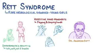
Rett syndrome is a neurodevelopmental disorder that primarily affects girls, characterized by normal early development followed by regression of acquired skills, especially speech and purposeful hand movements, with onset typically between 6–18 months of age.


Etiology

  • Genetic cause: Mutation in MECP2 gene (methyl-CpG-binding protein 2) on the X chromosome (Xq28)

  • Inheritance: Usually sporadic (de novo); rarely familial

  • Pathophysiology: Dysfunction of MECP2 protein → abnormal brain maturation and synaptic development


Epidemiology

  • Affects 1 in 10,000–15,000 female births

  • Lethal in males (most do not survive infancy unless mosaic or XXY)


Clinical Features

Phases of Disease

  1. Early Onset (6–18 months)

    • Normal development initially

    • Gradual loss of interest in surroundings

    • Loss of purposeful hand skills

    • Deceleration of head growth (acquired microcephaly)

  2. Rapid Destructive Phase (1–4 years)

    • Loss of speech and purposeful hand use

    • Stereotyped hand movements: hand-wringing, washing, clapping, or mouthing

    • Gait ataxia, truncal apraxia

    • Autistic-like behavior

  3. Plateau Phase (2–10 years)

    • Some improvement in social interaction and eye contact

    • Persistent motor problems and seizures

  4. Late Motor Deterioration (>10 years)

    • Progressive scoliosis, muscle wasting, rigidity, dystonia

    • Loss of ambulation in many cases


Other Features

  • Breathing abnormalities: hyperventilation, apnea during wakefulness

  • Seizures: common (up to 90%)

  • Bruxism, cold/purple extremities (autonomic dysfunction)

  • Sleep disturbances

  • Growth retardation


Investigations

  • Genetic testing: MECP2 mutation analysis (diagnostic)

  • EEG: slowing with epileptiform activity

  • MRI brain: may show nonspecific atrophy

  • Metabolic tests: normal (to rule out other causes)


Diagnosis

  • Clinical + confirmed MECP2 mutation

  • Diagnostic criteria include:

    • Regression after normal early development

    • Loss of purposeful hand skills and spoken language

    • Gait abnormalities

    • Stereotypic hand movements


Differential Diagnosis

  • Autism spectrum disorder

  • Angelman syndrome

  • Cerebral palsy (especially ataxic type)

  • Childhood disintegrative disorder


Management

  • No cure – supportive and multidisciplinary care

    • Physiotherapy & occupational therapy: maintain mobility

    • Speech therapy: communication support (eye-tracking devices)

    • Antiepileptics: for seizures

    • Nutritional support: adequate calories, manage feeding difficulties

    • Behavioral therapy: improve interaction

    • Orthopedic care: for scoliosis, contractures


Prognosis

  • Progressive but non-degenerative

  • Life expectancy: many survive into adulthood (40–50 years)

  • Main causes of death: sudden unexplained death, pneumonia, cardiac arrhythmias


Mnemonic (Key features)“RETT”

  • R = Regression (speech, hand skills)

  • E = Episodic breathing abnormalities

  • T = Typical hand movements (wringing, washing)

  • T = Tiny head (acquired microcephaly)

Hepatitis Full lecture PPT: Hepatitis introduction, causes, viral hepatitis, treatment of hepatitis and liver function test interpretation

Introduction

Viral hepatitis in children is a primary inflammation of the liver caused by at least five specific hepatotropic viruses (A, B, C, D, and E). While often milder in pediatric patients than in adults, it can progress to acute liver failure or chronic liver disease, particularly with types B and C.

I. Classification and Etiological Profile of Primary Viral Hepatitis

The five main viruses are distinguished by their genomic structure and mode of transmission:

  • Hepatitis A Virus (HAV): An RNA virus identical to enteroviruses; transmitted primarily via the feco-oral route through contaminated food or water. It never causes chronic infection.
  • Hepatitis B Virus (HBV): A DNA virus (Dane particle) transmitted parenterally, sexually, or vertically (perinatal). It is the most common cause of acute and chronic hepatitis worldwide.
  • Hepatitis C Virus (HCV): An RNA virus primarily transmitted via percutaneous blood exposure (IV drug use, transfusions before 1991) and vertically (5-6% risk).
  • Hepatitis D Virus (HDV): A defective RNA virus that requires co-infection or superinfection with HBV to replicate, as it uses the HBV lipoprotein envelope.
  • Hepatitis E Virus (HEV): An RNA virus transmitted enterally (water-borne), similar to HAV; it is a major cause of high mortality in pregnant women.

II. High-Yield Incubation Periods and Transmission Routes

VirusIncubation PeriodMain Route of Transmission
HAV28–42 daysFeco-oral (“The vowels go through the bowels”)
HBV60–150 daysParenteral, Sexual, Vertical (Perinatal)
HCV30–60 daysParenteral (Blood exposure), Vertical
HDV60–80 daysParenteral (Requires HBV co-infection)
HEV25–60 daysFeco-oral (Often water-borne epidemics)

Exam Point: “The Window Period” In HBV infection, the “window period” occurs when HBsAg has disappeared but Anti-HBs has not yet appeared. During this time, Anti-HBc IgM is the only marker of acute infection.

III. Detailed Clinical Presentation and Extrahepatic Features

Symptoms in children are often non-specific and vary by age:

  • Prodromal Phase: Fever, malaise, anorexia, nausea, vomiting, and right upper quadrant abdominal pain.
  • Icteric Phase: Subside of fever and anorexia, followed by jaundice (1–3 days after prodrome), dark urine, and pale stools.
  • Physical Findings: Tender hepatomegaly is common; splenomegaly occurs in 30% of cases.
  • HBV Extrahepatic Manifestations: High-yield exam points include serum sickness-like syndrome, polyarteritis nodosa (PAN), and membranous glomerulonephritis.
  • Chronic Hepatitis: Defined as continuing inflammation for \(\ge\)3–6 months; markers include persistently raised transaminases.

IV. Interpretation of Serological Markers (Crucial for MD Exams)

Diagnostic confirmation relies heavily on serology:

  • HAV: Diagnosis by Anti-HAV IgM (acute); Anti-HAV IgG indicates past infection and lifelong immunity.
  • HBV Complex Serology:
    • HBsAg: Indicates current infection (acute or chronic).
    • Anti-HBs: Indicates immunity (either via vaccine or recovered infection).
    • Anti-HBc IgM: Indicates acute/recent infection (useful in the window period).
    • Anti-HBc IgG: Indicates past or chronic infection.
    • HBeAg: Correlates with high viral replication and high infectivity.
    • Anti-HBe: Indicates lower infectivity.
  • HCV: Screen with Anti-HCV; confirm with HCV RNA PCR (detectable 1–2 weeks post-exposure).

V. Management and Pediatric Treatment Protocols

Treatment is primarily supportive for acute cases, but chronic cases require targeted therapy:

  • Supportive Care: Bed rest during jaundice, high carbohydrate diet, and avoidance of fats and hepatotoxic drugs (e.g., paracetamol, chlorpromazine).
  • Chronic HBV: Preferred treatments include Entecavir (\(\ge\)2 years) or Tenofovir (\(\ge\)12 years). Interferon-alfa is also an option for children 1–18 years.
  • Chronic HCV: Revolutionized by Direct-Acting Antivirals (DAAs). Recommended for all children \(\ge\)3 years. Regimens like Sofosbuvir/Ledipasvir (Harvoni) or Glecaprevir/Pibrentasvir (Mavyret) are highly effective.
  • Fulminant Hepatitis: Requires ICU admission, management of cerebral edema (Mannitol), and evaluation for liver transplantation.

VI. Prevention and Post-Exposure Prophylaxis (PEP)

  • Hepatitis A Vaccine: Two-dose series starting at 12 months.
  • Hepatitis B Vaccine: Routine three-dose series at birth, 1–2 months, and 6 months.
  • Perinatal HBV PEP: If a mother is HBsAg-positive, the neonate must receive HBIG (0.5 mL) and the first HBV vaccine dose within 12 hours of birth at separate sites.
  • Hygiene: Improving water supply and personal hygiene is the mainstay for preventing enteral (A and E) types.

High-Yield Laboratory “Rule of Thumb”: In acute viral hepatitis, ALT is typically > AST. If AST is twice as high as ALT, consider other etiologies like alcoholic hepatitis or hemolysis. Very high levels (>1000 U/L) are characteristic of acute viral or toxic injury.

Exit mobile version