Top disease that cause dyspnoea| What are the differential diagnoses of dyspnea?

Introduction of dyspenea: what is dyspena or shortness of breath

  • What is dyspnea?
  •  Shortness of breath causes and ddx?

Table of contents(toc)

Introduction of dyspnoea

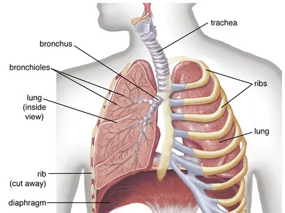
Dyspena is a condition in which a person feels difficulty breathing.

It is a subjective experience expressed   by the patient. The casuse or origin of this may come formvarying reasons including physiological, psychological, social, and environemntal factors.
Thus produced discomfort now is tackled by our physiological response as change in physiological parameters or behavioral modofications including shortnes of breathing. 

Dyspnoea can be classified as acute or chronic. 

Acute dyspnoea develops withing hours to days while chronic develops over weeks to lnger duration. The causes of acute and  chronic dyspnoea might be different depending upon the duration. 
Pulse oxymeter

After careful history taking and examination of the patient having dyspnoea we should make a good list of differential disagnoses. The various differential diagnoses of dyspnoea are summerized in the following list. 

The source of causes dyspnea, shortness of breath can be listed as:

  1. Cardiovascular and realted to use of oxygen
  2. Resporatory and chest wall pathology
  3. Psychogenic

Differential diagnoses of dyspnoea/shortness of breath:

1. Causes of acute SOB

  • Cardiovascular

  1. Cardiogenic
  2. Impaired oxygen delivery
  3. Impaired oxygen use

  • Respiratory causes 

  1. Upper airway
  2. Lower aiway causes

  • Psychogenic

  1. panic disorder
  2. conversion disorder
  3. drug withdrawal

2. Causes of Chronic SOB

  • Cardiovascular cause

  1. CHF
  2. Pericarditis
  3. Anemia

  • Respiratory causes

  1. Pathologies of chest wall, bronchoalveolar system, airways and lung parenchyma

The diseases can be summarised as below:

1. Cardiovascular

  • Acute MI
  • CHF/ LH failure
  • Aortic/Mitral stenosis
  • Aortic/Mitral Regurgitation
  • Arrhythmia
  • Cardiac tamponade
  • Constrictive pericarditis
  • Left sided obstructive lesions (atrial myxoma)
  • Elevated pulmonary venous pressure

2. Respiratory

    i) Airway
  • Asthma
  • COPD
  • Upper airway obstruction like foreign body, anaphylaxis, mucus plugging
    ii) Parenchymal disease
  • ARDS
  • Pneumonia
  • ILD
    iii) Pulmonary vascular disease
  • PE
  • Pulmonary HTM
  • Pulmonary vasculitis
    iv)Pleural disease
  • Pneumothorax
  • Pleural effusion

3. Neuromuscular and chest wall disorers

  • C-spine injury
  • Poliomyelitis
  • Myesthenia gravis
  • Gullian-Barre syndrome
  • Kyphoscoliosis

4. Anxiety, psychosomatic

5. Hematological and metabolic

  • Anemia
  • Acidosis
  • Hypercapnia
Thank you for reading the article. I hope this will be useful for you. Please write us your queries at here.

छुटेका बालबालिकाहरूलाई खोप किन र कसरी लगाउने? Nepal national immunization program for missed vaccine

राष्ट्रिय खोप तालिका: खोप सम्बन्धि जानकारी

Table of Contents(toc)

Nepal national immunization program to vaccinate the children that missed vaccine

राष्ट्रिय खोप तालिका बारे जानकारी पाउन यहाँ क्लिक गर्नुहोस्।

नेपाल को खोप तालिका बारे छोटो जानकारी  (Rastriya Khop talika Nepal)

  • नेपालमा बच्चा जन्मे देखि दुई वर्षको उमेर सम्म विभिन्न १३ रोग विरुद्धको खोप निःशुल्क लगाइन्छ।
  • यी खोपहरु सरकारी स्वास्थ्य संस्था हरु मा निःशुल्क पाइन्छ।
  • गर्भवती महिला लाई दुई डोज टिटानस विरुद्ध को खोप निःशुल्क लगाइन्छ।
  • कुकुर र रेबिज सार्न सक्ने जनावरले तोकेको व्यक्तिलाई समेत रेबिज विरुद्धको खोप विभिन्न संस्थाहरुमा निःशुल्क लगाइन्छ।
BCG vaccinating a child; SC subcuticular

खोप बारे विशेष जानकारी vaccine information

अहिले नेपाल सरकारले माथि उल्लेखित अवधिमा खोप लगाउन छुटेका बालबालिकाहरूलाई समेत खोप लगाएर रोगहरू बाट जोगाउन पांच वर्षको उमेरका बच्चा हरु लाई समेत खोप लगाउने निर्णय गरेको छ।
यो खोप वैशाख १५ र ३१ गते नेपाल सरकार अन्तर्गत का स्वास्थ्य संस्था हरु मा निःशुल्क लगाइन्छ।
दुई वर्ष को उमेर सम्म खोप लगाइसकेका बच्चा हरु लाई यो अवधिमा थप खोप लगाउन आवश्यक छैन। 

खोप लगाउन किन जरुरी छ? Why is vaccine important in Nepal

खोपले बालबालिका हरु लाई १३ थरी रोगहरूबाट जोगाउँछ। यो रोगहरू निम्नानुसार छन्।
२. रोटाभाइरसबाट लाग्ने पखाला
५. लहरेखोकि
६. धनुष्टंकार
७. दादुरा
८. रुबेला
९. जापानिज इन्सेफ्लाइटिस
१०. टाइफाइड
११. भ्यागुते रोग
१२. हेपाटाइटिस बि 
१३. हेमोफिलसले हुने सन्क्रमण

छुटेका बालबालिकालाइ खोप लगाउने खोप तालिका missed vaccine dose table

MCQs regarding Pulmonary tuberculosis Health exams preparation for Loksewa and NHPC 2024

MCQs regarding Pulmonary tuberculosis Health exams preparation 

Introduction

I hope you are doing well and reading this. As you are aware, we will be using Google Forms to administer an upcoming multiple-choice test. I’m writing to respectfully ask that you take the test and do your best.

Tips and tricks

It is quick and easy to gauge your level of subject knowledge by taking the test using Google Forms. As many tests are now taken online, it’s also a fantastic chance to practice test-taking techniques.

Before you proceed…

Before beginning the test, please be sure you read and comprehend the instructions and to have a reliable internet connection. Please do not hesitate to contact me for help if you run into any technical issues.

 MCQ on Pulmonary tuberculosis Google forms


Final Note on PTB

Please comment your score below
Thank you for your submision. 

Tags(please ignore): Health exams Exam preparation Medical exams Health test Study tips Health education Health knowledge Exam stress Health science Health information Exam strategies Health conditions Test anxiety Health promotion Exam resources Health resources Health research Exam techniques Health coaching Exam success PSC exam preparation Lok Sewa exam tips License exam study material Exam syllabus Exam pattern Previous year question papers Mock exams General knowledge for exams Exam eligibility criteria Exam registration Exam dates Exam centers Exam results Exam analysis Exam cutoff marks Exam books and guides Exam coaching Exam time management Exam revision techniques Exam success stories Public service commission exam Civil service exam preparation PSC exam syllabus PSC exam pattern PSC exam eligibility PSC exam registration PSC exam dates PSC exam centers PSC exam results PSC exam preparation tips Lok Sewa Aayog exam preparation Lok Sewa Aayog exam syllabus Lok Sewa Aayog exam pattern Lok Sewa Aayog exam eligibility Lok Sewa Aayog exam registration Lok Sewa Aayog exam dates Lok Sewa Aayog exam centers Lok Sewa Aayog exam results License exam preparation License exam syllabus License exam pattern License exam eligibility License exam registration License exam dates License exam centers License exam results License exam preparation tips General knowledge for PSC, Lok Sewa, and license exams Current affairs for exams Mental ability for exams

Respiratory system for medical students and paramedics by Dr chaitanya and Analog Health exams preparation

 You can view the PPT down below:

Respiratory system and related disorders COPD for medical and paramedical students health loksewa preparation and MCQs

Introduction to respiratory system

  • Respiration: process of gaseous exchange between an organism and its environment.

  • Internal respiration: Gaseous exchange between tissues and blood.

  • External respiration: Gaseous exchange between the body and the environment taking place in the lungs.

The external respiration constitutes processes of inspiration and expiration.

Structure of respratory system

  • External (Pulmonary) respiration: absorption of O2 and removal of CO2 from the body.

  • Internal (tissue) respiration: utilization of O2 and production of CO2 by cells and the gaseous exchanges between the cells and their fluid medium.

  • What is the normal respiratory rate in humans: 16-20 times per minute in adults. 

  • Children breathes more superficially and therefore have a higher respiratory rate.

Which of the following is the smallest functional unit of the lung?

A. Bronchiole
B. Alveolus
C. Bronchus
D. Pleura

Structure of Respiration System

:arynx anatomy

Larynx

AKA voice box 
Short section of the airway that connects the laryngopharynx and trachea.
Located in the anterior portion of the neck, just inferior to the hyoid bone and superior to trachea.
:arynx internal anatomy

Epiglottis:

 a flap of elastic cartilage that acts as a switch between the trachea and the esophagus.
Inferior to the epiglottis, is the thyroid cartilage, often known as Adam’s apple, visible in adult males.

Trachea 

AKA windpipe
5 inch long tube made up of C-shaped hyaline cartilage rings.
Connects the larynx to the bronchi and allows the air to pass through the neck and into the thorax.
Function: to provide a clear airway for air to enter and exit the lungs.

Lungs 

A pair of large, spongy organs found in the thorax lateral to the heart and superior to the diaphragm.
Each lung is surrounded by a pleural membrane that provides the lung with space to expand.
The left lungs are slightly different that right in size and shape due to the heart pointing in the left side of the body.
2 lobes in the left and 3 lobes in the right.
Processs of air exchange gas echange in alveoli

Which of the following structures is NOT part of the upper respiratory tract?

A. Nasopharynx
B. Larynx
C. Trachea
D. Pharynx

What is the function of the epiglottis?

A. To regulate the flow of air into the lungs
B. To produce mucus to moisten the airway
C. To protect the airway during swallowing
D. To exchange oxygen and carbon dioxide

What is the role of surfactant in the lungs? 

A. To transport oxygen and carbon dioxide
 B. To protect the lungs from infection
 C. To decrease surface tension in the alveoli
 D. To regulate air pressure during inhalation

Chronic Obstructive Pulmonary Disease

A board classification of disorder including emphysema, chronic bronchitis and asthma. 

Irreversible condition associated with dyspnea on exertion and reduced airflow in or out of the lung.

4th non communicable disease in the world according to recent study. 
Risk factors of COPD

Emphysema:

Abnormal permanent enlargement of the air space distal to the terminal bronchioles is accompanied by destruction of bronchioles.

Chronic Bronchitis:

Presence of productive cough for 3 or more months in each of 2 successive years in a patient whom other causes of chronic cough have been excluded.

Asthma:

Intermittent, reversible, obstructive airway disease in which trachea and bronchi respond in a hyperactive way to certain stimuli.

Which of the following respiratory disorders is characterized by inflammation and narrowing of the airways? 

A. Asthma
 B. Chronic obstructive pulmonary disease (COPD)
 C. Pneumonia
 D. Lung cance

Clinical manifestationsof COPD symptoms and signs of COPD

  • Age groups >40 years 
  • Productive cough 
  • Decreased exercise tolerance
  • Wheezing 
  • Shortness of breathe 
  • Prolonged expiration 
  • Use of accessory breathing muscles 
  • Pursed lip breathing 
  • Central Cyanosis 
  • Barrel chest 
  • Weight loss  
  • Polycythemia (Hb>18 gm/dl)
  • Flapping tremor 
  • On percussion: hyper resonance 
  • On auscultation: vesicular breathe sound with prolonged expiration
  • Rhonchi 
  • Crepitation if associated with chest infection 
  • FEV1 <80%

Which of the following is a common symptom of respiratory distress?

A. Cyanosis
B. Hypotension
C. Hyperthermia
D. Bradycardia

How is Diagnosis of COPD made COPD diagnosis method

Following tests and investigations are done to diagnose a person as having COPD:
  1. History taking and physical examination 
  2. Pulmonary function tests 
  3. Arterial blood gas levels 
  4. Spirometry 
  5. Chest X-ray 
  6. ECG, Echo 
  7. Alpha 1 antitrypsin screening for the patients with strong family history of COPD.

What is the most common cause of chronic bronchitis?

A. Smoking
B. Air pollution
C. Genetic predisposition
D. Infection

What is the treatment of COPD? chronic bronchitis, emphysema

  1. Risk factor reduction 
  • Pollution, smoking, dust, smoke, firewood
  • Promoting smoking cessation 
  • Decreasing exposure to polluted environment 
  • Oxygen therapy 
  • Increase protein and calorie intake 
  • Chest physiotherapy, postural drainage and breathing exercise 
  • Nebulization 
  • Medicines 
  • Anti-microbial agent 
  • Bronchodilators 
  • Beta antagonists 
  • Anticholinergics 
  • Corticosteroids
  • What are the nusing care required for COPD and chest disease patients?

    • Improving gaseous exchange 
    • Maintaining fluid balance 
    • Managing nutritional needs 
    • Removing bronchial secretions
    • Breathing exercises and respiratory training
    • Providing mouth care , maintaining skin and joint integrity 
    • Achieving thermoregulation 

    You can see the powerpoint presentation below:

    Bonus MCQ below PPT:

    All my links in one place here

    Lungs structure and lobes

    Which of the following respiratory muscles is responsible for normal breathing during rest?

    A. Diaphragm
    B. Intercostal muscles
    C. Sternocleidomastoid muscle
    D. Scalene muscles

    What is the most common cause of chronic obstructive pulmonary disease (COPD)?

    A. Smoking
    B. Air pollution
    C. Genetic predisposition
    D. Infection
    Answer: A (Smoking)

    Which of the following is a common symptom of pneumonia?

    A. Wheezing
    B. Chest pain
    C. Coughing up blood
    D. Rapid breathing (tachypnea)
    Answer: D (Rapid breathing)

    What is the hallmark symptom of asthma?

    A. Coughing up phlegm
    B. Wheezing
    C. Chest pain
    D. Shortness of breath at rest
    Answer: B (Wheezing

    Thank you so much for your time

    Content by: Dr Chaitanya Joshi

    Asthma and related MCQs by Dr chaitanya Joshi

    Asthma lecture ppt and related multiple choice question for NHPC, Nursing, Nclex and PSC 2024

    Asthma inhaler MDI device
    Table of contents (toc)
    Health exams preparation study tips health science Coaching syllabus wise MCQ test loksewa psc license

    Introduction of asthma

    Asthma is a Chronic inflammatory condition of airway Characterized by
      • 1. airway limitation: reversible
      • 2. airway hyperresponsiveness
      • 3. inflammation of bronchi T cell mediated, eosinophilic, hypertrophy, mucus plugging

    Classification of asthma

    Extrinsic asthma

    • Atopy (allergy)
    • Positive skin prick test
    • Childhood asthma a/w eczema

    Intrinsic asthma

    • Middle age
    • No cause
    • May show positive skin prick test
    • External stimuli sensitization like toulene, NSAIDs,  beta blockers etc
    • Etiology and precipitating factors

    Clinical features of asthma

    • Typical recurrent episodes with normal life in between
    • Wheezing
    • SOB
    • Chest tightness
    • Cough
    • Classical diurnal pattern with symptoms worse in morning and night
    • Cough and wheeze disturb sleep – nocturnal asthma

    Diagnosis of asthma

    • Mostly clinical diagnosis
    • Symptoms improvement following bronchodilator and corticosteroid
    • Diurnal variation on 3 days a week for 2 weeks on PEF diary
    • PEF decrease after 6 min of exercise
    • Investigations 
    • PFT
    • Exercise test
    • Histamine or methacholine bronchial provocation test
    • Corticosteroids trial
    • Exhaled NO test
    • Blood and sputum workup
    • CXR
    • Skin prick test for allergen

    Management of asthma- treatment of asthma

    Symptoms control for asthma

    • Restore normal lifestyle
    • Reduce risk of recurrent and severe attacks
    • Enable normal growth and nutrition
    • Family education and counselling
    • Avoid under or overtreatment

    Extrinsic factor control for asthma

    • Pet
    • Dust
    • Smoke
    • Medicine like NSAID, antibiotics
    • Allergens
    • Pollens 
    • Pharmacological treatment
    • Step down therapy
    • Once control established, dose of corticosteroid titrated to the lowest dose 
    • Decreasing dose of ICS around 25-50% every 3 months, a reasonable strategy for many

    AE of asthma

    • Aka acute severe asthma
    • Treatment done at ER or on the way
    • Bronchodilators, nebulization, high dose steroids etc.
    • May even lead to death if not treated on time
    • Pack year calculation

    Flapping tremor

    • Please refer to the slides

    Fine tremor

    • Please refer to the slides

    Drugs used for asthma

    • Please refer to the slides

    Modified MRC scale of SOB

    • Please refe to the slides below

    COPD types

    Two classical phenotypes

    • Pink puffers:

      • Thin, breathless and maintain a normal PaCO2 until the late stage of disease Emphysema, Compensated by hyperventilation, so ABG is normal

    • Blue bloaters: 

      • Hypercapnia> Early stage, Chronic bronchitis, Due to obstruction Co2 retention, may develop oedema and secondary polycythaemia

    Barrel chest

    Please refer to the slides

    Multiple Choice Questions  related to respiratory system

    • What is asthma attack?

      • When the lungs is filled with water 
      • When airways tighten and the lungs don’t get enough air
      • When the heart beats too fast 
      • When the heart and lungs are working too hard 
    Ans: b (When airways tighten and the lungs don’t get enough air)

    • Which of the following is not the characteristic of asthma? 
      • Increase in IgG immunoglobulins 
      • Airway hyperresponsiveness 
      • Infiltration of eosinophils into the airways 
      • Increased mucus production

    Ans: a (Increase in IgG immunoglobulins)

    • The immediate response of asthma involves:

      • • Mast cell degranulation
      • • Binding of antigen to IgE on macrophages
      • • Release of cytokines
      • • Activation of cholinergic nerves

    Ans: a (Mast cell degranulation)

    • Chronic asthma is associated with:

      • • Activation of T lymphocytes
      • • Reduced function of goblet cells
      • • Activation of eosinophils
      • • Decreased permeability of submucosal capillaries

    Ans: c (Activation of eosinophils)

    • Which test is NOT used during diagnosis of asthma?

      • • Diffusing capacity
      • • Peak expiratory flow rate
      • • Bronchodilator challenge test
      • • Skin prick test

    Ans: a (Diffusing capacity)

    • A diagnosis of severe asthma should be made after about how many months of uncontrolled asthma?

      • • 1 month
      • • 3-6 months
      • • 6-9 months
      • • More than 9 months

    Ans: b (3-6 months)

    • When a patient is diagnoses with severe asthma, which of the following is recommended?

      • • Referral to specialist
      • • Increased dosage of patient’s current medication
      • • Education about risk factors and triggers such as smoking
      • • Treatment of comorbidities and triggers such as smoking

    Ans: a (Referral to specialist)

    • Which drug is most commonly prescribed preventer therapy in asthma?

      • • Beta2 adrenoreceptor agonists
      • • Xanthine such as theophylline
      • • Muscarinic receptor antagonists
      • • Inhaled steroids

    Ans: d (Inhaled steroids)

    • Once a patient is diagnosed with asthma and referred to specialist, what does GINA recommend next step?

      • • Referral to mental health professional
      • • Increased dosage of patient’s current medication
      • • Assessment of patient’s inflammatory phenotype
      • • Hospitalization for intense observation
    Ans: c (Assessment of patient’s inflammatory phenotype)

    •  Acute management of asthma include all of the following, EXCEPT: 

      • A high concentration of oxygen to achieve oxygen saturation of >90%
      • Short acting beta-2- agonists 
      • A slow infusion of aminophylline with monitoring blood values 
      • A rapid infusion of dopamine 
    Ans: d (A rapid infusion of dopamine)
    Thank you!

    Asthma and COPD lecture slide and Video Lecture

    A doctor viewing x-ray of asthma patient

    Thank you very much

    Pneumothorax and pleural effusion by dr chaitanya with pathophysiology of pleural effusion 2024

    Pneumothorax and pleural effusion- diagnosis and management with MCQs

    Table of Contents(toc)
    left sided pneumothorax


    PNEUMOTHORAX

    Pneumothorax is the presence of air in the pleural space.

    Two types

    • Traumatic pneumothorax
    • Spontaneous pneumothorax
    Traumatic pneumothorax 
    • Results from penetrating or non-penetrating chest injuries.
    Iatrogenic
    • Transthoracic needle aspiration
    • Thoracentesis
    • The insertion of central intravenous catheters
    Spontaneous 

    • Primary Pneumothorax

    1. No evidence of overt lung disease.
    2. Due to rupture of a small subpleural emphysematous bulla or pleural bleb, or the pulmonary end of a pleural adhesion
    3. Occur almost exclusively in smokers
    4. one-half of patients will have a recurrence

    • Secondary Pneumothorax

    1. Underlying lung disease, most commonly COPD and TB;
    2. also seen in asthma, lung abscess, pulmonary infarcts, bronchogenic carcinoma, all forms of fibrotic and cystic lung disease

    Tension pneumothorax

    • Results from a wound in the chest wall which acts as a valve that permits air to enter the pleural cavity but prevents its escape
    • Intrapleural pressure rises to well above atmospheric levels. 
    • The pressure causes mediastinal displacement towards the opposite side, with compression of the opposite normal lung 
    • Impairment of systemic venous return, causing cardiovascular compromise

    Closed pneumothorax

    • Communication between the airway and the pleural space seals off
    • mean pleural pressure remains negative
    • reabsorption of air, re-expansion, infection is uncommon

    Open pneumothorax

    • Communication fails to seal and air continues to pass freely
    • Occur following rupture of an emphysematous bulla, tuberculous cavity or lung abscess into the pleural space
    • Infection is common

    Clinical Features of tension pneumothorax

    • sudden-onset unilateral pleuritic chest pain or breathlessness
    • larger pneumothorax (> 15% of the hemithorax) results in decreased or absent breath sounds
    • combination of absent breath sounds and resonant percussion note is diagnostic of pneumothorax.

    • Inspection

      • Tachypnoea (pain, deflation reflex)

    • Palpation

      • ↓Expansion

    • Percussion

      • Resonant or hyper-resonant

    • Auscultation

      • Absent breath sounds

    Tension pneumothorax also causes

    • Deviation of trachea to opposite side
    • Tachycardia and hypotension
    • Cyanosis 

    Investigations for pneumothorax

    • Chest X-ray

      • Sharply defined edge of the deflated lung with complete translucency
      • Extent of any mediastinal displacement and reveal any pleural fluid or underlying pulmonary disease.

    • CT scan
      •  In doubt, distinguish bullae from pleural air.

    Treatment of tension pneumothorax

    • Primary pneumothorax with lung edge < 2 cm from the chest wall, not breathless requires no intervention.

    Percutaneous needle aspiration of air 

    • In young patients with a moderate or large spontaneous primary pneumothorax
    • Intercostal tube drainage
    • In those over 50 years old and those with respiratory compromise

    Surgical pleurodesis 

    • Recommended following a second pneumothorax
    • should be considered following the first episode of secondary pneumothorax if low respiratory reserve makes recurrence hazardous. 
    • Pleurodesis can be achieved by pleural abrasion or parietal pleurectomy at thoracotomy or thoracoscopy.
    • Prevents recurrences

    Post Chest Tube monitoring: what to look for after keeping a chest tube

    • After 24 hours;  if lungs reinflated and no bubblingremove drain
    • Continuing bubbling after 5- 7days  is indication of surgery
    • High flow O2 supplement- may speed resolution
    • If bubbling stops before full inflation of lung then tube is either blocked , kinked or displaced
    • Pleural Effusion 

    Pleural effusion:

    Pleural Fluid

    • Excessive accumulation of fluid in the pleural space. 
    • Detected clinically, when ≥ 500 mL is present.
    • Produced and absorbed at a rate of 15mL per day
    • Each pleural space contains 10mL of fluid

    The estimated prevalence of pleural effusion is 320 cases per 100,000 people.

    Causes of pleural effusion:

    Pathophysiology of pleural effusion:

    Fluid enters the pleural space:

    • from the capillaries in the parietal pleura
    • from the peritoneal cavity via small holes in the diaphragm
    • Fluid removed via the lymphatics situated in the parietal pleura. 

    lymphatics have capacity to absorb 20 times more fluid than is normally formed.

    Pleural fluid accumulates when:

    pleural fluid formation >pleural fluid absorption. 

    Exudative or Transudative

    Alteration of hydrostatic and/or oncotic factors Increases the formation or decreases the reabsorption Transudate

    Direct or cytokine induced pleural membrane and/or vascular damage Increased capillary permeability Exudate

    Types of pleural effusion:

    Transudative effusion

    1. Congestive Heart failure
    2. Cirrhosis 
    3. Pulmonary embolization 
    4. Neoplastic syndrome
    5. Constrictive pericarditis
    6. Hypothyroidism

    Exudative effusion:

    1. Bacterial pneumonia (common)
    2. Tuberculosis
    3. Carcinoma of the bronchus
    4. Pulmonary infarction
    5. Autoimmune 
    6. -Rheumatic diseases
    7. – SLE 

    Clinical features of pleural effuion:

    • Shortness of breath
    • Chest pain, especially on breathing in deeply (pleurisy, or pleuritic pain)
    • Fever
    • Cough

    How Patient Presents in pleural effusion?

    History in pleural effusion

    • Pain on inspiration and coughing
    • Often  asymptomatic
    • Pleurisy 

    Examination finding in pleural effusion:

    1. Chest wall movement : Reduced in affected side 
    2. trachea and mediastinum shift away from affected side
    3.  With massive effusion (>1000ml)]
    4. Percussion note: Stony dull 
    5. Breath sounds: Vesicular
    6. (Reduced or absent)
    7. Vocal resonance: Reduced or absent

    On Chest Examination of pleural effusion following findings  can be found:

    • Bronchial breathing may be present
    • Expansion ↓
    • Percussion stony dull
    • Air entry ↓
    • Vocal resonance ↓
    • With massive effusion (>1000ml) trachea and mediastinum shift away from affected side

    Diagnostic Approach in pleural effusion

    Light’s criteria for Exudative Effusion

    • Pleural fluid protein: serum protein >0.5
    • Pleural fluid LDH: serum LDH >0.6
    • Pleural fluid LDH > 2/3 upper limit of normal for serum
    • Investigations 
    • Serum
    • CBC
    • LDH
    • protein
    • Coagulation studies (PT, PTT, INR )

    Pleural fluid analysis for pleural effusion:

    • Note color and consistency
    • Chemistries: Protein, albumin, LDH, glucose, pH
    • Cell count with differential
    • Microbiological stains and culture 
    • Cytology

    Pleural fluid finding in effusion

    Lateral decubitus x ray in pleural effusion:

    • Demonstrates fluidity
    • Suggest for thoracentesis if fluid layer > 1cm. 
    • Transudates
    • Usually resolve with treatment of underlying cause
    • Therapeutic thoracentesis in persistent larger effusion
    • Pleurodesis, shunts, or placement of a indwelling pleural catheter for palliation

    Treatment of pleural effuusion:

    • Simple Parapneumonic Effusion

      • Antiobiotics
      • Selected base on causing organism
      • Generally anaerobic coverage
      •   ( clindamycine, imipenem, extended spectrum penicillin )
      • Close observation
    • Complicated Parapneumonic Effusion
      • Antiobiotics
      • Early thoracostomy tube drainage
      • Surgical decortication if extensive pleural thickening, fibrous organization, and /or multiple loculations.
      • Malignant Pleural Effusion
      • 2nd most common type of exudative pleural effusion (lung carcinoma, breast carcinoma, & lymphoma)
      • Diagnosis: cytology of the pleural fluid
      • If cytology is negative, thoracoscopy is done if malignancy is suspected

    Management  of pleural effusion

    • Symptomatic treatment 
    • Tube thoracostomy ( sclerosing agents : doxycycline )
    • Plurodesis when there is rapid reaccumulation of fluid
    • Insertion of indwelling pleural catheter

    Complications of pleural effsuion if untreated

    1. empyema
    2. constrictive fibrosis
    3. Sepsis
    4.  lung cancer

    Nursing care for pleural effusion and chest tube inserted patient

    • Observe for infection, local inflammation
    • Check dressing
    • Check if tube is blocked
    • Mobilization care
    • Check for amount and color of drainage
    • Pain management
    • Ensure breathing and lung reexpansion in case of collapse
    • Prevent clamping
    • Ensure positioning

    References

    • Davidson’s Principles and Practice of Medicine 21st edition
    • Harrison’s Principle of Internal Medicine, 18th edition

    MCQs related to pleral effusion:

    What is pleural effusion?

    a) Accumulation of air in the pleural space

    b) Collection of fluid in the pleural space

    c) Infection of the pleural cavity

    Answer: b) Collection of fluid in the pleural space

    What is pneumothorax?

    a) Collection of fluid in the pleural space

    b) Accumulation of air in the pleural space

    c) Infection of the pleural cavity

    Answer: b) Accumulation of air in the pleural space

    Which condition is more likely to cause chest pain?

    a) Pleural effusion

    b) Pneumothorax

    c) Both conditions can cause chest pain

    Answer: c) Both conditions can cause chest pain

    Which condition is more likely to cause shortness of breath?

    a) Pleural effusion

    b) Pneumothorax

    c) Both conditions can cause shortness of breath

    Answer: c) Both conditions can cause shortness of breath

    Which of the following imaging studies is most commonly used to diagnose pleural effusion?

    a) Chest x-ray

    b) Computed tomography (CT)

    c) Magnetic resonance imaging (MRI)

    Answer: a) Chest x-ray

    Which of the following conditions can be diagnosed by thoracentesis?

    a) Pleural effusion

    b) Pneumothorax

    c) Both conditions can be diagnosed by thoracentesis

    Answer: a) Pleural effusion

    Which of the following imaging studies is most commonly used to diagnose pneumothorax?

    a) Chest x-ray

    b) Computed tomography (CT)

    c) Magnetic resonance imaging (MRI)

    Answer: a) Chest x-ray

    Which of the following conditions can be treated with a chest tube?

    a) Pleural effusion

    b) Pneumothorax

    c) Both conditions can be treated with a chest tube

    Answer: c) Both conditions can be treated with a chest tube

    What is the primary treatment for small, uncomplicated pneumothorax?

    a) Observation

    b) Chest tube insertion

    c) Surgery

    Answer: a) Observation

    What is the primary treatment for large, complicated pneumothorax?

    a) Observation

    b) Chest tube insertion

    c) Surgery

    Answer: b) Chest tube insertion

    Which of the following is a potential complication of pleural effusion?

    a) Respiratory failure

    b) Cardiac arrest

    c) Seizures

    Answer: a) Respiratory failure

    Which of the following is a potential complication of pneumothorax?

    a) Respiratory failure

    b) Cardiac arrest

    c) Seizures

    Answer: a) Respiratory failure

    Which of the following conditions is more common in individuals with chronic obstructive pulmonary disease (COPD)?

    a) Pleural effusion

    b) Pneumothorax

    c) Both conditions are equally common in individuals with COPD

    Answer: b) Pneumothorax

    Which of the following conditions is more common in individuals with heart failure?

    a) Pleural effusion

    b) Pneumothorax

    c) Both conditions are equally common in individuals with heart failure

    Answer: a) Pleural effusion

    What is the most important factor in determining the severity of pneumothorax?

    a) The size of the pneumothorax

    b) The cause of the pneumothorax

    c) The location of the pneumothorax

    Answer: a) The size of the pneumothorax

    Tags(please ignore): Health exams Exam preparation Medical exams Health test Study tips Health education Health knowledge Exam stress Health science Health information Exam strategies Health conditions Test anxiety Health promotion Exam resources Health resources Health research Exam techniques Health coaching Exam success PSC exam preparation Lok Sewa exam tips License exam study material Exam syllabus Exam pattern Previous year question papers Mock exams General knowledge for exams Exam eligibility criteria Exam registration Exam dates Exam centers Exam results Exam analysis Exam cutoff marks Exam books and guides Exam coaching Exam time management Exam revision techniques Exam success stories Public service commission exam Civil service exam preparation PSC exam syllabus PSC exam pattern PSC exam eligibility PSC exam registration PSC exam dates PSC exam centers PSC exam results PSC exam preparation tips Lok Sewa Aayog exam preparation Lok Sewa Aayog exam syllabus Lok Sewa Aayog exam pattern Lok Sewa Aayog exam eligibility Lok Sewa Aayog exam registration Lok Sewa Aayog exam dates Lok Sewa Aayog exam centers Lok Sewa Aayog exam results License exam preparation License exam syllabus License exam pattern License exam eligibility License exam registration License exam dates License exam centers License exam results License exam preparation tips General knowledge for PSC, Lok Sewa, and license exams Current affairs for exams Mental ability for exams

    Smoking and alcohol use? the benefits of smoking the harms of smoking 2024?

    Smoking and alcohol use? the benefits of smoking and the harms of smoking?

    Table of Contents(toc)

    Introduction

    Smoking and alcohol use are two of the most prevalent and dangerous health risks in the world today. Both habits can have a devastating impact on a person’s physical and mental health, leading to a wide range of diseases and conditions that can be life-threatening.

    The Harms of Smoking

    There are several harms of smoking that include personal, health, family , social and nation wise harms. It not only harms teh user but also the whole bnation and the global effect is there.

    Effect of smoking

    Cigarette smoking is a leading cause of preventable death worldwide. Smoking can cause a wide range of health problems, including lung cancer, heart disease, stroke, and chronic obstructive pulmonary disease (COPD). Smoking also affects the reproductive system, leading to fertility issues in both men and women. It can also harm unborn babies, causing low birth weight and a range of other problems.

    No Smoking

    Smoking and lung cancer

    One of the most significant risks of smoking is lung cancer. According to the American Lung Association, smoking causes 85% of lung cancer cases in the United States. In addition to lung cancer, smoking can also cause other types of cancer, including throat, bladder, and pancreatic cancer. Smoking can also lead to emphysema, a condition in which the air sacs in the lungs are damaged, making it difficult to breathe.

    Cardiovascular risks of smoking

    Smoking also damages the cardiovascular system, increasing the risk of heart attack and stroke. Nicotine in tobacco smoke raises blood pressure and heart rate, while carbon monoxide reduces the amount of oxygen that can be carried in the blood, making it more difficult for the heart to pump blood.
    The Harms of Alcohol Use

    Alcohol use is also a significant health risk, particularly when consumed in excess. Alcohol consumption can cause liver disease, pancreatitis, and a range of other health problems. Chronic alcohol use can also lead to high blood pressure, heart disease, and stroke.

    Excessive alcohol use can also lead to alcoholism, a condition in which a person becomes physically and psychologically dependent on alcohol. Alcoholism can lead to a range of problems, including social isolation, relationship issues, and financial difficulties.

    Alcohol drinks

    Physical health risks of smoking

    In addition to the physical health risks, alcohol use can also have a significant impact on mental health. Alcohol use can increase the risk of depression and anxiety, and it can also lead to other mental health problems, such as bipolar disorder.

    No smoking sign

    Conclusion

    Beer in a glass

    Smoking and alcohol use are two of the most significant health risks facing people today. Both habits can have a devastating impact on a person’s physical and mental health, leading to a wide range of diseases and conditions that can be life-threatening. If you smoke or drink alcohol, it is important to take steps to quit or reduce your use. Talk to your healthcare provider for guidance and support on quitting smoking and cutting back on alcohol use. Your health and well-being are too important to risk.

    Comprehensive Abortion Care in Nepal 2024

    What is Comprehensive Abortion Care?

    Table of Contents(toc)

    Introduction

    Comprehensive Abortion Care (CAC) is a patient-centered, comprehensive approach to abortion that includes services ranging from counseling and medical procedures to post-abortion care and support. It prioritizes women’s health, safety, and well-being while protecting their reproductive rights. CAC addresses not only the medical elements of abortion, but also the social, emotional, and psychological issues that influence a woman’s decision-making process. This article discusses the significance of comprehensive abortion treatment and its components.

    Understanding Comprehensive Abortion Care

    Abortion Care that is Comprehensive goes beyond the typical focus on medical procedures. It recognizes that women’s needs and experiences vary, and that these must be taken into account while providing abortion services. CAC’s basic components are as follows:

    Safe and Legal Abortion Services:

    CAC guarantees that women have safe and legal abortion methods available to them. Abortions are performed in a clinical setting by qualified healthcare professionals in accordance with medical standards and guidelines. Legal frameworks must protect women’s right to choose abortion without fear of legal prosecution.

    Pre-Abortion Counseling:

    Proper counseling is crucial to ensure that women make informed decisions about their reproductive health. It involves providing accurate information about abortion methods, potential risks, and available support services. Counseling also addresses emotional and psychological concerns, helping women feel confident in their choices.

    Medical Procedures:

    Proper counseling is essential to ensuring that women make educated reproductive health decisions. It entails disseminating correct information regarding abortion methods, dangers, and available support services. Counseling also tackles emotional and psychological issues, allowing women to feel more confidence in their decisions.

    Post-Abortion Care:

    Comprehensive care goes beyond the actual abortion. Follow-up consultations to check on physical healing and treat any issues are part of post-abortion care. To assist women in overcoming any potential emotional difficulties, counseling and emotional support are also available.

    Contraceptive Services:

    Access to contraceptive advice and services is a component of CAC in order to avoid future unwanted pregnancies. Based on their health, lifestyle, and reproductive objectives, this assists women in selecting an effective contraceptive technique.

    Respect for Women’s Autonomy and Rights:

    Women’s right to make their own decisions about their reproductive health is upheld by CAC. It acknowledges that every woman has a special situation, and that her decisions should be respected without bias or pressure.

    Importance of Comprehensive Abortion Care

    Here is teh importance of CAC explained in detail.

    Promoting Women’s Health and Well-being:

    Women’s health and safety are given top priority by CAC, lowering the likelihood of unsafe and unauthorized abortions. Abortion is a safe medical procedure with few risks when done by qualified specialists.

    Reducing Maternal Mortality and Morbidity:

    Abortions performed unsafely play a substantial role in maternal mortality and morbidity rates around the world. By guaranteeing that women have access to secure abortion services, CAC plays a crucial part in lowering these dangers.

    (ads1)

    Supporting Reproductive Rights:

    An essential reproductive right is the availability of comprehensive abortion care. Women are given more control over their bodies and destinies thanks to CAC, which promotes gender equality and women’s empowerment.

    Addressing Stigma and Emotional Well-being:

    The emotional and psychological effects of abortion are acknowledged by CAC. It aids women in overcoming any stigma or emotional difficulties they may experience by offering counseling and support.

    Preventing Unintended Pregnancies:

    Contraceptive services are a part of CAC, which supports family planning and reproductive health by assisting women in preventing future unplanned pregnancies.

    Happy family

    Conclusion

    Comprehensive abortion care includes counseling, support, and empowerment in addition to medical treatments. It is a comprehensive strategy that defends women’s rights, respects their autonomy, and advances their health. To protect women’s health, lower maternal mortality, and advance reproductive justice, access to CAC must be ensured. In order to guarantee that women have the support and tools they need to make educated decisions about their reproductive health, it is critical to promote and emphasize comprehensive abortion care as societies change.

    Juka (round worm) जुकाको औषधि र उपचार नेपालीमा

    जुका भनेको के हो? Juka bhaneko ke ho?

    Abdominal pain

    Table of Contents (toc)

    Juka in English जुकालाई अंग्रेजिमा के भनिन्छ?

    जुका एक परजीवी जनावरको संक्रमणले गर्दा पेटमा हुने समस्या हो।
    जुकालाई अङ्ग्रेजीमा राउण्ड वर्म “round worm” भनिन्छ। यो aschelminthes ग्रुपमा पर्ने एक परजीवी हो। जुकाले पेटमा गएर आन्द्राको भित्री भागमा घाउ बनाउँछ र त्यहाँबाट रगत र पौष्टिक तत्व चुस्ने गर्दछ।

    Juka ko symptoms जुकाका लक्षणहरू

    सुरुसुरुमा जुका परेको बेला कुनै पनि लक्षण नदेखिन सक्छन्। बिस्तारै पेट बाउँडिने, वाकवाकी लाग्ने, बान्ता आउने, खाना नपच्ने, पखाला लाग्ने आदि लक्षण देखिन सक्छन्। 
    धेरै जुका परेको खण्डमा यी लक्षण झनै कडा हुन्छन् र कहिलेकाहीं आन्द्रा बन्द हुने र अपरेसन गर्न पर्ने अवस्था समेत आउन सक्छ।
     Juka

    pet ma juka parnu पेटमा जुका पर्नुका लक्षणहरु:

    पेटमा जुका पर्नुको अर्थ आन्द्रामा जुका पर्नु हो। जुका फोहोर हात, खानेकुराबाट सरेर मानिसको मुखमा पुग्छ र निलिएर पेट सम्म पुग्छ। यदि सो जुका को अण्डा पेटको पाचन प्रणालीले मार्न सकेन भने त्यो आन्द्रामा पुग्छ र त्यहा बढ्न थाल्छ। त्यसपछि लक्षणहरु देखा पर्दछन। 

    Juka in stomach symptoms, symptoms of juka जुकाका लक्षणहरु

    जुका परेमा निम्न लक्षणहरु देखा पर्न सक्छन:
    1. पेट दुख्ने
    2. वा्न्ता आउने, वाकवाकि लाग्ने
    3. पखाला लाग्ने
    4. खाना नरुच्ने
    5. कब्जियत हुने
    6. बच्चको केसमा, रुने, कराउने
    7. कमजोरि हुने
    8. थकान महसुस हुने
    9. खाना नपच्ने
    10. रगतको कमि हुने

    Juka ko aushadhi, juka ko dabai

    जुका लागेको भएमा डाक्टरले तलका मध्ये कुनै औषधि दिन सक्नुहुन्छ:
    1. Albendazoole
    2. Mebendazole
    3.  Pyrantel pamoate for pergnant ladies

    Juka ko aushadhi in english

    एल्बेनडाजोल, मेबेन्डाजोल र पाइरेनटल पामोएट लाई जुकाको उपचारको लागि प्रयोग गर्न सकिन्छ।

    Juka ko medicine name

    Albendazoole, Mebendazole, Pyrantel pamoate for pergnant ladies

    Namle juka in english Called?

    नाम्ले जुकालाई English मा tape worm ( टेप वोर्म) भनिन्छ। 

    अन्य प्रकारका जुकाहरु: Other worm types

    मानव सरीरमा अन्य थुप्रै प्रकारका परजीवी जुका जस्ता प्राणीको संक्रमण हुने गर्दछ। जस्तै::
    1. नाम्ले जुका
    2. चुर्ना जुका
    3. कलेजि जुका
    4. हात्तिपाइले
    5. अंकुसे जुका
    अंग्रेजीमा: 
    1. Ascariasis
    2. Hookworm Infection
    3. Pinworm Infection (Enterobiasis)
    4. Trichinosis
    5. Tapeworm Infections
    6. Liver Fluke Infection (Fascioliasis)
    7. Schistosomiasis (Bilharzia)
    8. Strongyloidiasis
    9. Trichuriasis
    10. Dracunculiasis (Guinea Worm Disease)

    Churna juka in English called?

    चुर्ना जुका लाई अंग्रेजीमा  Pinworm भनिन्छ।
    Exit mobile version