Blog

What to look for to assess work of breathing in infants and neonates? – Pediatrics Notes

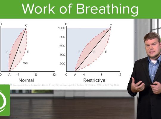
Work of Breathing (WOB) — clinical parameters

Work of breathing is assessed mainly by respiratory effort, chest movement, and signs of respiratory distress.

According to Nelson Textbook of Pediatrics, work of breathing (WOB) refers to the effort required to breathe—i.e., the amount of respiratory muscle activity needed to move air into and out of the lungs.

Clinically, Nelson assesses increased WOB by looking for signs of respiratory distress, particularly:

  • Tachypnea
  • Nasal flaring
  • Retractions/recessions — intercostal, subcostal, suprasternal
  • Grunting
  • Accessory muscle use
  • Head bobbing in infants
  • Paradoxical/see-saw breathing
  • Changes in respiratory pattern or effort

Exam definition

Work of breathing = the effort required to generate ventilation and move air through the respiratory system.

Increased WOB means the child must generate greater-than-normal respiratory effort to maintain adequate ventilation.

High-yield distinction:
WOB ≠ respiratory rate alone. A child can have severe increased WOB even with a relatively normal RR, particularly when respiratory muscle fatigue is developing.

Parameters to assess work of breathing

ParameterWhat to look forSignificance
Respiratory rateTachypnea for ageEarly sign of increased WOB
Nasal flaringAlae nasi widening during inspiration↓ upper-airway resistance
Retractions / recessionSuprasternal, intercostal, subcostal, substernalIncreased negative intrathoracic pressure
GruntingExpiratory gruntMaintains end-expiratory pressure; important neonatal distress sign
Head bobbingEspecially infantsSevere respiratory effort
Tracheal tugDownward movement of trachea on inspirationIncreased inspiratory effort
See-saw/paradoxical breathingChest moves inward while abdomen moves outward, or vice versaSevere distress/fatigue
Accessory muscle useSCM, scalene, abdominal musclesIncreased WOB
Abnormal chest movementAsymmetry, poor expansionAirway/lung pathology
Breath soundsWheeze, stridor, crackles, diminished/absent air entryIdentifies cause/severity
Expiratory phaseProlonged expirationParticularly obstructive disease
GruntingEspecially in neonatesSuggests significant respiratory distress
PostureTripod/sniffing positionCompensatory increase in airway patency
Ability to speak/feedFull sentences vs words; feeding interruption in infantsFunctional measure of WOB
Mental statusIrritability → lethargy → exhaustionLate/severe respiratory compromise
OxygenationSpO₂, cyanosisConsequence rather than direct measure of WOB

Quick severity framework

Mild ↑ WOB

  • Tachypnea
  • Mild nasal flaring
  • Mild intercostal/subcostal recession
  • Child alert and feeding reasonably well

Moderate ↑ WOB

  • Marked retractions
  • Nasal flaring
  • Accessory muscle use
  • Grunting
  • Difficulty feeding/speaking
  • Persistent tachypnea

Severe ↑ WOB / impending failure

  • Severe recession
  • Head bobbing
  • Paradoxical/see-saw breathing
  • Poor air entry or “quiet chest”
  • Exhaustion, altered consciousness
  • Apnea/bradypnea
  • Cyanosis despite oxygen

Important: A falling respiratory rate is not necessarily improvement. In a tiring child, tachypnea may progress to a normal or low RR as respiratory muscles fatigue—often a dangerous sign.

For bedside pediatric assessment, the highest-yield things to document are RR + retractions + nasal flaring + grunting + accessory muscle use + air entry + SpO₂ + mental status/feed/speech ability.

Are you stuck with Diagnosis? Here is prolonged fever + hepatosplenomegaly + persistent leukocytosis diagnoses summarized!

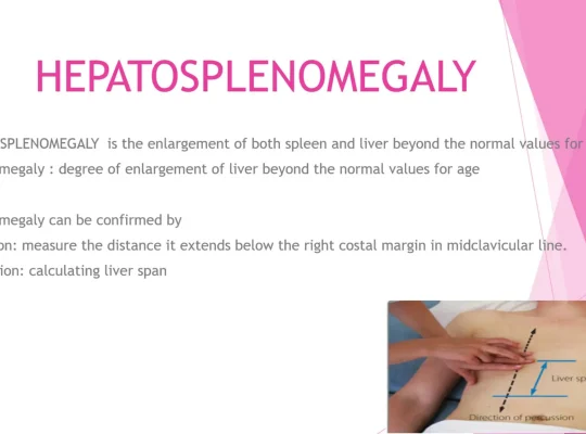
For a child with prolonged fever + hepatosplenomegaly + persistent leukocytosis, I would use the following as a bedside differential/workup table, with particular attention to infections relevant to Nepal and hematologic disease.

1. Infectious causes

DifferentialHistory: ask forExamination cluesKey investigations
TuberculosisContact, chronic cough, weight loss, night sweats, poor appetite; household exposureCervical/generalized nodes, wasting, respiratory signs, hepatosplenomegalyCXR; TST/IGRA; Xpert MTB/RIF/Ultra from appropriate specimen; culture; node FNAC/biopsy if present
Enteric feverProlonged fever, contaminated food/water, travel/outbreak exposure, abdominal symptomsToxicity may be mild; abdominal tenderness, hepatosplenomegaly; relative bradycardia is unreliable in childrenBlood culture before antibiotics; CBC, LFT; stool culture in selected cases
MalariaResidence/travel to endemic Terai areas, mosquito exposure, intermittent/chills patternPallor, jaundice, splenomegaly, hepatomegaly; thrombocytopeniaThick + thin smear + malaria RDT; repeat smear if high suspicion
Visceral leishmaniasisProlonged fever, travel/residence in endemic region, weight lossMassive splenomegaly, hepatomegaly, pallor, wastingrK39/other serology; CBC; confirmatory parasitologic testing when indicated
BrucellosisCattle/goat/sheep exposure, raw/unpasteurized milk, occupational exposure, undulating feverHSM, lymphadenopathy, arthritis; sometimes nonspecificBlood cultures; Brucella serology/PCR where available
EBVSick contacts, sore throat, fatiguePosterior cervical nodes, pharyngitis/tonsillar enlargement, HSMCBC differential, atypical lymphocytes; EBV VCA IgM/IgG ± EBNA; LFT
CMVSick contacts; congenital/perinatal history in younger childHSM, lymphadenopathy; prolonged feverCMV IgM/IgG ± PCR in selected cases; LFT
Scrub typhusRural/forest exposure, mite exposure, recent outdoor activityEschar, rash, lymphadenopathy; hepatosplenomegaly; thrombocytopeniaScrub typhus IgM/ELISA ± PCR
LeptospirosisFloodwater/freshwater, rodents, animal urineConjunctival suffusion, jaundice, renal involvement, myalgiaCBC, renal/LFT; Leptospira PCR/IgM
DengueMosquito exposure, acute febrile illnessRash, bleeding, abdominal tenderness, hepatomegaly; usually thrombocytopeniaCBC trend; NS1/PCR early, IgM later
Disseminated fungal infectionImmunodeficiency, prolonged antibiotics, steroid/immunosuppressive therapyHSM, lymphadenopathy, skin/mucosal lesionsBlood cultures, fungal biomarkers/cultures; imaging/tissue diagnosis
HIVMaternal risk, transfusion, recurrent/opportunistic infections, growth failureGeneralized nodes, HSM, oral candidiasis, wastingHIV Ag/Ab testing; confirmatory algorithm; viral load/CD4 where appropriate
ToxoplasmosisCat/feces exposure, undercooked meat; immunocompromised stateCervical nodes, HSM occasionallyToxoplasma IgM/IgG ± PCR in selected cases
ToxocariasisDogs/cats, soil exposure, picaHepatomegaly, fever, eosinophilia, sometimes ocular diseaseCBC with AEC, Toxocara serology
Hydatid diseaseDog/sheep exposureUsually mass/organ-specific findings rather than feverUSG/CT; Echinococcus serology

2. Hematologic / malignant causes

DifferentialHistoryExaminationInvestigations
ALLFever, fatigue, weight loss, bone/joint pain, bruising, recurrent infectionsPallor, petechiae, lymphadenopathy, HSM, bone tendernessCBC + differential + PBS, retic, LDH/uric acid; bone marrow + flow cytometry if suspected
AMLFever, fatigue, bleeding, infections, bone painPallor, petechiae, HSM, gingival hypertrophy, chloromasCBC/PBS; marrow morphology + flow cytometry/cytogenetics/molecular studies
LymphomaFever, weight loss, night sweats, pruritus; node enlargementFirm/painless nodes, mediastinal signs, HSMCBC, LDH/uric acid; CXR/USG/CT as indicated; excisional lymph-node biopsy
CMLFatigue, weight loss, early satiety, abdominal discomfortMarked splenomegaly, hepatomegaly; pallorCBC differential + PBS; BCR-ABL1 testing, marrow/cytogenetics
JMMLUsually young child; fever, recurrent infections, pallor, rashSplenomegaly, lymphadenopathy, pallor, skin lesionsCBC showing persistent monocytosis, PBS; marrow; molecular testing (e.g. RAS-pathway abnormalities)
Leukemoid reactionInfection/inflammation symptomsUnderlying infectious focus; may have HSMSerial CBC, PBS; neutrophilia/left shift; investigate underlying cause
Hemolytic anemiaJaundice, dark urine, episodic symptoms, drugs/infection/family historyPallor, jaundice, splenomegalyRetic, indirect bilirubin, LDH, haptoglobin, DAT, ± Hb electrophoresis/G6PD

3. Inflammatory / immune causes

DifferentialHistoryExaminationInvestigations
Systemic JIAFever ≥2 weeks, arthritis, rash; fever often quotidianArthritis, evanescent salmon rash, lymphadenopathy, HSMCBC, ESR/CRP, ferritin, LFT; diagnosis is clinical after exclusion
MAS secondary to sJIAPersistent fever, acute deteriorationHSM, rash, bleeding/neurologic changesFerritin, TG, fibrinogen, CBC, AST/ALT, D-dimer
HLHPersistent fever, family history/recurrent episodes, infection triggerSplenomegaly, HSM, lymphadenopathy, neurologic/skin findingsFerritin, TG, fibrinogen, CBC, LFT; sCD25/NK function/genetics in appropriate cases
Kawasaki diseaseFever ≥5 days, irritabilityConjunctival injection, oral changes, rash, extremity changes, cervical nodeCRP/ESR, CBC, LFT, urinalysis; echocardiography
SLEFever, fatigue, rash, photosensitivity, arthralgia, oral ulcersRash, arthritis, alopecia, lymphadenopathy/HSM, hypertensionCBC, ESR/CRP, ANA, dsDNA, C3/C4, urinalysis/proteinuria

4. Important noninfectious causes of HSM

CauseHistory/exam cluesInvestigations
Gaucher diseaseChronic massive HSM, pallor, bone pain/crises, growth issuesβ-glucocerebrosidase enzyme assay ± genetics
Niemann-Pick diseaseHSM + developmental regression/neurologic signsEnzyme/genetic testing
Glycogen storage diseaseHepatomegaly, growth failure, hypoglycemia; fever usually suggests a separate processGlucose, lactate, LFT, metabolic/genetic testing
Chronic liver disease/portal hypertensionJaundice, ascites, bleeding, pruritus, liver disease historyLFT, INR, albumin, viral/autoimmune/metabolic workup, USG Doppler

Particularly important with persistent leukocytosis

Don’t just record the WBC. Break it down:

CBC patternDifferential to prioritize
Neutrophilia + left shiftBacterial infection, occult abscess, leukemoid reaction, inflammatory disease
Very high neutrophils + basophilia + myelocytes/metamyelocytesCML
Persistent monocytosisJMML, TB, brucellosis, chronic infections
Absolute lymphocytosisEBV/CMV, pertussis, viral infection, hematologic disease
EosinophiliaHelminths, Toxocara, drug/allergic disease
BlastsAcute leukemia until proven otherwise
Leukocytosis + anemia + thrombocytopeniaStrongly consider marrow disease/leukemia
Leukocytosis with normal morphology and active childInfection/inflammation or leukemoid reaction more likely, but persistent marked leukocytosis requires further evaluation

A practical initial panel for this child

Given your specific presentation, I would start with:

CBC + differential + platelets + repeat PBS
→ CRP/ESR ± PCT
→ reticulocyte count + MCV/RDW + ferritin
→ LFT/bilirubin/LDH
→ malaria RDT + thick/thin smear
→ blood culture
→ urine R/M + culture
→ USG abdomen

Then based on findings/exposure:

TB testing | EBV/CMV | Brucella | scrub typhus | leptospira | VL/rK39 | stool/parasite testing | Toxocara | HIV | HLH/MAS panel | BCR-ABL1/marrow studies.

For persistent leukocytosis + HSM specifically, I would make sure the report gives the absolute neutrophil, lymphocyte, monocyte, eosinophil and basophil counts—not just total WBC. That single detail can substantially narrow this differential.

Pediatric Nutrition — Practice MCQs Free For You All

Section 1: Questions

Q1. According to WHO/AAP recommendations, exclusive breastfeeding should be continued for the first:

A. 3 months

B. 4 months

C. 6 months

D. 12 months

Q2. Deficiency of which vitamin is the classic cause of nutritional rickets in infants?

A. Vitamin A

B. Vitamin C

C. Vitamin D

D. Vitamin K

Q3. What is the recommended daily vitamin D supplementation dose for exclusively and partially breastfed infants?

A. 100 IU/day

B. 200 IU/day

C. 400 IU/day

D. 1000 IU/day

Q4. Vitamin K is administered at birth primarily to prevent which condition?

A. Neonatal jaundice

B. Vitamin K deficiency bleeding (hemorrhagic disease of the newborn)

C. Necrotizing enterocolitis

D. Neonatal sepsis

Q5. A child presents with generalized edema, hypoalbuminemia, a fatty liver, and skin/hair changes, but has been receiving adequate calories with very low protein intake. This is most consistent with:

A. Marasmus

B. Kwashiorkor

C. Vitamin A deficiency

D. Scurvy

Q6. Marasmus differs from kwashiorkor in that marasmus is characterized by:

A. Prominent edema with preserved muscle mass

B. Severe wasting of fat and muscle without edema, due to deficiency of both calories and protein

C. Normal weight-for-height with fatty liver

D. Isolated micronutrient deficiency only

Q7. In a well infant, which laboratory marker is the most sensitive early indicator of depleted iron stores before anemia develops?

A. Serum ferritin

B. Hemoglobin

C. Mean corpuscular volume (MCV)

D. Reticulocyte count

Q8. Exclusively breastfed term infants are generally recommended to start iron supplementation or iron-rich complementary foods by what age?

A. 1 month

B. 4 months

C. 9 months

D. 12 months

Q9. A malnourished child presents with dry conjunctivae, Bitot spots, and corneal xerosis progressing toward keratomalacia. This is due to deficiency of:

A. Vitamin A

B. Vitamin B1 (thiamine)

C. Vitamin D

D. Vitamin E

Q10. A child with poor dietary vitamin C intake presents with gum swelling/bleeding, perifollicular hemorrhages, and corkscrew body hairs. The diagnosis is:

A. Scurvy

B. Rickets

C. Pellagra

D. Beriberi

Q11. Perioral and acral erythematous, scaly/vesiculobullous dermatitis with diarrhea and alopecia in an infant is classically associated with deficiency of:

A. Zinc

B. Copper

C. Selenium

D. Iodine

Q12. An infant on prolonged fat-restricted parenteral nutrition without adequate lipid develops a dry, scaly rash, poor growth, and thrombocytopenia. This is most consistent with deficiency of:

A. Essential fatty acids (linoleic acid)

B. Vitamin E

C. Biotin

D. Vitamin B12

Q13. During nutritional rehabilitation of a severely malnourished child, the electrolyte abnormality most characteristic of refeeding syndrome is:

A. Hyperphosphatemia

B. Hypophosphatemia

C. Hypernatremia

D. Hypercalcemia

Q14. In chronic protein-energy malnutrition, which anthropometric parameter is affected LAST (most preserved)?

A. Weight

B. Height/length

C. Head circumference

D. Mid-upper arm circumference

Q15. Failure to thrive is most commonly identified on growth charts by:

A. A single weight measurement at the 25th percentile

B. Weight persistently below the 3rd–5th percentile or crossing down two major percentile lines over time

C. Head circumference above the 95th percentile

D. Height above the 90th percentile

Q16. Periconceptional supplementation with which micronutrient reduces the risk of neural tube defects?

A. Folate (folic acid)

B. Vitamin B12

C. Iodine

D. Vitamin D

Q17. An exclusively breastfed infant of a strict vegan mother (with no B12 supplementation) presents with pallor, developmental regression, and macrocytic anemia. The most likely deficiency is:

A. Vitamin B12 (cobalamin)

B. Folate

C. Iron

D. Vitamin B6

Q18. WHO growth standards use weight-for-age, length/height-for-age, weight-for-length/height, and BMI-for-age primarily to assess a child’s:

A. Immunization status

B. Nutritional status and growth pattern

C. Developmental milestones

D. Bone age

Q19. The first-line dietary management for an infant with confirmed cow’s milk protein allergy who is not exclusively breastfed is:

A. Soy-based formula

B. Extensively hydrolyzed formula (or amino acid–based formula if severe/refractory)

C. Whole cow’s milk diluted with water

D. Goat’s milk formula

Q20. Childhood obesity is defined as a BMI-for-age and sex at or above which percentile on standard growth charts?

A. 50th percentile

B. 75th percentile

C. 85th percentile

D. 95th percentile

Section 2: Answer Key, Explanations & High-Yield Pearls

Q1. According to WHO/AAP recommendations, exclusive breastfeeding should be continued for the first:

Correct Answer: C. 6 months

Explanation: Exclusive breastfeeding (no other liquids or solids, only breast milk) is recommended for the first 6 months of life, with continuation of breastfeeding alongside complementary foods up to 1 year of age or beyond.

High-Yield Pearl: Complementary feeding should begin around 6 months — introduced too early, it displaces breast milk and increases infection risk; too late, it risks micronutrient deficiency (especially iron and zinc).

Q2. Deficiency of which vitamin is the classic cause of nutritional rickets in infants?

Correct Answer: C. Vitamin D

Explanation: Vitamin D deficiency impairs intestinal calcium and phosphate absorption, leading to defective mineralization of growing bone (rickets) and, in adults, osteomalacia.

High-Yield Pearl: Exclusively breastfed infants, dark-skinned infants, and those with minimal sun exposure are at highest risk — hence universal vitamin D supplementation from birth.

Q3. What is the recommended daily vitamin D supplementation dose for exclusively and partially breastfed infants?

Correct Answer: C. 400 IU/day

Explanation: All breastfed and partially breastfed infants should receive 400 IU/day of oral vitamin D beginning in the first few days of life, continued until the infant is weaned to at least 1 L/day (about 1 quart) of vitamin D–fortified formula or whole milk.

High-Yield Pearl: Formula-fed infants taking less than 1 L/day of fortified formula also need supplementation — fortified formula alone rarely meets the requirement until intake is substantial.

Q4. Vitamin K is administered at birth primarily to prevent which condition?

Correct Answer: B. Vitamin K deficiency bleeding (hemorrhagic disease of the newborn)

Explanation: Newborns have low vitamin K stores, poor placental transfer, and sterile gut flora (which normally synthesizes vitamin K), predisposing them to bleeding. A single intramuscular dose of vitamin K at birth prevents early, classic, and late forms of vitamin K deficiency bleeding.

High-Yield Pearl: Late VKDB (2–12 weeks) can present with intracranial hemorrhage and is strongly associated with exclusive breastfeeding without vitamin K prophylaxis — oral regimens are less reliable than a single IM dose.

Q5. A child presents with generalized edema, hypoalbuminemia, a fatty liver, and skin/hair changes, but has been receiving adequate calories with very low protein intake. This is most consistent with:

Correct Answer: B. Kwashiorkor

Explanation: Kwashiorkor results from severe protein deficiency in the presence of relatively adequate energy (calorie) intake. It produces edema (from hypoalbuminemia), hepatomegaly with fatty infiltration, skin depigmentation/desquamation, and sparse, discolored (‘flag sign’) hair.

High-Yield Pearl: The edema of kwashiorkor can mask underlying wasting and make the child appear less malnourished than they are — always check for pitting edema and serum albumin when assessing severe malnutrition.

Q6. Marasmus differs from kwashiorkor in that marasmus is characterized by:

Correct Answer: B. Severe wasting of fat and muscle without edema, due to deficiency of both calories and protein

Explanation: Marasmus is a global energy/protein deficiency leading to severe loss of subcutaneous fat and muscle wasting (‘skin and bones’ appearance) without the edema seen in kwashiorkor.

High-Yield Pearl: Marasmic kwashiorkor (mixed form, with both wasting and edema) carries the highest mortality risk among severe acute malnutrition presentations.

Q7. In a well infant, which laboratory marker is the most sensitive early indicator of depleted iron stores before anemia develops?

Correct Answer: A. Serum ferritin

Explanation: Serum ferritin reflects total body iron stores and falls before hemoglobin or MCV change, making it the earliest marker of iron depletion (though it can be falsely elevated as an acute-phase reactant during inflammation).

High-Yield Pearl: Iron deficiency progresses in stages: depleted stores (↓ferritin) → iron-deficient erythropoiesis (↓transferrin saturation) → iron deficiency anemia (↓Hgb, microcytosis) — hemoglobin alone misses the earliest stage.

Q8. Exclusively breastfed term infants are generally recommended to start iron supplementation or iron-rich complementary foods by what age?

Correct Answer: B. 4 months

Explanation: Breast milk iron content, while well absorbed, is low, and fetal iron stores typically last about 4–6 months. The AAP recommends iron supplementation (1 mg/kg/day) for exclusively breastfed infants starting at 4 months until iron-rich complementary foods (such as iron-fortified cereal or pureed meats) are reliably introduced.

High-Yield Pearl: Preterm infants have lower iron stores at birth and need earlier, higher-dose iron supplementation (typically starting by 2 weeks to 1 month of age).

Q9. A malnourished child presents with dry conjunctivae, Bitot spots, and corneal xerosis progressing toward keratomalacia. This is due to deficiency of:

Correct Answer: A. Vitamin A

Explanation: Vitamin A deficiency causes xerophthalmia — a spectrum from night blindness and conjunctival xerosis to Bitot spots (foamy, keratinized conjunctival patches) and, if untreated, corneal ulceration/keratomalacia leading to irreversible blindness.

High-Yield Pearl: Vitamin A deficiency also impairs immune function and increases mortality from measles and diarrheal disease — WHO recommends vitamin A supplementation with measles treatment in deficient populations.

Q10. A child with poor dietary vitamin C intake presents with gum swelling/bleeding, perifollicular hemorrhages, and corkscrew body hairs. The diagnosis is:

Correct Answer: A. Scurvy

Explanation: Vitamin C is a cofactor for collagen hydroxylation; its deficiency (scurvy) impairs collagen synthesis, producing gingival bleeding, perifollicular hemorrhages, corkscrew hairs, and subperiosteal hemorrhage causing bone pain and pseudoparalysis in infants.

High-Yield Pearl: Infantile scurvy classically presents with irritability and pseudoparalysis of the legs from painful subperiosteal hemorrhage, often mistaken for non-accidental trauma.

Q11. Perioral and acral erythematous, scaly/vesiculobullous dermatitis with diarrhea and alopecia in an infant is classically associated with deficiency of:

Correct Answer: A. Zinc

Explanation: Zinc deficiency (acquired or the inherited form, acrodermatitis enteropathica) causes a characteristic triad of periorificial/acral dermatitis, diarrhea, and alopecia, along with growth failure and impaired immune function.

High-Yield Pearl: Zinc deficiency should be considered in infants with unexplained perioral dermatitis plus poor growth — treatment with oral zinc produces dramatic skin improvement within days.

Q12. An infant on prolonged fat-restricted parenteral nutrition without adequate lipid develops a dry, scaly rash, poor growth, and thrombocytopenia. This is most consistent with deficiency of:

Correct Answer: A. Essential fatty acids (linoleic acid)

Explanation: Essential fatty acid (linoleic/linolenic acid) deficiency occurs with prolonged fat-free parenteral nutrition or severe fat malabsorption, producing dry scaly dermatitis, alopecia, poor wound healing, growth failure, and thrombocytopenia.

High-Yield Pearl: Essential fatty acid deficiency can develop within 1–2 weeks in infants on fat-free parenteral nutrition because of their high metabolic demand and limited fat stores — hence lipid emulsions are started early in neonatal PN.

Q13. During nutritional rehabilitation of a severely malnourished child, the electrolyte abnormality most characteristic of refeeding syndrome is:

Correct Answer: B. Hypophosphatemia

Explanation: Refeeding after prolonged starvation triggers an insulin surge that drives phosphate (along with potassium and magnesium) intracellularly, causing severe hypophosphatemia, which can lead to cardiac dysfunction, respiratory failure, and hemolysis.

High-Yield Pearl: In severe acute malnutrition, nutritional rehabilitation must be introduced cautiously and gradually with close monitoring/replacement of phosphate, potassium, and magnesium — aggressive early refeeding is a preventable cause of death.

Q14. In chronic protein-energy malnutrition, which anthropometric parameter is affected LAST (most preserved)?

Correct Answer: C. Head circumference

Explanation: In progressive chronic malnutrition, weight is affected first (wasting), followed by linear growth/height (stunting) if malnutrition persists, with head circumference relatively spared until malnutrition is severe and prolonged, since brain growth is preferentially protected.

High-Yield Pearl: Weight-for-height identifies acute wasting, while height-for-age identifies chronic stunting — using both classifies malnutrition as acute, chronic, or acute-on-chronic.

Q15. Failure to thrive is most commonly identified on growth charts by:

Correct Answer: B. Weight persistently below the 3rd–5th percentile or crossing down two major percentile lines over time

Explanation: Failure to thrive (poor weight gain/growth faltering) is defined using serial measurements — either weight sustained below the 3rd–5th percentile for age, or a downward crossing of two or more major percentile lines on the growth chart, rather than a single low value.

High-Yield Pearl: A single low percentile can simply reflect a small, constitutionally normal child — the trend over serial visits is what distinguishes true growth faltering from normal variation.

Q16. Periconceptional supplementation with which micronutrient reduces the risk of neural tube defects?

Correct Answer: A. Folate (folic acid)

Explanation: Folate is essential for DNA synthesis and neural tube closure, which occurs very early in pregnancy. Periconceptional folic acid supplementation (started before conception and continued through early pregnancy) significantly reduces the risk of neural tube defects such as spina bifida and anencephaly.

High-Yield Pearl: Women with a prior pregnancy affected by a neural tube defect are advised a much higher folic acid dose (4 mg/day) starting at least one month before conception, compared with the standard 0.4 mg/day for average-risk pregnancies.

Q17. An exclusively breastfed infant of a strict vegan mother (with no B12 supplementation) presents with pallor, developmental regression, and macrocytic anemia. The most likely deficiency is:

Correct Answer: A. Vitamin B12 (cobalamin)

Explanation: Maternal vitamin B12 stores are the sole source for an exclusively breastfed infant; strict maternal vegan diets without B12 supplementation lead to low breast milk B12, producing infantile megaloblastic anemia along with irritability, hypotonia, and developmental regression/failure to thrive.

High-Yield Pearl: Neurologic manifestations of infantile B12 deficiency can be more prominent and may precede or occur without significant anemia — a high index of suspicion is needed in breastfed infants of vegan mothers.

Q18. WHO growth standards use weight-for-age, length/height-for-age, weight-for-length/height, and BMI-for-age primarily to assess a child’s:

Correct Answer: B. Nutritional status and growth pattern

Explanation: These four anthropometric indices, plotted on standardized growth curves, are used together to evaluate whether a child’s growth and nutritional status are appropriate, and to detect wasting, stunting, underweight, or overweight/obesity.

High-Yield Pearl: The WHO standards (based on breastfed infants from multiple countries) are preferred for children under 2 years as a ‘growth standard,’ while CDC/WHO hybrid or national references are commonly used for older children.

Q19. The first-line dietary management for an infant with confirmed cow’s milk protein allergy who is not exclusively breastfed is:

Correct Answer: B. Extensively hydrolyzed formula (or amino acid–based formula if severe/refractory)

Explanation: Extensively hydrolyzed protein formulas are first-line for most infants with cow’s milk protein allergy; amino acid–based (elemental) formulas are reserved for infants with severe presentations (e.g., anaphylaxis, eosinophilic esophagitis, or failure to improve on hydrolyzed formula).

High-Yield Pearl: Soy formula is not recommended as first-line therapy for cow’s milk protein allergy because a significant proportion of affected infants (especially with non-IgE-mediated disease) also react to soy protein.

Q20. Childhood obesity is defined as a BMI-for-age and sex at or above which percentile on standard growth charts?

Correct Answer: D. 95th percentile

Explanation: In children and adolescents, obesity is defined as BMI ≥ 95th percentile for age and sex; a BMI between the 85th and <95th percentile is classified as overweight.

High-Yield Pearl: Unlike in adults, fixed BMI cutoffs cannot be used in children because body composition changes with age — percentile-based, age- and sex-specific charts are required for correct classification.

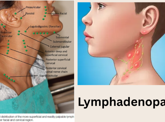
Significant lymph node enlargement: Definition, Meaning and Interpretation (MD Level)

Introduction

The definition of significant lymph node enlargement depends on the anatomical site and the patient’s age. The following table summarizes commonly accepted clinical cutoffs.

Cut off of Size for adult and children

Lymph Node RegionChildrenAdultsComments
Cervical>2 cm>1 cmSmall (<1 cm) cervical nodes are common in healthy children.
Axillary>1 cm>1 cmPersistent enlargement warrants evaluation.
Inguinal>1.5 cm>1.5 cmOften enlarged due to minor skin trauma or infections.
Epitrochlear>0.5 cm>0.5 cmAny palpable node >0.5 cm is considered abnormal.
SupraclavicularAny palpable nodeAny palpable nodeAlways considered abnormal; investigate for malignancy or serious infection.
PoplitealAny palpable nodeAny palpable nodeUsually abnormal.
MediastinalAny enlarged node on imagingAny enlarged node on imagingRequires further evaluation based on imaging findings.

Pediatric notes (Nelson Pediatrics)

  • Cervical nodes ≤1 cm are common and usually normal.
  • Inguinal nodes ≤1.5 cm may be normal.
  • Axillary nodes ≤1 cm may be normal.
  • Palpable supraclavicular or epitrochlear nodes are abnormal regardless of age.

Red flags regardless of size

Evaluate urgently if lymph nodes are:

  • Supraclavicular
  • Hard, fixed, or matted
  • Rapidly enlarging
  • Persisting >4–6 weeks without improvement
  • Associated with fever, weight loss, night sweats, hepatosplenomegaly, or generalized lymphadenopathy
  • 2 cm in the cervical region, especially if persistent or progressive

These cutoffs are consistent with standard pediatric references such as Nelson Textbook of Pediatrics and widely used clinical guidelines.

The Blood That Baffled Science for 50 Years is Solved Now

Hey there, I have an amazing tooltip !

MAL Blood Group  ·  Published in Blood Journal  ·  2024

In 1972, a routine blood test on a pregnant woman yielded something deeply puzzling: her red blood cells were missing a surface molecule that existed on every other known human blood sample of the time. Doctors noted the anomaly, filed it away, and moved on. They had no framework to explain it.

For the next five decades, that molecular absence lingered as an open question in the world of hematology — a quiet mystery buried in the archives of transfusion medicine. Then, in 2024, a team of researchers from the United Kingdom and Israel finally cracked it, identifying an entirely new human blood group system and solving one of the field’s longest-standing puzzles.

More Than Just A and B

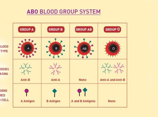
Most of us learned about blood types in school: A, B, AB, and O, with a positive or negative Rh factor tacked on. But that picture is far from complete. Human blood cells are coated in a complex landscape of proteins and sugars called antigens — and scientists use these antigen patterns to classify blood into different “group systems.” The ABO and Rh systems are simply the most medically well-known of a much larger family.

To date, researchers have identified 47 such blood group systems in humans. Most of the major ones were catalogued in the early 20th century, but new systems continue to emerge — typically rare, typically tied to a small number of individuals worldwide, and typically uncovered when something goes unexpectedly wrong during a blood transfusion.

Quick Facts — Human Blood Group Systems

  • Humans have 47 recognised blood group systems, not just ABO and Rh.
  • Blood group systems are defined by distinct antigen molecules on the surface of red blood cells.
  • Over 99.9% of people carry the AnWj antigen — meaning those without it are extraordinarily rare.
  • The newly identified system is called MAL, after the myelin and lymphocyte protein that hosts the AnWj antigen.
  • Patients who are AnWj-negative risk severe immune reactions if given AnWj-positive blood during a transfusion.

The AnWj Antigen — A Puzzle Without a Gene

The molecule missing from that 1972 patient’s blood was eventually given a name: the AnWj antigen. Scientists confirmed that more than 99.9 percent of people carry it — making those without it extraordinarily rare. But despite decades of awareness, no one could identify the gene responsible for producing it. Without a genetic explanation, it was impossible to develop a reliable test to screen for AnWj-negative patients before transfusions — leaving those individuals vulnerable to potentially severe immune reactions if given incompatible blood.

That gap was both a scientific frustration and a genuine clinical risk. Transfusion reactions can range from mild discomfort to life-threatening organ damage. For patients whose blood type falls outside known systems, matching compatible donors is extraordinarily difficult — often relying on informal networks and a great deal of luck.

It represents a huge achievement, and the culmination of a long team effort, to finally establish this new blood group system and be able to offer the best care to rare, but important, patients.— Louise Tilley, Hematologist, NHS Blood and Transplant

The Breakthrough: The MAL Gene

The team — led by senior research scientist Louise Tilley of NHS Blood and Transplant, with colleagues from the University of Bristol and collaborators in Israel — spent nearly two decades piecing together the puzzle. Their breakthrough came when they traced the AnWj antigen to a specific protein: the myelin and lymphocyte protein, encoded by the MAL gene.

When both copies of a person’s MAL gene carry mutated versions, the AnWj antigen is absent from their blood entirely. This is the inherited form of AnWj-negativity — and it is what that 1972 patient almost certainly had. Importantly, the researchers also found that in some cases, the MAL gene can be functionally suppressed by other underlying conditions, such as certain blood cancers or autoimmune disorders. This means that a newly AnWj-negative result in a patient could sometimes be a flag pointing toward a deeper, undiagnosed illness.

The study, published in Blood, the journal of the American Society of Hematology, confirmed that all AnWj-negative patients studied shared the same mutation pattern — and crucially, that no other cell abnormalities or diseases were linked to the inherited form of the condition.

A 50-Year Timeline

Why This Matters Beyond the Lab

For the handful of individuals worldwide who are AnWj-negative, this discovery is potentially life-changing. Previously, their rare blood type made surgical procedures, childbirth, or any situation requiring a transfusion a logistical and medical challenge. Identifying compatible donors without a genetic test was a matter of laborious manual screening — if it was possible at all.

Now, with the MAL gene identified, blood banks and hospitals can develop targeted genetic tests to identify AnWj-negative patients proactively — before a transfusion crisis occurs. It also allows clinicians to distinguish between patients who are AnWj-negative due to an inherited mutation (a stable, lifelong condition) and those whose MAL expression has been suppressed by illness, which may require a very different medical response.

Tilley acknowledged the difficulty of the research: “The work was difficult because the genetic cases are very rare.” Rare cases, by definition, generate little data, making statistical patterns harder to detect and genetic links harder to confirm. That the team persisted across two decades speaks to both the scientific importance of the question and the very real human stakes attached to it.

The Bigger Picture: Blood Is Still Surprising Us

The MAL discovery arrives on the heels of another recent milestone. In 2022, researchers described the Er blood group system — yet another rare system affecting a small number of people globally. Together, these findings are a reminder that human biology, even in something as fundamental as blood, continues to yield surprises. Each new blood group system identified is not merely a scientific footnote; it represents a population of patients who were previously invisible to modern medicine’s tools.

Understanding these rare variants matters because it builds the infrastructure for safer, more personalised transfusion medicine. It means fewer unexplained reactions, fewer desperate searches for compatible donors, and more patients receiving care that is genuinely matched to their biology.

Half a century after a pregnant woman’s unusual blood sample puzzled a doctor somewhere in the world, science finally has an answer.

The MAL blood group system is now the 47th addition to our understanding of human blood — a testament to the slow, painstaking, but ultimately rewarding work of science. And for the extraordinarily rare individuals who carry this invisible distinction in their veins, it may mean the difference between a routine transfusion and a medical emergency.

Based on research published in Blood, American Society of Hematology, 2024. Research led by Louise Tilley, NHS Blood and Transplant & University of Bristol.

Sometimes, the most important discoveries begin with a single anomaly that refuses to be forgotten.

PCOS? No Here is the New Name of so Common multisystem Syndrome Renamed in 2026

Polycystic Ovary Syndrome (PCOS / PCOD) — High-Yield Notes

Based on international evidence-based guidelines, Endocrine Society, ACOG, and peer-reviewed literature. (NCBI)


Definition

  • PCOS = common endocrine-metabolic disorder in reproductive-age women
  • Characterized by:
    • Hyperandrogenism
    • Ovulatory dysfunction
    • Polycystic ovarian morphology
  • Associated with:
    • Insulin resistance
    • Obesity
    • Infertility
    • Metabolic syndrome

Etiopathogenesis

Multifactorial Disorder

  • Genetic predisposition
  • Environmental factors
  • Hormonal dysregulation
  • Insulin resistance

Core Pathophysiology

1. Increased LH secretion

  • ↑ GnRH pulse frequency → preferential LH secretion
  • ↑ LH stimulates theca cells
  • ↑ androgen production

2. Insulin resistance

  • Present in many patients (even lean PCOS)
  • Hyperinsulinemia:
    • Stimulates ovarian androgen synthesis
    • Suppresses SHBG production in liver
    • ↑ free testosterone

3. Follicular arrest

  • Failure of dominant follicle maturation
  • Multiple immature follicles accumulate

4. Hyperandrogenism

  • Causes:
    • Hirsutism
    • Acne
    • Alopecia
    • Menstrual irregularity

Diagnostic Criteria (Rotterdam Criteria)

Diagnosis requires 2 out of 3 after excluding other causes: (NCBI)

A. Ovulatory Dysfunction

  • Oligomenorrhea
  • Amenorrhea
  • Anovulation

Menstrual abnormalities

  • Cycle >35 days
  • <8 cycles/year

B. Hyperandrogenism

Clinical

  • Hirsutism
  • Acne
  • Androgenic alopecia

Biochemical

  • ↑ Total testosterone
  • ↑ Free testosterone
  • ↑ DHEAS

C. Polycystic Ovarian Morphology (USG)

  • ≥20 follicles per ovary OR
  • Ovarian volume >10 mL

Classic appearance

  • “String of pearls”

Important Diagnostic Point


Differential Diagnoses to Exclude

Endocrine causes

  • Hypothyroidism
  • Hyperprolactinemia
  • Cushing syndrome
  • Congenital adrenal hyperplasia
  • Androgen-secreting tumors

Others

  • Acromegaly
  • Premature ovarian insufficiency

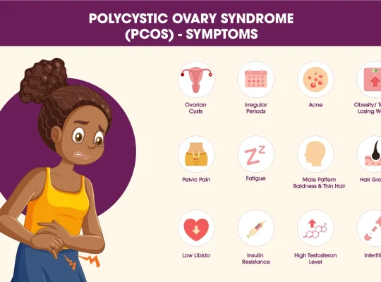
Clinical Features

Menstrual

  • Oligomenorrhea
  • Amenorrhea
  • Irregular cycles
  • Infertility

Hyperandrogenic Features

  • Hirsutism
  • Acne
  • Alopecia
  • Seborrhea

Metabolic Features

  • Obesity
  • Central obesity
  • Insulin resistance
  • Acanthosis nigricans

Reproductive Features

  • Subfertility/infertility
  • Recurrent miscarriage

Psychological Associations

  • Anxiety
  • Depression
  • Eating disorders
  • Poor body image

Investigations

Hormonal Tests

  • Total/free testosterone
  • DHEAS
  • LH, FSH
  • Prolactin
  • TSH
  • 17-hydroxyprogesterone

Metabolic Screening

  • Fasting glucose
  • HbA1c
  • Lipid profile
  • OGTT (high-risk patients)

Imaging

  • Pelvic ultrasonography

Typical Laboratory Findings

  • ↑ LH:FSH ratio (>2:1 sometimes)
  • ↑ Testosterone
  • ↑ Insulin
  • ↓ SHBG

Complications

Reproductive

  • Infertility
  • Anovulation
  • Pregnancy complications

Metabolic


Cardiovascular

  • Increased long-term CV risk

Endometrial

  • Endometrial hyperplasia
  • Endometrial carcinoma
    • Due to chronic unopposed estrogen

Management

1. Lifestyle Modification (First-line)

  • Weight reduction
  • Exercise
  • Calorie restriction
  • Low glycemic diet

Benefits

  • Improves ovulation
  • Reduces insulin resistance
  • Improves fertility

2. Menstrual Irregularity Management

Combined Oral Contraceptive Pills (COCPs)

  • First-line for nonfertility symptoms
  • Benefits:
    • Regular cycles
    • ↓ androgen production
    • Improves acne/hirsutism

3. Hirsutism & Acne

Antiandrogens

  • Spironolactone
  • Finasteride
  • Flutamide (rare due to hepatotoxicity)

Important

  • Use contraception with antiandrogens

4. Insulin Resistance

Metformin

  • Improves insulin sensitivity
  • May restore ovulation
  • Useful in:
    • Obesity
    • Prediabetes
    • Metabolic syndrome

5. Infertility Treatment

First-line Ovulation Induction

  • Letrozole (preferred)
  • Clomiphene citrate

Others

  • Gonadotropins
  • IVF if resistant

Pregnancy Risks in PCOS

  • Gestational diabetes
  • Pregnancy-induced hypertension
  • Preeclampsia
  • Preterm birth

Adolescent PCOS

  • Diagnosis difficult soon after menarche
  • Physiologic irregular cycles common
  • Ultrasound less reliable in adolescents (NCBI)

High-Yield Exam Pearls

  • Most accepted criteria = Rotterdam criteria
  • Need 2 out of 3 criteria
  • PCOS is a diagnosis of exclusion
  • Most common cause of anovulatory infertility
  • Insulin resistance is central mechanism
  • Chronic anovulation → unopposed estrogen → endometrial cancer risk
  • First-line treatment = lifestyle modification
  • First-line ovulation induction = letrozole
  • COCPs are first-line for menstrual symptoms
  • “String of pearls” appearance on USG

Very Short Summary

PCOS is a common endocrine disorder characterized by:

  • Hyperandrogenism
  • Irregular ovulation
  • Polycystic ovaries

Main problems:

  • Irregular periods
  • Infertility
  • Hirsutism
  • Obesity
  • Insulin resistance

Treatment:

  • Lifestyle change
  • COCPs
  • Metformin
  • Letrozole for fertility

Renaming PCOS to PMOS

Yes — there has been a major recent international change in terminology.

New Name for PCOS

The condition previously called PCOS (Polycystic Ovary Syndrome) has officially been renamed:

PMOS

Polyendocrine Metabolic Ovarian Syndrome

This was announced in 2026 after a 14-year international consensus effort involving:

  • Endocrinologists
  • Gynecologists
  • Researchers
  • Patient advocacy groups
  • More than 50 global medical organizations (The Guardian)

Why Was the Name Changed?

Experts felt the term “PCOS” was misleading because:

  • Many patients do not actually have ovarian cysts
  • The disorder affects multiple body systems, not only ovaries
  • The old name caused:
    • Delayed diagnosis
    • Confusion
    • Stigma
    • Under-recognition of metabolic disease

The new term “PMOS” better reflects:

  • Endocrine dysfunction
  • Metabolic abnormalities
  • Hormonal imbalance
  • Reproductive effects (The Guardian)

Full Form Breakdown

P — Polyendocrine

Multiple hormone systems are involved

M — Metabolic

Strong association with:

  • Insulin resistance
  • Obesity
  • Diabetes
  • Dyslipidemia

O — Ovarian

Ovarian dysfunction and ovulatory problems remain important

S — Syndrome

Collection of related clinical features


Important Clinical Point

Diagnostic criteria remain essentially the same.

The name changed, but the underlying disorder and diagnostic approach remain based on established international criteria. (The Cut)


Key Facts

  • Affects approximately 1 in 8 women worldwide
  • Estimated >170 million affected globally
  • Strongly associated with:
    • Infertility
    • Metabolic syndrome
    • Type 2 diabetes
    • Cardiovascular risk
    • Mental health disorders (endocrine.org)

Transition Timeline

International organizations plan gradual adoption of the term PMOS in:

  • Clinical guidelines
  • Research papers
  • Medical education
  • Public awareness campaigns

Implementation is expected over the next few years. (The Guardian)

Acute Hepatic Failure in Children (Pediatric Acute Liver Failure): Complete Clinical Guide

What is Acute Hepatic Failure?

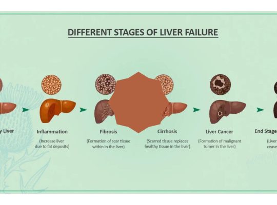
Acute Hepatic Failure (AHF), also called Pediatric Acute Liver Failure (PALF), is a rapidly progressive liver dysfunction occurring in a child without pre-existing chronic liver disease, leading to severe impairment of liver synthetic function and encephalopathy.

It is a medical emergency associated with:

  • Massive hepatocellular injury
  • Coagulopathy
  • Hepatic encephalopathy
  • Multi-organ dysfunction
  • High mortality without timely management or liver transplantation

According to major pediatric references including AAP, Nelson Textbook of Pediatrics, and ISPGHAN, early recognition and aggressive supportive care are critical for survival.


Definition of Pediatric Acute Liver Failure

Pediatric acute liver failure is defined by:

Essential Criteria

  • No evidence of chronic liver disease
  • Acute liver injury
  • Coagulopathy not corrected by vitamin K

Coagulation Criteria

  • INR >1.5 with encephalopathy
    OR
  • INR >2 without encephalopathy

Why Acute Hepatic Failure is Dangerous

The liver performs critical functions:

  • Glucose regulation
  • Protein synthesis
  • Clotting factor production
  • Ammonia detoxification
  • Drug metabolism
  • Immune regulation

When the liver suddenly fails:

  • Toxins accumulate
  • Cerebral edema develops
  • Severe bleeding can occur
  • Shock and renal failure may follow

Epidemiology of Pediatric Acute Liver Failure

  • Rare but life-threatening condition
  • Significant cause of PICU admissions
  • Common indication for pediatric liver transplantation
  • Mortality remains high despite advances

Common Age Groups

  • Infants: metabolic and viral causes
  • Older children/adolescents: drugs, autoimmune hepatitis, Wilson disease

Etiology of Acute Hepatic Failure in Children

acute liver failure causes

1. Viral Hepatitis

Common Viral Causes

  • Hepatitis A
  • Hepatitis B
  • Hepatitis E
  • HSV (especially neonates)
  • EBV
  • CMV
  • Adenovirus
  • Enteroviruses

Important Point

In developing countries including Nepal and South Asia:

  • Hepatitis A and E remain major causes

2. Drug-Induced Liver Injury (DILI)

Common Drugs

  • Acetaminophen (Paracetamol)
  • Antitubercular drugs
  • Valproate
  • Antiepileptics
  • Herbal medications

Acetaminophen Toxicity

Most common cause in many developed countries.

Toxic metabolite:

  • NAPQI

Normally detoxified by glutathione.


3. Metabolic Disorders

Especially important in infants.

Major Causes

  • Galactosemia
  • Tyrosinemia
  • Mitochondrial disorders
  • Fatty acid oxidation defects
  • Wilson disease
  • Neonatal hemochromatosis

4. Autoimmune Hepatitis

Can present dramatically with:

  • Jaundice
  • Coagulopathy
  • Encephalopathy

Look for:

  • ANA
  • ASMA
  • Elevated IgG

5. Ischemic and Toxic Causes

  • Shock liver
  • Sepsis
  • Mushroom poisoning
  • Toxins

6. Indeterminate Causes

A substantial number of pediatric cases remain unexplained despite extensive workup.


Pathophysiology of Acute Hepatic Failure

Hepatocyte Injury

Massive hepatocyte necrosis leads to:

  • Failure of detoxification
  • Reduced clotting factor synthesis
  • Metabolic instability

Hyperammonemia

Ammonia accumulates due to impaired hepatic detoxification.

This causes:

  • Astrocyte swelling
  • Cerebral edema
  • Increased intracranial pressure

Coagulopathy

Liver cannot synthesize:

  • Factors II
  • V
  • VII
  • IX
  • X

Result:

  • Severe bleeding tendency

Immune Dysfunction

Patients become highly susceptible to:

  • Sepsis
  • Fungal infections

Clinical Features of Acute Hepatic Failure

Early Symptoms

  • Nausea
  • Vomiting
  • Malaise
  • Fever
  • Abdominal pain
  • Poor feeding
  • Irritability

Liver-Specific Findings


Features of Hepatic Encephalopathy

Stage I

  • Irritability
  • Sleep disturbances
  • Behavioral changes

Stage II

  • Confusion
  • Drowsiness
  • Asterixis

Stage III

  • Stupor
  • Hyperreflexia

Stage IV

  • Coma

Signs of Cerebral Edema

  • Hypertension
  • Bradycardia
  • Unequal pupils
  • Abnormal posturing

This is a life-threatening emergency.


Diagnostic Evaluation of Acute Hepatic Failure

Initial Laboratory Workup

Liver Function Tests

  • AST/ALT
  • Bilirubin
  • Albumin
  • ALP
  • GGT

Synthetic Function

  • PT/INR
  • Fibrinogen

Metabolic Evaluation

  • Blood glucose
  • Lactate
  • Serum ammonia
  • ABG

Viral Studies

  • HAV IgM
  • HBsAg
  • Anti-HBc IgM
  • HEV serology
  • HSV PCR

Autoimmune Tests

  • ANA
  • ASMA
  • Anti-LKM
  • IgG

Metabolic Tests

  • Ceruloplasmin
  • Urine succinylacetone
  • Plasma amino acids
  • Urine organic acids

Imaging

Ultrasound Abdomen with Doppler

Useful for:

  • Liver size
  • Vascular patency
  • Ascites
  • Chronic liver disease exclusion

Important ICU Monitoring

Continuous monitoring of:

  • Mental status
  • Blood glucose
  • ICP signs
  • Urine output
  • Electrolytes
  • INR
  • Ammonia

Management of Acute Hepatic Failure

Core Principles

  • PICU admission
  • Aggressive supportive care
  • Prevent cerebral edema
  • Treat underlying cause
  • Early transplant referral

Stabilization

Airway

Intubate if:

  • Grade III/IV encephalopathy
  • Airway compromise

Circulation

Maintain:

  • Adequate perfusion
  • MAP
  • Renal function

Avoid fluid overload.


Management of Hypoglycemia

Frequent glucose monitoring is mandatory.

Treatment:

  • Dextrose bolus
  • Continuous glucose infusion

Cerebral Edema Management

General Measures

  • Head elevation to 30°
  • Avoid neck compression
  • Minimize stimulation

Osmotherapy

Mannitol

  • 0.5–1 g/kg IV

OR

Hypertonic Saline

Target serum sodium:

  • 145–150 mEq/L

Ammonia Reduction

Lactulose

Reduces ammonia absorption from gut.

Renal Replacement Therapy

Indicated for:

  • Severe hyperammonemia
  • Renal failure

Coagulopathy Management

Important principle:

  • Do NOT correct INR routinely unless bleeding or procedure planned.

Options

  • Vitamin K
  • FFP
  • Cryoprecipitate
  • Platelets

Infection Control

High suspicion for:

  • Bacterial infections
  • Fungal sepsis

Empiric antibiotics are often used in critically ill patients.


Etiology-Specific Treatment

Acetaminophen Toxicity

N-acetylcysteine (NAC)

Acts by:

  • Replenishing glutathione
  • Improving hepatic perfusion

N-acetylcysteine restores glutathione and reduces NAPQI toxicity\text{N-acetylcysteine restores glutathione and reduces } NAPQI \text{ toxicity}N-acetylcysteine restores glutathione and reduces NAPQI toxicity


HSV Hepatitis

  • IV acyclovir

Autoimmune Hepatitis

  • Corticosteroids

Wilson Disease

Usually requires urgent liver transplantation.


Nutrition in Acute Hepatic Failure

Key Principles

  • Early enteral nutrition preferred
  • Avoid prolonged fasting
  • Adequate calories essential

Protein

Previously restricted heavily, but modern pediatric guidelines recommend:

  • Avoid excessive restriction
  • Individualize according to encephalopathy severity

Liver Transplantation in Acute Hepatic Failure

Indications

  • Progressive encephalopathy
  • Refractory coagulopathy
  • Severe acidosis
  • Persistent hyperammonemia
  • Multi-organ failure

Poor Prognostic Factors

  • INR worsening
  • Severe encephalopathy
  • Cerebral edema
  • Renal failure
  • Rising bilirubin
  • Persistent lactic acidosis

Complications of Acute Hepatic Failure

Neurologic

  • Cerebral edema
  • Seizures
  • Herniation

Hematologic

  • Bleeding
  • DIC

Renal

Infectious

  • Sepsis
  • Fungal infections

Prognosis

Outcome depends on:

  • Etiology
  • Speed of recognition
  • Availability of transplant
  • Degree of encephalopathy

Better Prognosis

  • Hepatitis A
  • Acetaminophen toxicity (early NAC)

Worse Prognosis

  • Wilson disease
  • Indeterminate PALF
  • Severe cerebral edema

High-Yield Exam Points on Acute Hepatic Failure

Most Important Diagnostic Marker

  • Elevated INR

Most Common Cause in Developed Countries

  • Acetaminophen toxicity

Major Cause of Death

  • Cerebral edema and sepsis

Drug of Choice in Acetaminophen Toxicity

  • N-acetylcysteine

Key Emergency

  • Raised intracranial pressure

Acute Hepatic Failure Flowchart

Acute Liver Injury

Coagulopathy (INR ↑)

Evaluate Etiology

PICU Supportive Care

Prevent Cerebral Edema

Treat Specific Cause

Assess for Liver Transplant

Frequently Asked Questions (FAQs)

Is acute hepatic failure reversible?

Yes. Some causes recover completely with early management, while others require liver transplantation.

What is the most dangerous complication?

Cerebral edema leading to brain herniation.

Why is ammonia elevated?

The failing liver cannot convert ammonia into urea effectively.

Can children survive without liver transplant?

Yes, depending on etiology and severity. Hepatitis A-related PALF often recovers spontaneously.


Key Takeaway

Acute hepatic failure in children is a rapidly progressive and potentially fatal condition requiring:

  • Early diagnosis
  • Intensive monitoring
  • Prevention of cerebral edema
  • Etiology-directed therapy
  • Timely liver transplantation evaluation

Rapid recognition and evidence-based critical care significantly improve survival outcomes.

5 Critical Mistakes Pediatricians Make During Neonatal Resuscitation (And How to Avoid Them)


Introduction

Neonatal resuscitation is one of the most time-sensitive and high-stakes procedures in pediatrics. Despite structured guidelines like the American Academy of Pediatrics and the Neonatal Resuscitation Program, errors still occur—even among experienced clinicians.

These mistakes can lead to preventable morbidity and mortality. Understanding them is essential not only for pediatricians but also for residents, nurses, and delivery room staff.

In this article, we’ll break down 5 common mistakes pediatricians make during neonatal resuscitation and how to correct them using evidence-based practice.


1. Delayed Initiation of Positive Pressure Ventilation (PPV)

❌ The Mistake

Failure to initiate Positive Pressure Ventilation (PPV) within the “Golden Minute” when the neonate is apneic, gasping, or has a heart rate <100 bpm.

⚠️ Why It Matters

  • Ventilation—not chest compressions—is the most critical intervention
  • Delays increase risk of:
    • Hypoxic-ischemic injury
    • Bradycardia progression
    • Poor neurological outcomes

✅ Best Practice

  • Assess breathing and heart rate immediately after birth
  • Start PPV within 60 seconds (“Golden Minute”)
  • Use pulse oximetry early

2. Inadequate Mask Seal and Poor Ventilation Technique

❌ The Mistake

  • Improper mask size or seal
  • Incorrect head positioning
  • Ineffective ventilation despite apparent effort

⚠️ Why It Matters

  • Most failed resuscitations are due to ineffective ventilation
  • Leads to persistent hypoxia and bradycardia

✅ Best Practice (MR SOPA Approach)

Follow the MR SOPA corrective steps:

  • Mask adjustment
  • Reposition airway
  • Suction mouth and nose
  • Open mouth
  • Pressure increase
  • Airway alternative (ET tube or LMA)

3. Premature Initiation of Chest Compressions

❌ The Mistake

Starting chest compressions before ensuring adequate ventilation

⚠️ Why It Matters

  • Neonatal cardiac arrest is usually respiratory in origin
  • Without proper ventilation:
    • Compressions are ineffective
    • Oxygen delivery remains inadequate

✅ Best Practice

  • Ensure effective ventilation for at least 30 seconds
  • Start compressions only if:
    • HR <60 bpm despite effective PPV

4. Incorrect Oxygen Use (Too Much or Too Little)

❌ The Mistake

  • Starting all neonates on 100% oxygen
  • Failure to titrate oxygen using pulse oximetry

⚠️ Why It Matters

  • Hyperoxia → oxidative stress, especially in preterms
  • Hypoxia → organ damage

✅ Best Practice

  • Term babies: start with 21% oxygen (room air)
  • Preterm babies: start with 21–30% oxygen
  • Adjust based on preductal SpO₂ targets

5. Poor Team Communication and Role Assignment

❌ The Mistake

  • Lack of clear leadership
  • Unassigned roles
  • Ineffective communication during resuscitation

⚠️ Why It Matters

  • Leads to:
    • Delayed interventions
    • Duplicate or missed actions
    • Increased stress and errors

✅ Best Practice

  • Assign roles before delivery:
    • Airway manager
    • Compressor
    • Medication nurse
    • Team leader
  • Use closed-loop communication
  • Conduct pre-resuscitation briefing

Pro Tips for Better Neonatal Resuscitation

  • Always prepare equipment before delivery
  • Anticipate high-risk deliveries
  • Use checklists
  • Practice simulation training regularly
  • Follow updates from American Heart Association

Conclusion

Even skilled pediatricians can make errors during neonatal resuscitation—but most are preventable. The key lies in:

  • Mastering ventilation techniques
  • Following structured protocols
  • Practicing teamwork and communication

By avoiding these common mistakes, clinicians can significantly improve neonatal outcomes and reduce mortality.

FAQs

What is the most common mistake in neonatal resuscitation?

The most common mistake is ineffective ventilation due to poor mask seal or technique.

When should chest compressions be started in neonates?

Only when the heart rate is below 60 bpm after 30 seconds of effective ventilation.

Why is oxygen titration important in newborn resuscitation?

Both hypoxia and hyperoxia are harmful, especially in preterm infants.

20 Common MCQs in The Spleen: Your Body’s Hidden Guardian

Introduction

While most people can easily point to their heart or stomach, the spleen remains a bit of a mystery. Tucked away in the upper left side of your abdomen, this fist-sized organ quietly performs some of the body’s most critical “housekeeping” and security tasks.

Where is it Located?

The spleen is located in the left upper quadrant of the abdomen, shielded by the 9th, 10th, and 11th ribs. It sits just below the diaphragm and behind the stomach. In a healthy adult, it is usually about the size of a small avocado or a clenched fist and cannot be felt through the skin.

What Does the Spleen Actually Do?

Think of your spleen as a multi-purpose facility that serves two primary systems: the immune system and the blood (hematologic) system.

  • The Blood Filter: One of its main jobs is to act as a quality control center for your blood. As blood flows through the spleen, it identifies and removes old, malformed, or damaged red blood cells.
  • Immune Surveillance: It is the largest organ in the lymphatic system. It produces and stores white blood cells (lymphocytes) and antibodies that spring into action to fight off bacteria and viruses.
  • Emergency Reservoir: The spleen acts as a backup tank, storing about one-third of the body’s platelets and a significant reserve of red blood cells. In the event of severe bleeding or physical stress, the spleen can contract to squeeze this extra blood into your circulation.
  • Recycling Center: When it breaks down old red blood cells, it carefully recycles the iron, sending it back to the bone marrow to help create new hemoglobin.

A Tale of Two Pulps

Inside the spleen’s tough outer capsule, there are two distinct types of tissue, each with a specific specialty:

  1. Red Pulp: This makes up about 80% of the organ and is responsible for the filtering and storage of blood.
  2. White Pulp: This tissue is part of the immune system. It produces white blood cells that produce antibodies to target specific infections.

Can You Live Without It?

Yes, you can live without a spleen—a condition known as asplenia. If the spleen is removed (a surgery called a splenectomy), other organs like the liver and bone marrow take over many of its duties. However, because the spleen is so vital for fighting certain types of bacteria, people without one are at a much higher risk for infections and must stay up-to-date on specific vaccinations.

Keeping Your Spleen Healthy

While many spleen issues are secondary to other conditions (like liver disease or certain cancers), you can support yours by:

  • Preventing Injury: Wear protective gear during contact sports to avoid a ruptured spleen, which is a medical emergency.
  • Hydration & Diet: Drinking plenty of water and eating nutrient-rich foods supports the lymphatic system overall.
  • Infection Control: Managing infections like mononucleosis (which can cause the spleen to enlarge, or “splenomegaly”) is key to preventing long-term damage.

Spleen MCQs (NCLEX Style)

Spleen MCQs (NCLEX Style)

1. The spleen is located in:
A. Right hypochondrium
B. Left hypochondrium
C. Epigastrium
D. Umbilical region

Click to view answer

Answer: B. Left hypochondrium
Explanation: The spleen lies in the LUQ under ribs 9–11.


2. The spleen becomes palpable when enlarged to:
A. 1.5 times
B. 2 times
C. 3 times
D. 5 times

Click to view answer

Answer: C. 3 times
Explanation: Clinically palpable when significantly enlarged.


3. Most common cause of splenomegaly worldwide:
A. Leukemia
B. Malaria
C. Cirrhosis
D. TB

Click to view answer

Answer: B. Malaria
Explanation: Very common in endemic regions.


4. Most common cause of splenic rupture:
A. Infection
B. Tumor
C. Trauma
D. Congenital

Click to view answer

Answer: C. Trauma


5. The spleen develops from:
A. Endoderm
B. Mesoderm
C. Ectoderm
D. Neural crest

Click to view answer

Answer: B. Mesoderm


6. Arterial supply of spleen:
A. Hepatic artery
B. SMA
C. Splenic artery
D. IMA

Click to view answer

Answer: C. Splenic artery


7. Splenic artery arises from:
A. Aorta
B. SMA
C. Celiac trunk
D. Renal artery

Click to view answer

Answer: C. Celiac trunk


8. Spleen is attached to kidney by:
A. Gastrosplenic ligament
B. Splenorenal ligament
C. Falciform ligament
D. Coronary ligament

Click to view answer

Answer: B. Splenorenal ligament


9. Which is NOT a function of spleen?
A. RBC destruction
B. Immunity
C. Platelet storage
D. Insulin secretion

Click to view answer

Answer: D. Insulin secretion


10. Spleen stores:
A. Lymphocytes only
B. RBC only
C. Platelets and RBC
D. Plasma

Click to view answer

Answer: C. Platelets and RBC


11. Hypersplenism causes:
A. Leukocytosis
B. Pancytopenia
C. Polycythemia
D. Thrombocytosis

Click to view answer

Answer: B. Pancytopenia


12. Common indication for splenectomy:
A. Iron deficiency anemia
B. ITP
C. Diabetes
D. Hypertension

Click to view answer

Answer: B. ITP


13. Functional asplenia is seen in:
A. Thalassemia
B. Sickle cell disease
C. Leukemia
D. Hemophilia

Click to view answer

Answer: B. Sickle cell disease


14. Most serious complication after splenectomy:
A. Bleeding
B. Thrombosis
C. Infection
D. Anemia

Click to view answer

Answer: C. Infection (OPSI)


15. Most common organism in OPSI:
A. Streptococcus pneumoniae
B. E. coli
C. Pseudomonas
D. Klebsiella

Click to view answer

Answer: A. Streptococcus pneumoniae


16. Essential vaccine before splenectomy:
A. Hep B
B. Rabies
C. Pneumococcal
D. BCG

Click to view answer

Answer: C. Pneumococcal


17. Left shoulder pain in splenic injury is:
A. Murphy sign
B. Cullen sign
C. Kehr sign
D. Rovsing sign

Click to view answer

Answer: C. Kehr sign


18. Most commonly injured organ in blunt trauma:
A. Liver
B. Spleen
C. Kidney
D. Pancreas

Click to view answer

Answer: B. Spleen


19. Howell-Jolly bodies indicate:
A. Liver disease
B. Splenic dysfunction
C. Iron deficiency
D. Infection

Click to view answer

Answer: B. Splenic dysfunction


20. Massive splenomegaly is seen in:
A. Appendicitis
B. CML
C. Asthma
D. Diabetes

Click to view answer

Answer: B. CML

Complete Definition of Acute Liver Failure (According to Nelson)

📘 Standard Pediatric Definition (Nelson Textbook of Pediatrics)

👉 Acute Liver Failure (ALF) is defined as:

Evidence of acute liver injury in a child with no pre-existing chronic liver disease, accompanied by hepatic-based coagulopathy (INR ≥1.5 with encephalopathy OR INR ≥2.0 without encephalopathy), not corrected by vitamin K.


🔑 Key Components to Remember

1. Acute liver injury

  • Elevated transaminases (AST/ALT)
  • Recent onset (days to weeks)

2. No prior chronic liver disease

  • Important to differentiate from acute-on-chronic liver failure

3. Coagulopathy (core criterion)

  • INR ≥1.5 + encephalopathy
    OR
  • INR ≥2.0 without encephalopathy
  • Must be unresponsive to vitamin K

4. Encephalopathy (may be absent in children)

  • Unlike adults, pediatric ALF does NOT require encephalopathy for diagnosis

🧠 Exam Pearls (Very Important)

  • Coagulopathy is mandatory
  • Encephalopathy is NOT mandatory in pediatrics
  • Always mention vitamin K non-correction

🧾 One-line Answer for Exams

👉 “Acute liver failure is acute hepatic injury without prior liver disease, with INR ≥1.5 with encephalopathy or ≥2 without encephalopathy, not corrected by vitamin K.”

Exit mobile version