Blog

Crohn’s Disease — MD-Level Note (based on Harrison’s Principles of Internal Medicine)


Video on Chron’s Disease (sensitizer)

🔬 Overview & Definition

Crohn’s disease (CD) is a chronic, relapsing inflammatory bowel disease (IBD) characterized by:

  • Transmural inflammation
  • Segmental (“skip”) involvement
  • Can affect any part of GI tract (mouth → anus), most commonly:
    • Terminal ileum ± colon

🧬 Etiopathogenesis

1. Genetic Susceptibility

  • Strong association with:
    • NOD2 (CARD15) mutation
  • Other genes: ATG16L1, IL23R
  • Family clustering common

2. Immune Dysregulation

  • Predominantly Th1 and Th17 mediated response
  • ↑ Cytokines:
    • TNF-α, IL-12, IL-23
  • Impaired regulatory T-cell function

3. Microbiome Interaction

  • Dysbiosis with abnormal response to gut flora
  • Loss of tolerance to commensals

4. Environmental Factors

  • Smoking (↑ risk, worse prognosis)
  • NSAIDs, infections, diet

🧠 Pathology

Gross Features

  • Skip lesions
  • Cobblestone appearance
  • Strictures (“string sign”)
  • Creeping fat

Microscopy

  • Transmural inflammation
  • Non-caseating granulomas (not always present)
  • Lymphoid aggregates
  • Fissuring ulcers → fistula formation

📍 Distribution Patterns

  • Ileocolonic (most common)
  • Isolated ileal
  • Isolated colonic
  • Upper GI involvement (rare but important)

⚠️ Clinical Features

Intestinal Symptoms

  • Chronic diarrhea (may be non-bloody)
  • Abdominal pain (RLQ common)
  • Weight loss, malnutrition
  • Fever during flares

Complications

  • Strictures → obstruction
  • Fistulas:
    • Enteroenteric
    • Enterocutaneous
    • Perianal (hallmark)
  • Abscess formation

Extraintestinal Manifestations

  • Joints: peripheral arthritis, ankylosing spondylitis
  • Skin: erythema nodosum, pyoderma gangrenosum
  • Eyes: uveitis, episcleritis
  • Hepatobiliary: PSC (less common than in UC)

🔎 Diagnosis

1. Endoscopy (Gold Standard)

  • Patchy inflammation
  • Aphthous ulcers → deep linear ulcers
  • Cobblestone mucosa

2. Imaging

  • MR enterography preferred
  • CT for complications
  • Barium:
    • String sign

3. Histology

  • Transmural inflammation
  • Granulomas (supportive, not mandatory)

4. Laboratory Findings

  • ↑ CRP, ESR
  • Anemia (iron deficiency, chronic disease)
  • Hypoalbuminemia
  • Fecal calprotectin ↑

🧾 Differential Diagnosis

  • Ulcerative colitis
  • Intestinal tuberculosis (important in Nepal)
  • Infectious enterocolitis
  • Ischemic colitis

🧑‍⚕️ Disease Classification

Montreal Classification

  • Age at diagnosis (A1–A3)
  • Location (L1–L4)
  • Behavior:
    • B1: inflammatory
    • B2: stricturing
    • B3: penetrating

💊 Management (Step-Up vs Top-Down Approach)

1. Induction Therapy

Mild–Moderate

  • Budesonide (ileocecal disease)
  • 5-ASA (limited role in CD)

Moderate–Severe

  • Systemic corticosteroids

2. Maintenance Therapy

  • Thiopurines (azathioprine, 6-MP)
  • Methotrexate
  • Biologics:
    • Anti-TNF:
      • Infliximab
      • Adalimumab
    • Anti-integrin:
      • Vedolizumab
    • Anti-IL-12/23:
      • Ustekinumab

3. Surgical Management

  • Not curative (unlike UC)
  • Indications:
    • Obstruction
    • Fistula
    • Abscess
    • Refractory disease

🚨 Complications

  • Short bowel syndrome
  • Malabsorption (B12 deficiency)
  • Colorectal cancer (less than UC but still increased risk)
  • Osteoporosis (steroid use)

📊 Prognosis

  • Chronic relapsing course
  • Majority require surgery at some point
  • Smoking cessation improves outcomes significantly

🧠 High-Yield Harrison Pearls

  • Transmural + skip lesions = Crohn’s
  • Perianal disease strongly suggests CD
  • Granulomas: specific but not sensitive
  • Surgery is not curative
  • Anti-TNF revolutionized management

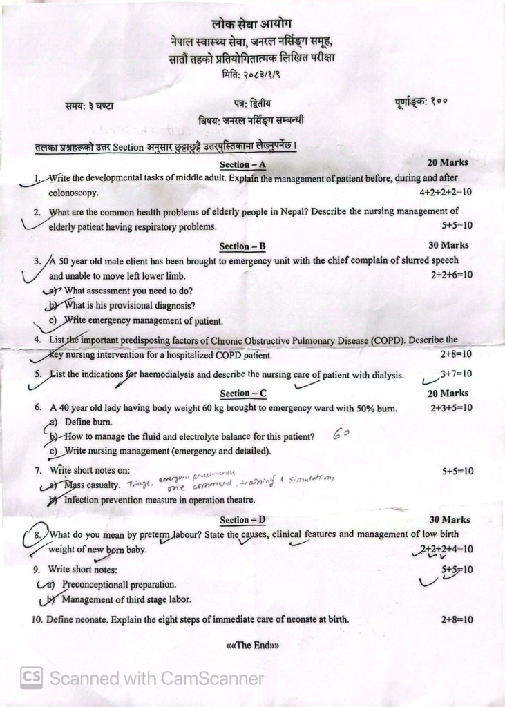
Nepal Nursing 7th level Question 2083 (past nursing officer question)


लोक सेवा आयोग

नेपाल स्वास्थ्य सेवा, जनरल नर्सिङ समूह,

सातौं तहको प्रतियोगितात्मक लिखित परीक्षा

मिति: २०८३/१/९

समय: ३ घण्टा | पत्र: द्वितीय | पूर्णाङ्क: १००

विषय: जनरल नर्सिङ सम्बन्धी

तलका प्रश्नहरूको उत्तर Section अनुसार छुट्टाछुट्टै उत्तरपुस्तिकामा लेख्नुपर्नेछ।

Section – A (20 Marks)

  1. Write the developmental tasks of middle adult. Explain the management of patient before, during and after colonoscopy. (4+2+2+2=10)
  2. What are the common health problems of elderly people in Nepal? Describe the nursing management of elderly patient having respiratory problems. (5+5=10)

Section – B (30 Marks)

  1. A 50 year old male client has been brought to emergency unit with the chief complain of slurred speech and unable to move left lower limb. (2+2+6=10)a) What assessment you need to do?b) What is his provisional diagnosis?c) Write emergency management of patient.
  2. List the important predisposing factors of Chronic Obstructive Pulmonary Disease (COPD). Describe the key nursing intervention for a hospitalized COPD patient. (2+8=10)
  3. List the indications for haemodialysis and describe the nursing care of patient with dialysis. (3+7=10)

Section – C (20 Marks)

  1. A 40 year old lady having body weight 60 kg brought to emergency ward with 50% burn. (2+3+5=10)a) Define burn.b) How to manage the fluid and electrolyte balance for this patient? [Note: ’60’ is written next to this]c) Write nursing management (emergency and detailed).
  2. Write short notes on: (5+5=10)a) Mass casualty. [Handwritten addition: Triage, emergency preparedness, one command, training & simulation]b) Infection prevention measure in operation theatre.

Section – D (30 Marks)

  1. What do you mean by preterm labour? State the causes, clinical features and management of low birth weight of new born baby. (2+2+2+4=10)
  2. Write short notes: (5+5=10)a) Preconceptionall preparation.b) Management of third stage labor.
  3. Define neonate. Explain the eight steps of immediate care of neonate at birth. (2+8=10)

5 Very Important Abdominal (GI) anomalies in Down syndrome

🚻 Genitourinary (GU) anomalies in Down syndrome

Renal / urinary tract anomalies:

  • Hydronephrosis
  • Vesicoureteral reflux (VUR)
  • Posterior urethral valves (PUV) (in males)
  • Structural renal anomalies (less frequent than cardiac/GI)

External genital anomalies:

  • Cryptorchidism (undescended testes)
  • Hypospadias
  • Smaller genitalia (common phenotypic feature, not a malformation)

🧠 Clinical Pearls (Exam gold)

  • If a neonate with Down syndrome has bilious vomiting → think duodenal atresia first
  • If there is delayed meconium → rule out Hirschsprung disease
  • Always screen:
    • Echocardiography (most important—AV canal defects common)
    • Abdominal ultrasound if symptoms suggest GU involvement

🔑 High-yield GI associations:

  • Duodenal atresia
    • Classic “double bubble” sign on X-ray
    • Presents with early bilious vomiting
  • Hirschsprung disease
    • Failure to pass meconium, abdominal distension
    • Due to absence of ganglion cells in distal colon
  • Annular pancreas
    • Can cause duodenal obstruction
  • Imperforate anus (less common than in VACTERL but reported)

⚠️ Pattern to remember

  • Down syndrome = GI obstruction + Hirschsprung risk
  • GU anomalies are less consistent but still clinically relevant

Kawasaki Disease: Complications & Prognosis

Complications:

  • Macrophage Activation Syndrome (MAS):
    • Life-threatening hyperinflammation
    • Labs: hyperferritinemia, coagulopathy, thrombocytopenia
    • May mimic MIS-C → needs aggressive immunosuppression
  • Coronary Artery Abnormalities (CAA):
    • Giant aneurysms → myocardial infarction, angina, sudden death
    • Moderate aneurysms → may use dual antiplatelet therapy (aspirin + clopidogrel)
    • Large/giant aneurysms → anticoagulation (warfarin, LMWH) + aspirin
    • Acute thrombosis → thrombolytic therapy
    • Rarely: aneurysm rupture
  • Long-term sequelae of CAA:
    • Coronary stenosis, inducible ischemia → may require CABG or catheter interventions
    • Regressed aneurysms → myointimal thickening, abnormal vascular function

Prognosis:

  • Majority recover fully; timely treatment reduces CAA risk to <5%
  • Recurrence of acute KD: 1–3%
  • Fatality: <1%
  • CAA outcomes:
    • ~50% regress to normal diameter within 1–2 years
    • Giant aneurysms less likely to regress → higher risk thrombosis/stenosis
    • Revascularization or rarely heart transplant may be needed
    • Adult survival with history of giant aneurysms: ~90% at 30 years
  • Children without CAA have normal long-term outcomes
  • Lifestyle & preventive counseling recommended for all KD patients

FAQs

What is Kawasaki disease?

Kawasaki disease is an acute, self-limited vasculitis of medium-sized arteries, particularly the coronary arteries, occurring mainly in children under 5 years.
Key point: It is an important cause of acquired heart disease in children.

What are the classic clinical features?

The classic features are:
Fever ≥5 days
Bilateral non-purulent conjunctival injection
Oral changes: red/cracked lips, strawberry tongue
Polymorphous rash
Changes in extremities: erythema/edema of hands and feet, later periungual desquamation
Cervical lymphadenopathy, usually ≥1.5 cm

Mnemonic: CRASH
Conjunctivitis
Rash
Adenopathy
Strawberry tongue
Hands/feet changes

What is the most important complication?

The major complication is coronary artery involvement, including:
Coronary artery dilatation
Coronary artery aneurysms
Myocarditis
Arrhythmias
Myocardial infarction
High-yield: Kawasaki disease is the leading cause of acquired coronary artery disease in children in many developed countries.

How is Kawasaki disease treated?

First-line treatment:
IVIG (intravenous immunoglobulin)
Aspirin
IVIG is given as 2 g/kg IV, ideally within the first 10 days of illness.
Aspirin is initially given at an anti-inflammatory dose according to local protocol, followed by a low-dose antiplatelet regimen.
Important: Early IVIG significantly reduces the risk of coronary artery aneurysms.

How is Kawasaki disease diagnosed?

Diagnosis is primarily clinical.
Classic Kawasaki disease: fever for ≥5 days plus 4 of the 5 principal clinical features.
There is no single diagnostic test.
Investigations supporting the diagnosis may include:
↑ CRP and ESR
Leukocytosis
Thrombocytosis, especially in the subacute phase
Sterile pyuria
Elevated liver enzymes
Echocardiography to assess coronary arteries

How to calculate sodium deficit in dehydration?

Hyponatremic Dehydration (Na <130 mEq/L)

Here you calculate sodium deficit explicitly:Na⁺ deficit (mEq)=(135−Serum Na)×0.6×weight (kg)\textbf{Na⁺ deficit (mEq)} = (135 – \text{Serum Na}) \times 0.6 \times \text{weight (kg)}Na⁺ deficit (mEq)=(135−Serum Na)×0.6×weight (kg)

👉 135 mEq/L is taken as desired Na


Example

10 kg child, Na = 125 mEq/L(135−125)×0.6×10=10×6=60 mEq(135 – 125) \times 0.6 \times 10 = 10 \times 6 = \textbf{60 mEq}(135−125)×0.6×10=10×6=60 mEq


Important

  • Correct slowly over 24–48 hrs
  • Max rise: ≤8–10 mEq/L/day
  • Give via DNS / NS + added Na as needed

Isonatremic Dehydration (Na 130–150)

No sodium deficit calculation needed

Instead calculate:

  • Fluid deficit = % dehydration × weight × 10

Example:

  • 10% dehydration in 10 kg child →
    = 10 × 10 × 10 = 1000 mL deficit

Give:

  • Deficit + maintenance + ongoing loss
  • Usually NS / RL initially

Hypernatremic Dehydration (Na >150)

👉 Do NOT calculate sodium deficit
Instead calculate free water deficit:Water deficit (L)=0.6×wt×(Na145−1)\textbf{Water deficit (L)} = 0.6 \times \text{wt} \times \left(\frac{\text{Na}}{145} – 1\right)Water deficit (L)=0.6×wt×(145Na​−1)


Example

10 kg child, Na = 1600.6×10×(160/145−1)≈6×0.103=0.62 L0.6 \times 10 \times (160/145 – 1) \approx 6 \times 0.103 = \textbf{0.62 L}0.6×10×(160/145−1)≈6×0.103=0.62 L


Key Pediatric Pearls (Exam Gold)

  • Hyponatremia → calculate Na deficit
  • Hypernatremia → calculate water deficit
  • Isonatremia → calculate fluid deficit only

Correction Rules

  • Hyponatremia: ↑ Na ≤8 mEq/day
  • Hypernatremia: ↓ Na ≤10–12 mEq/day
  • Rapid correction → ODS (hypo) / cerebral edema (hyper)

What is enesthitis and how is it treated?

Introduction

Enthesitis is the painful inflammation of an enthesis, the site where ligaments or tendons attach to bone. Common in spondyloarthritis (e.g., psoriatic arthritis), it typically causes pain, stiffness, and tenderness, often in the heels, elbows, or hips. Treatment includes NSAIDs, biologics, rest, and physical therapy to manage symptoms and prevent potential joint damage. 

Symptoms of Enthesitis

  • Pain: Often described as severe or burning, specifically at tendon insertion sites, such as the Achilles tendon or bottom of the foot.
  • Stiffness: Increased difficulty moving the affected joint.
  • Tenderness: Sensitivity to touch.
  • Swelling: While not always present, swelling can occur in the soft tissue surrounding the site.
  • Impact on Mobility: Chronic cases can limit mobility and cause damage to adjacent bone and joints.

Causes and Risk Factors

  • Inflammatory Arthritis: Most frequently associated with psoriatic arthritis (PsA), ankylosing spondylitis (AS), and other forms of spondyloarthritis (SpA).
  • Physical Stress/Overuse: Repeated physical activity causing strain at the attachment site.
  • Immune System Dysfunction: Pro-inflammatory cytokine activity (specifically IL-17 and TNF-) is a major contributor to this inflammatory cascade.

Treatment Approaches

  • Medication: Non-steroidal anti-inflammatory drugs (NSAIDs) are the first line of treatment. For more chronic cases, biologics targeting TNF or IL-17 are often used.
  • Physical Interventions: Rest, immobilization, and gentle stretching.
  • Injections: Local corticosteroid injections, though used with caution near tendons.
  • Lifestyle Changes: Maintaining a healthy weight to reduce pressure on joints. CreakyJoints

Common Sites

Enthesitis can occur throughout the body, with more than 100 potential locations. Common sites include:

  • Achilles tendon (heel)
  • Plantar fascia (bottom of the foot)
  • Elbows (epicondylitis)
  • Hips
  • Patellar tendon (knee)

JIA, Classifications, Type and Diagnostic Criteria

JIA = Juvenile Idiopathic Arthritis
A chronic inflammatory arthritis of unknown cause beginning before age 16 and lasting ≥ 6 weeks, after exclusion of other causes.


✅ Definition (Juvenile Idiopathic Arthritis)

  • Arthritis in ≥1 joint
  • Onset < 16 years
  • Duration ≥ 6 weeks
  • Other causes excluded (infection, malignancy, trauma, connective tissue diseases)

🧬 ILAR Classification (Most used worldwide)

1️⃣ Oligoarticular JIA

Most common type (~50%)

Criteria

  • ≤4 joints involved in first 6 months

Subtypes

  • Persistent: remains ≤4 joints
  • Extended: becomes >4 joints after 6 months

Features

  • Large joints (knee most common)
  • Asymmetric
  • ANA positive common
  • Risk of uveitis

2️⃣ Polyarticular JIA (RF negative)

Criteria

  • ≥5 joints in first 6 months
  • RF negative

Features

  • Small joints of hands/feet
  • Symmetrical
  • Chronic course

3️⃣ Polyarticular JIA (RF positive)

Criteria

  • ≥5 joints
  • RF positive (≥2 tests, 3 months apart)

Features

  • Similar to adult rheumatoid arthritis
  • Severe, erosive disease
  • Adolescents (girls common)

4️⃣ Systemic JIA (Still disease)

Criteria

  • Arthritis with or preceded by fever ≥2 weeks
  • Daily (quotidian) fever for ≥3 days
    PLUS ≥1:
  • Evanescent salmon-pink rash
  • Hepatosplenomegaly
  • Lymphadenopathy
  • Serositis

Features

  • High ferritin
  • Risk of MAS (macrophage activation syndrome)

Criteria
Arthritis + enthesitis
OR arthritis/enthesitis + ≥2:

  • Sacroiliac tenderness/inflammatory back pain
  • HLA-B27 positive
  • Male >6 years
  • Acute anterior uveitis
  • Family history of spondyloarthropathy

Features

  • Lower limb arthritis
  • May progress to ankylosing spondylitis

6️⃣ Psoriatic Arthritis

Criteria
Arthritis + psoriasis
OR arthritis + ≥2:

  • Dactylitis
  • Nail pitting/onycholysis
  • Psoriasis in first-degree relative

7️⃣ Undifferentiated JIA

  • Does not fit above categories
    OR fits more than one category

🧪 Diagnostic Criteria & Workup

🔹 Clinical Diagnosis (Primary)

No single confirmatory test.

🔹 Essential Features

✔ Chronic joint swelling or limitation
✔ Morning stiffness
✔ Pain improves with activity
✔ Reduced range of motion


🔬 Laboratory Findings (Supportive)

TestSignificance
CBCanemia, leukocytosis (systemic JIA)
ESR / CRPinflammation
ANAoligoarticular JIA, uveitis risk
RFpolyarticular RF+
Anti-CCPerosive disease predictor
Ferritinvery high in systemic JIA
HLA-B27ERA subtype

🩻 Imaging

Early

  • Ultrasound → synovitis, effusion

Later

  • X-ray → joint space narrowing, erosions, growth abnormalities
  • MRI → early synovitis & sacroiliitis

🚨 Important Diagnostic Exclusions

Before diagnosing JIA, rule out:

  • Septic arthritis
  • Tuberculosis
  • Leukemia
  • SLE
  • Rheumatic fever
  • Trauma
  • Hemophilia

⚠️ Red Flags suggesting other diagnosis

  • Severe bone pain at night → leukemia
  • High fever with toxicity → infection
  • Weight loss, pallor → malignancy
  • Acute monoarthritis → septic arthritis

🧠 Exam Pearls (High Yield)

✔ Oligoarticular = most common
✔ Systemic JIA = quotidian fever + rash
✔ ANA+ girls → high uveitis risk
✔ RF+ polyarticular → resembles adult RA
✔ ERA → HLA-B27 boys, lower limb arthritis
✔ Screen for uveitis regularly even if asymptomatic

How to approach a child with Obesity in Pediatric OPD?

First principle:
👉 Most overweight infants are exogenous (overfeeding).
👉 Investigations are needed only if there are red flags for endocrine, genetic, or metabolic causes.


1️⃣ Step 1: Confirm Overweight / Obesity

Anthropometry

  • Weight-for-length (WHO growth charts)
  • BMI (if >2 years; not for infants)
  • Head circumference
  • Mid-upper arm circumference (optional)

Definitions (WHO)

  • > +2 SD weight-for-length → Overweight
  • > +3 SD → Obese

2️⃣ When to Investigate?

Send investigations if:

  • Rapid weight gain
  • Short length/height (↓ linear growth)
  • Dysmorphic features
  • Developmental delay
  • Hypotonia
  • Organomegaly
  • Hyperphagia
  • Family history of endocrine/genetic disorders
  • Signs of hypothyroidism, Cushing, etc.

If thriving, normal length, normal development → usually no labs required.


3️⃣ Baseline Investigations (If Indicated)

InvestigationWhy Send It
CBCBaseline health
Fasting blood glucoseInsulin resistance (rare in infancy but possible in severe obesity)
Serum insulin (if strong suspicion)Hyperinsulinemia
Lipid profileIf severe obesity or family history
LFT (ALT, AST)NAFLD screening (rare but possible in severe cases)
Thyroid profile (TSH, Free T4)Rule out hypothyroidism
Serum cortisol (8 AM)If Cushing features
IGF-1If growth failure

4️⃣ Endocrine Causes to Rule Out

A. Hypothyroidism

  • TSH
  • Free T4

Clues:

  • Constipation
  • Large tongue
  • Hypotonia
  • Poor linear growth

B. Cushing Syndrome (Very Rare in Infants)

  • 8 AM cortisol
  • Low-dose dexamethasone suppression test (if needed)

Clues:

  • Moon face
  • Hypertension
  • Growth failure
  • Thin skin

C. Hyperinsulinism

  • Fasting insulin
  • Blood glucose

5️⃣ Genetic / Syndromic Evaluation

If:

  • Hypotonia
  • Developmental delay
  • Dysmorphism
  • Hyperphagia

Consider:

  • Karyotype
  • Microarray
  • Referral to genetics

Examples:

  • Prader-Willi syndrome
  • Beckwith-Wiedemann syndrome

6️⃣ Metabolic Screening (If Suspicion)

If:

  • Hepatomegaly
  • Hypoglycemia
  • Recurrent vomiting
  • Developmental delay

Send:

  • Serum ammonia
  • Lactate
  • Tandem mass spectrometry
  • Urine organic acids

7️⃣ If Severe Obesity (> +3 SD)

Consider screening for:

  • Lipid profile
  • LFT (NAFLD)
  • Blood pressure monitoring
  • HbA1c (if strong suspicion)

8️⃣ What NOT to Routinely Send

❌ Insulin levels in every overweight baby
❌ Extensive metabolic panels without red flags
❌ Hormone panels without growth failure


9️⃣ Practical Clinical Algorithm (Exam-Friendly)

Normal length + normal development + formula overfeeding → NO LABS

Overweight + short length → TSH + Free T4

Overweight + hypotonia + hyperphagia → genetic workup

Overweight + moon face + hypertension → cortisol evaluation


🔟 For Your Clinical Practice in Nepal

In most cases in our setup:

  • It is formula concentration error or early complementary feeding.
  • Counseling on feeding practice is more important than investigations.

Here is Treatment: Perforated Acute Otitis Media (AOM with tympanic membrane perforation)

Perforated Acute Otitis Media (AOM with tympanic membrane perforation) is treated slightly differently from uncomplicated AOM because the perforation allows topical therapy to reach the middle ear.


1. First-line Treatment

A. Topical Antibiotic Ear Drops (Most Important)

Use quinolone ear drops because they are safe in perforated TM.

  • Ofloxacin ear drops
    • Dose: 5 drops in affected ear twice daily
    • Duration: 7–10 days

OR

  • Ciprofloxacin ear drops
    • Dose: 4–5 drops twice daily
    • Duration: 7–10 days

Avoid aminoglycoside drops (e.g., Gentamicin, Neomycin) because they can be ototoxic if TM is perforated.


2. Oral Antibiotics (if indicated)

Give systemic antibiotics if:

  • Moderate/severe infection
  • Fever
  • Young child (<2 years)
  • Bilateral disease
  • Systemic symptoms

First line:

  • Amoxicillin
    • 80–90 mg/kg/day divided BID
    • Duration 7–10 days

If severe infection or recent amoxicillin use:

  • Amoxicillin‑clavulanate
    • 90 mg/kg/day (amoxicillin component)

3. Analgesics

  • Paracetamol 10–15 mg/kg every 6 hours
    OR
  • Ibuprofen 10 mg/kg every 8 hours

4. Local Care

  • Keep ear dry (no water entry).
  • Do not plug ear tightly.
  • Gentle ear toilet/suction if discharge excessive.

5. Follow-up

  • Re-examine after 1–2 weeks.
  • Most perforations heal spontaneously within 2–4 weeks.
  • If persistent perforation >6 weeks → ENT referral.

6. Red Flags (Refer ENT)

  • Persistent otorrhea >2 weeks
  • Suspected mastoiditis
  • Hearing loss
  • Recurrent perforations

Example Pediatric Prescription

  • Ofloxacin ear drops: 5 drops BD × 7 days
  • Amoxicillin: 80–90 mg/kg/day divided BD × 7 days
  • Paracetamol: 10–15 mg/kg every 6 hr PRN pain

Cerebral Palsy: Complete Clinical Guide (Causes, Types, Diagnosis and Management)

  • Nelson Textbook of Pediatrics
  • First Aid for the USMLE Step 1
  • Cloherty and Stark’s Manual of Neonatal Care

Introduction


Cerebral palsy (CP) is the most common cause of permanent motor disability in childhood. It results from injury or abnormal development of the immature brain, leading to abnormalities of movement, posture, and coordination.

Despite the term palsy, cerebral palsy is not a progressive disease—the brain injury is static. However, symptoms may change as the child grows.

The worldwide prevalence is approximately 2–3 per 1000 live births, and the condition is more common in premature infants and low-birth-weight neonates.


Overview of Cerebral Palsy

Definition

Cerebral palsy is defined as:

A group of permanent disorders of movement and posture causing activity limitation, attributed to non-progressive disturbances in the developing fetal or infant brain.

Key Characteristics

FeatureDescription
NatureNon-progressive brain injury
OnsetEarly childhood
Primary problemMotor dysfunction
Associated problemsCognitive, sensory, and behavioral issues

Pathophysiology

According to First Aid for the USMLE Step 1, cerebral palsy results from injury to motor control systems of the developing brain.

Brain Areas Involved

Brain StructureResulting Clinical Type
Motor cortexSpastic CP
Basal gangliaDyskinetic CP
CerebellumAtaxic CP
Multiple regionsMixed CP

Mechanisms of Brain Injury

Major mechanisms include:

  • Hypoxic-ischemic injury
  • White matter injury
  • Intracranial hemorrhage
  • Inflammation
  • Toxic injury (bilirubin toxicity)

Periventricular Leukomalacia (Common Mechanism in Preterm Infants)

Periventricular leukomalacia (PVL) is the most common neuropathologic lesion in premature infants who develop CP.

Pathogenesis

  1. Immature cerebral circulation
  2. Hypoxia or ischemia
  3. White matter injury near ventricles
  4. Damage to descending corticospinal tracts

Clinical Outcome

PVL is strongly associated with spastic diplegia.


Etiology of Cerebral Palsy

Modern research shows most CP originates before birth, rather than during delivery.

Causes by Timing of Brain Injury

TimingCauses
PrenatalBrain malformations, infections, genetic disorders
PerinatalPrematurity, birth asphyxia, intracranial hemorrhage
PostnatalInfection, trauma, stroke

Major Risk Factors

Based on Cloherty and Stark’s Manual of Neonatal Care.

Maternal FactorsNeonatal Factors
Maternal infectionPrematurity
Placental insufficiencyLow birth weight
PreeclampsiaNeonatal seizures
Multiple pregnancyIntraventricular hemorrhage

Classification of Cerebral Palsy

Types Based on Motor Pattern

Table: Major Types of Cerebral Palsy

TypeBrain RegionKey FeaturesFrequency
SpasticMotor cortexStiff muscles, hyperreflexia~70–80%
DyskineticBasal gangliaInvoluntary movements~6–10%
AtaxicCerebellumPoor balance and coordination~5–10%
MixedMultiple areasCombination of symptomsVariable

Spastic Cerebral Palsy

Most common type.

Pathophysiology

Damage to corticospinal tracts leads to:

  • Increased muscle tone
  • Hyperreflexia
  • Clonus

Distribution Patterns

TypeBody Areas Involved
HemiplegiaOne side of body
DiplegiaLegs > arms
QuadriplegiaAll limbs
MonoplegiaSingle limb

Dyskinetic Cerebral Palsy

Associated with basal ganglia injury.

Clinical Features

  • Dystonia
  • Chorea
  • Athetosis
  • Involuntary twisting movements

Important Cause

Severe neonatal jaundice causing
Kernicterus.


Ataxic Cerebral Palsy

Results from cerebellar damage.

Symptoms

SymptomDescription
AtaxiaUnsteady walking
Intention tremorTremor during movement
Poor coordinationDifficulty performing fine motor tasks
Wide-based gaitInstability while walking

Clinical Features of Cerebral Palsy

Symptoms depend on severity and brain area affected.

Early Warning Signs

AgeRed Flag
3 monthsPoor head control
6 monthsStiff or floppy muscles
9 monthsNot sitting
12 monthsEarly hand preference

Associated Conditions

Children with CP often have additional neurological problems.

ConditionFrequency
Epilepsy30–50%
Intellectual disability40–60%
Visual impairment20–40%
Speech disorderscommon
Hearing loss10–15%

Diagnosis

Diagnosis is mainly clinical, supported by imaging.

Diagnostic Evaluation

EvaluationPurpose
Developmental historyIdentify delays
Neurological examTone, reflexes
MRI brainIdentify structural lesion
EEGIf seizures present
Genetic testingIf atypical features

Neuroimaging Findings

Common MRI findings include:

  • Periventricular leukomalacia
  • Cortical malformations
  • Brain atrophy
  • Old infarction

Gross Motor Function Classification System (GMFCS)

This system classifies severity of CP.

LevelFunctional Ability
Level IWalks independently
Level IIWalks with limitations
Level IIIWalks with assistive device
Level IVLimited self mobility
Level VWheelchair dependent

Management of Cerebral Palsy

There is no cure, but multidisciplinary management improves function.


Multidisciplinary Treatment

TherapyRole
PhysiotherapyImprove mobility
Occupational therapyDaily living skills
Speech therapyCommunication
Special educationCognitive development

Pharmacological Treatment

Used mainly for spasticity management.

DrugMechanism
BaclofenGABA agonist
DiazepamMuscle relaxant
TizanidineAlpha-2 agonist
Botulinum toxinLocal spasticity control

Surgical Management

Indicated in severe deformities.

Examples include:

  • Tendon lengthening
  • Hip reconstruction
  • Selective dorsal rhizotomy
  • Spinal surgery for scoliosis

Prevention Strategies

Important preventive measures include:

StrategyBenefit
Antenatal carePrevent infections
Prevention of prematurityReduce PVL
Neonatal intensive carePrevent brain injury
Early jaundice treatmentPrevent kernicterus

Prognosis

Outcome depends on:

  • Severity of brain injury
  • Type of cerebral palsy
  • Associated neurological deficits
  • Access to rehabilitation

Many individuals with CP can live productive lives with appropriate therapy and support.


Clinical Pearls (High-Yield)

  • Spastic diplegia → periventricular leukomalacia
  • Dyskinetic CP → basal ganglia injury
  • Ataxic CP → cerebellar damage
  • Kernicterus → dyskinetic cerebral palsy

Conclusion

Cerebral palsy is a lifelong neurological disorder caused by early brain injury. Although the underlying brain damage is permanent, early diagnosis, multidisciplinary therapy, and supportive care can significantly improve functional outcomes and quality of life.


Exit mobile version