
Blog

Classification of Newborn Care: 3 Level Care for Future

Newborn care is broadly classified into three levels based on the complexity of care, monitoring, and interventions required.


1. Level I – Basic Newborn Care (Well Newborn Nursery)

Definition

Care provided to healthy term newborns and stable late preterm infants who require only routine monitoring.

Babies managed

  • Term newborns (≥37 weeks)
  • Birth weight ≥2500 g
  • Babies with no complications
  • Stable late preterm (≥35 weeks)

Services provided

  • Routine neonatal examination
  • Thermal care
  • Early breastfeeding support
  • Cord care
  • Monitoring:
    • Temperature
    • Feeding
    • Urination/stooling
  • Immunization (BCG, OPV, Hep B depending on country policy)
  • Screening tests
  • Parent education

Procedures allowed

  • Basic resuscitation
  • Oxygen by hood
  • IV fluids if needed briefly

Staffing

  • Pediatrician or trained medical officer
  • Nurses trained in newborn care

2. Level II – Special Care Newborn Unit (SCNU / Special Care Nursery)

Definition

Care for moderately ill newborns who require closer monitoring or short-term intensive support.

Babies managed

  • Preterm infants ≥32 weeks
  • Birth weight ≥1500 g
  • Neonates recovering from severe illness
  • Neonates needing short-term respiratory support

Indications

  • Respiratory distress
  • Neonatal jaundice requiring phototherapy
  • Sepsis (suspected)
  • Feeding difficulty
  • Hypoglycemia
  • Hypothermia
  • Apnea of prematurity

Services provided

  • Continuous monitoring
  • Phototherapy
  • IV fluids and medications
  • Tube feeding
  • Oxygen therapy
  • CPAP (in many Level II units)
  • Short-term mechanical ventilation (<24 hr)

Procedures

  • Umbilical catheterization
  • Lumbar puncture
  • Exchange transfusion (in some units)

Staffing

  • Pediatricians
  • Neonatal nurses
  • Access to laboratory and imaging

3. Level III – Neonatal Intensive Care Unit (NICU)

Definition

Provides comprehensive intensive care for very sick or extremely premature neonates.

Babies managed

  • Gestational age <32 weeks
  • Birth weight <1500 g (VLBW)
  • Extremely low birth weight (<1000 g)
  • Neonates requiring advanced respiratory support

Conditions managed

  • Severe respiratory distress syndrome
  • Birth asphyxia
  • Persistent pulmonary hypertension
  • Sepsis/septic shock
  • Major congenital anomalies
  • Surgical conditions
  • Severe prematurity complications

Services provided

  • Mechanical ventilation
  • High frequency ventilation
  • Surfactant therapy
  • Total parenteral nutrition (TPN)
  • Continuous cardiorespiratory monitoring
  • Central lines
  • Invasive procedures
  • Advanced imaging
  • Neonatal surgery (in Level III C)

Sub-classification (sometimes used)

  • Level III A – mechanical ventilation
  • Level III B – advanced ventilation + surgery
  • Level III C – ECMO capable units

Staffing

  • Neonatologist
  • Pediatric intensivists
  • Specialized neonatal nurses
  • Respiratory therapists
  • Full laboratory and imaging services

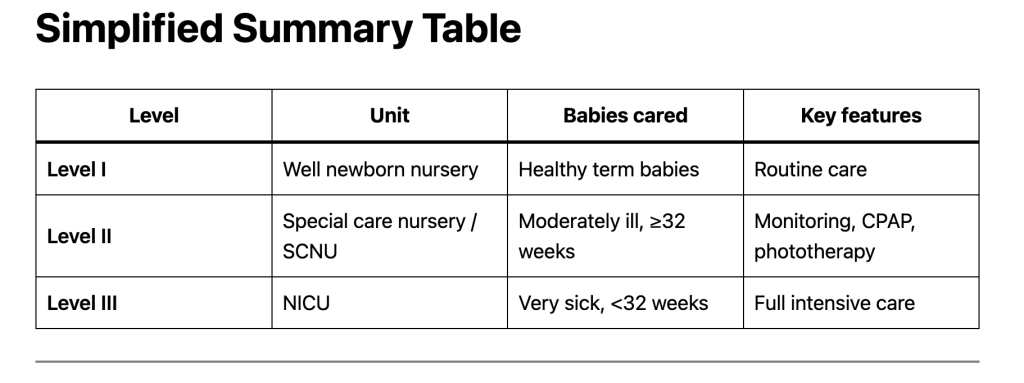
Simplified Summary Table

LevelUnitBabies caredKey features
Level IWell newborn nurseryHealthy term babiesRoutine care
Level IISpecial care nursery / SCNUModerately ill, ≥32 weeksMonitoring, CPAP, phototherapy
Level IIINICUVery sick, <32 weeksFull intensive care

Alternative Public Health Classification (Used in Many Countries)

  1. Home care
  2. Community level care
  3. Facility based newborn care
    • NBCC (Newborn Care Corner)
    • SNCU
    • NICU

Exam Pearls (Pediatrics / Neonatology)

  • Level I → routine care
  • Level II → moderate illness, ≥32 weeks
  • Level III → intensive care, <32 weeks
  • VLBW infants (<1500 g) should be managed in Level III NICU

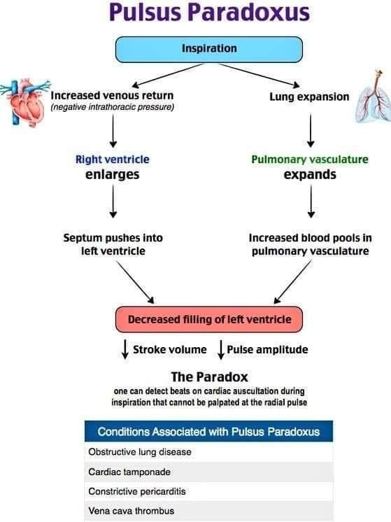
Pulsus Paradoxus: Definition, common cause, Pathology and Clinical relevance

Definition

Pulsus paradoxus is an exaggerated fall in systolic blood pressure (>10 mmHg) during inspiration.

Normally, systolic BP falls slightly during inspiration (<10 mmHg).
When the drop is >10 mmHg, it is called pulsus paradoxus.

Despite the name “paradoxus”, the pulse does not actually disappear; it only becomes weaker or unpalpable during inspiration.


Normal Physiology

During inspiration:

  • Intrathoracic pressure decreases
  • Venous return to the right ventricle increases
  • RV expands
  • Interventricular septum shifts slightly toward the left ventricle
  • LV filling decreases slightly
  • Systolic BP falls by <10 mmHg

Mechanism of Pulsus Paradoxus

In certain conditions:

  1. Excess RV filling or external compression
  2. RV expansion pushes interventricular septum toward LV
  3. LV filling markedly decreases
  4. LV stroke volume falls
  5. Systolic BP drops >10 mmHg during inspiration

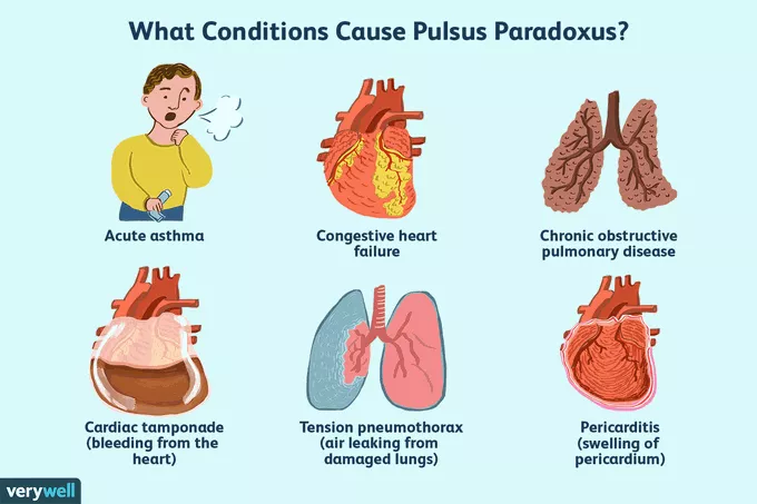
Causes of Pulsus Paradoxus

Cardiac Causes

  • Cardiac Tamponade (classic cause)
  • Constrictive Pericarditis
  • Large pericardial effusion

Respiratory Causes

  • Severe Asthma
  • Chronic Obstructive Pulmonary Disease exacerbation

Other Causes

  • Tension Pneumothorax
  • Massive pulmonary embolism
  • Severe hypovolemia
  • Upper airway obstruction

How to Measure Pulsus Paradoxus

Using a sphygmomanometer:

  1. Inflate cuff above systolic pressure.
  2. Slowly deflate.
  3. Note two pressures:
  • First pressure: Korotkoff sounds heard only during expiration
  • Second pressure: Sounds heard throughout inspiration and expiration

Difference between the two = Pulsus paradoxus

Example:

  • Expiration only = 120 mmHg
  • Throughout respiration = 105 mmHg

Pulsus paradoxus = 15 mmHg


Clinical Significance

A pulsus paradoxus >10 mmHg suggests serious cardiopulmonary disease, especially:

  • Cardiac tamponade
  • Severe asthma attack

Reverse Pulsus Paradoxus

Reverse pulsus paradoxus is a rare cardiovascular finding in which systolic blood pressure increases during inspiration, opposite to the normal drop seen in typical pulsus paradoxus. It is most commonly associated with conditions that alter intrathoracic pressure dynamics or ventricular interaction, such as positive pressure ventilation, hypertrophic obstructive cardiomyopathy (HOCM), isovolumetric ventricular pacing, and sometimes aortic regurgitation.

The mechanism usually involves enhanced left ventricular filling or reduced afterload during inspiration, leading to a paradoxical rise in systolic pressure. Clinically, it is less frequently encountered than classic pulsus paradoxus and is often identified in intensive care settings where patients are mechanically ventilated. Recognition of reverse pulsus paradoxus is important because it can provide clues about underlying cardiac physiology and ventilatory influences rather than indicating conditions like cardiac tamponade, which are linked to the traditional form.


Quick Exam Definition

Pulsus paradoxus is an inspiratory fall in systolic blood pressure greater than 10 mmHg.

6 Steps of How to Demonstrate / Measure Pulsus Paradoxus (Bedside Method)

Pulsus paradoxus is demonstrated using a blood pressure cuff and stethoscope.


Step-by-Step Procedure

1. Position the Patient

  • Patient should be lying supine or semi-recumbent
  • Ask the patient to breathe normally

2. Inflate the BP Cuff

  • Inflate the cuff 20–30 mmHg above systolic pressure

3. Slowly Deflate the Cuff

Deflate slowly at 2–3 mmHg per second while auscultating the brachial artery.


4. Identify the First Pressure

At first you will hear Korotkoff sounds only during expiration and they disappear during inspiration.

Record this pressure.

Example: 120 mmHg

Reason: During inspiration the systolic pressure falls below cuff pressure.


5. Continue Deflating

As the cuff pressure falls further, Korotkoff sounds will be heard throughout the respiratory cycle (during both inspiration and expiration).

Record this second pressure.

Example: 105 mmHg


6. Calculate Pulsus Paradoxus

Pulsus paradoxus=First pressure−Second pressure\text{Pulsus paradoxus} = \text{First pressure} – \text{Second pressure}Pulsus paradoxus=First pressure−Second pressure

Example:120−105=15 mmHg120 – 105 = 15\ mmHg120−105=15 mmHg

If >10 mmHg → Pulsus paradoxus present


Clinical Example

If:

  • Sounds heard only in expiration at 118 mmHg
  • Sounds heard throughout breathing at 102 mmHg

Then:118−102=16 mmHg118 – 102 = 16\ mmHg118−102=16 mmHg

Positive pulsus paradoxus


How to Demonstrate in Clinical Exam (Short Viva Answer)

You can say:

Pulsus paradoxus is demonstrated using a sphygmomanometer. The cuff is inflated above systolic pressure and slowly deflated. The first pressure at which Korotkoff sounds are heard only during expiration is noted. The pressure at which sounds are heard during both inspiration and expiration is then noted. The difference between these pressures is the pulsus paradoxus. A difference greater than 10 mmHg indicates pulsus paradoxus.


Important Clinical Associations

  • Cardiac Tamponade (classic)
  • Severe Asthma
  • Tension Pneumothorax
  • Constrictive Pericarditis

Exam Tip:
If a patient has hypotension + raised JVP + pulsus paradoxus, suspect cardiac tamponade (Beck’s triad).

Heart Failure: Classes and Stages


Introduction

Heart failure (HF) severity and progression are described using two complementary systems:

  1. NYHA Functional Classification → based on symptoms and activity limitation
  2. ACC/AHA Stages of Heart Failure → based on disease progression and structural changes

1. NYHA Functional Classification

Used in patients with established heart disease to assess symptom severity and functional limitation.

Class I

  • No limitation of physical activity
  • Ordinary physical activity does not cause symptoms
  • Comfortable at rest

Examples

  • Walking
  • Climbing stairs normally
  • Daily activities without dyspnea

Class II

  • Slight limitation of physical activity
  • Comfortable at rest
  • Ordinary activity causes symptoms

Symptoms may include:

  • Fatigue
  • Palpitations
  • Shortness of breath

Example

  • Dyspnea when climbing several flights of stairs

Class III

  • Marked limitation of physical activity
  • Comfortable at rest
  • Less than ordinary activity causes symptoms

Symptoms:

  • Dyspnea
  • Fatigue
  • Palpitations with mild activity

Example

  • Breathlessness when walking short distances

Class IV

  • Severe limitation
  • Symptoms even at rest
  • Unable to perform any physical activity without discomfort

Examples

  • Dyspnea at rest
  • Orthopnea
  • Severe fatigue

2. ACC/AHA Stages of Heart Failure

Describes development and progression of HF and emphasizes prevention.

Unlike NYHA classes, stages are progressive and irreversible.


Stage A – At Risk for Heart Failure

Patients without structural heart disease or symptoms but with risk factors.

Common risk factors:

  • Hypertension
  • Coronary artery disease
  • Diabetes mellitus
  • Metabolic syndrome
  • Obesity
  • Exposure to cardiotoxic drugs
  • Family history of cardiomyopathy

Goal:

  • Prevent development of structural heart disease

Stage B – Pre-Heart Failure

Patients without symptoms but with structural heart disease.

Examples:

  • Left ventricular hypertrophy
  • Previous myocardial infarction
  • Reduced ejection fraction
  • Valvular heart disease

Features:

  • No symptoms yet
  • Evidence of cardiac structural abnormalities

Goal:

  • Prevent symptomatic HF

Stage C – Symptomatic Heart Failure

Structural heart disease with current or previous symptoms.

Common symptoms:

  • Dyspnea
  • Fatigue
  • Reduced exercise tolerance
  • Fluid retention (edema)

Patients often correspond to NYHA Class II–III.

Goal:

  • Symptom control and prevention of hospitalization

Stage D – Advanced Heart Failure

  • Severe symptoms despite optimal medical therapy
  • Recurrent hospitalizations
  • Marked limitation of daily activities

Management may include:

  • Advanced therapies
  • Mechanical circulatory support
  • Heart transplant
  • Palliative care

Patients usually correspond to NYHA Class IV.


Key Differences Between NYHA and ACC/AHA

FeatureNYHA ClassificationACC/AHA Staging
BasisSymptoms and activity limitationDisease progression
ReversibilityCan improve or worsenUsually progressive
UseFunctional assessmentPreventive and therapeutic planning
RangeClass I–IVStage A–D

Clinical Correlation

Typical relationship:

ACC/AHA StageNYHA Class
Stage ANo class
Stage BClass I
Stage CClass II–III
Stage DClass IV

Exam Pearl

  • ACC/AHA = structural disease progression
  • NYHA = symptom severity

When is lactulose indicated in Wilson disease?

What is wilson disease?

Add lactulose if any of the following are present:

  • Overt hepatic encephalopathy
    • Altered sensorium
    • Irritability, sleep reversal
    • Asterixis
  • Minimal / impending HE
    • Poor school performance
    • Behavioral change
    • Subtle confusion
  • Advanced decompensated liver disease
    • High ammonia levels (if measured)
    • Severe portal hypertension with prior HE
  • Acute liver failure due to Wilson disease

👉 Dose (pediatrics):

  • 0.5–1 mL/kg/dose orally
  • Titrate to 2–3 soft stools/day

When lactulose is NOT needed

Do not add lactulose if the child has:

  • Wilson disease with hepatitis only
  • No encephalopathy
  • Normal mental status
  • Compensated chronic liver disease

Adding lactulose unnecessarily may cause:

  • Diarrhea
  • Electrolyte imbalance
  • Poor compliance

What should be prioritized instead

For Wilson disease with hepatitis, focus on:

Copper chelation

  • D-penicillamine (with pyridoxine)
  • OR Trientine

Zinc therapy (as maintenance or adjunct)

Supportive liver care

  • Low-copper diet
  • Salt restriction if ascites
  • Diuretics if needed
  • Fat-soluble vitamins if cholestasis

Exam-oriented takeaway (very important)

Lactulose is NOT a routine drug in Wilson disease.
It is used only for hepatic encephalopathy, not for hepatitis itself.

Postcoital Vaginal Bleeding in Woman: top 5 Causes, Evaluation, and When to Worry (is it dangerous?)

Postcoital bleeding (bleeding after sexual intercourse / vaginal bellding after sex) can be alarming, especially in young women. If a woman notices spotting only on the same day after sex, the cause is usually benign — but it should never be ignored.

This article explains the common causes, red flags, and when medical evaluation is necessary.


What Is Postcoital Bleeding?

Postcoital bleeding refers to vaginal bleeding that occurs immediately after sexual intercourse and is not related to menstruation.

It may present as:

  • Light spotting
  • Pink or brown discharge
  • Fresh red bleeding
  • Bleeding that stops within a few hours

Common Causes in a young Woman

In this age group, most causes are non-cancerous.


1. Cervical Ectropion (Most Common Cause)

Cervical ectropion occurs when the delicate inner cervical cells are exposed on the outer surface of the cervix.

These cells are fragile and bleed easily when touched during intercourse.

It is common in:

  • Young women
  • Women taking oral contraceptive pills
  • Pregnancy (due to high estrogen levels)

Typically:

  • Bleeding is mild
  • Occurs only after sex
  • No severe pain

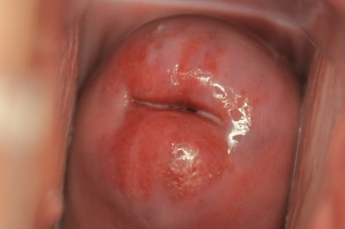
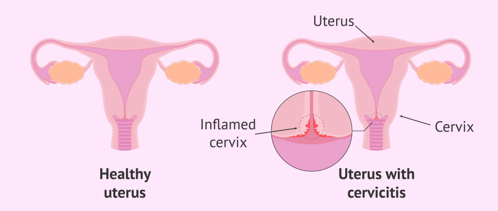
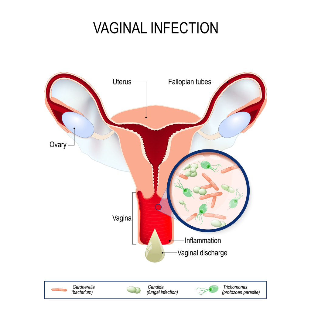
2. Cervicitis (Cervical Infection)

Inflammation of the cervix can cause contact bleeding.

Common sexually transmitted infections include:

  • Chlamydia trachomatis
  • Neisseria gonorrhoeae
  • Trichomonas vaginalis

Associated symptoms:

  • Abnormal vaginal discharge
  • Foul smell
  • Pain during intercourse
  • Burning urination

Young sexually active women are at higher risk.


3. Vaginal Infections (Vaginitis)

Infections make vaginal tissue inflamed and fragile.

Common causes:

  • Candida albicans
  • Trichomonas vaginalis

Symptoms:

  • Itching
  • Thick or foul discharge
  • Pain during sex

4. Mechanical Trauma

Minor tears may occur due to:

  • Rough intercourse
  • Inadequate lubrication
  • First sexual intercourse
  • Vaginal dryness

Bleeding is usually mild and short-lived.


Even light bleeding after sex can occur in early pregnancy due to a fragile cervix.

Important causes:

  • Implantation bleeding
  • Threatened miscarriage
  • Ectopic pregnancy

A urine pregnancy test should be done if periods are delayed.


Less Common but Important Causes

Although rare at 23 years of age:

Cervical Dysplasia

Often associated with:

  • Human papillomavirus

May present with recurrent postcoital bleeding.

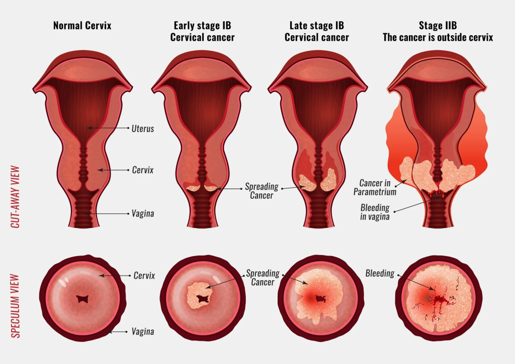
Cervical Cancer

Rare in young women but should be considered if:

  • Bleeding is persistent
  • There is unexplained weight loss
  • There is pelvic pain
  • Abnormal Pap smear

When Is It Likely Benign?

Postcoital bleeding is usually harmless if:

  • It occurs only on the day of intercourse
  • Bleeding is light
  • It stops within hours
  • There is no pain or foul discharge
  • It does not happen frequently

When to See a Doctor

Seek medical evaluation if:

  • Bleeding happens repeatedly
  • Bleeding becomes heavy
  • There is pelvic pain
  • There is abnormal discharge
  • Periods are missed
  • Bleeding occurs unrelated to intercourse

How Is It Evaluated?

A doctor may perform:

  • Detailed history
  • Speculum examination
  • Pregnancy test
  • STI testing (NAAT)
  • Vaginal swab
  • Pap smear (if due)
  • Colposcopy if cervix looks abnormal

Key Takeaway

In woman, postcoital bleeding occurring only on the same day after intercourse is most commonly due to cervical ectropion, mild infection, or minor trauma.

However, repeated episodes should always be medically evaluated to rule out infection and cervical pathology.

Early evaluation ensures reassurance in benign cases and timely treatment if needed.

MIS-C / MIS-P : Facts and more

Multisystem Inflammatory Syndrome in Children (Pediatric)

It’s a rare but serious inflammatory response, most commonly seen after a COVID-19 infection.

What it is:

An overactive immune response

Causes widespread inflammation affecting multiple organs

Common features:

Persistent fever

GI symptoms (abdominal pain, vomiting, diarrhea)

Rash, red eyes, swollen lips/hands

Heart involvement (myocarditis, low blood pressure)

Fatigue, headache

Timing:

Usually appears 2–6 weeks after a viral infection (classically COVID-19)

Treatment:

Hospital care
IVIG, steroids, anti-inflammatory meds

Most kids recover well with early treatment

If this is about a specific child or symptoms, I can explain warning signs, how it’s diagnosed, or how it differs from Kawasaki disease.

Gotcha — here’s a DM-level (doctor/medical) clinical note on MIS-C (Multisystem Inflammatory Syndrome in Children), written in a professional charting style.

Diagnosis:

Multisystem Inflammatory Syndrome in Children (MIS-C)

Definition:

MIS-C is a post-infectious hyperinflammatory syndrome occurring in pediatric patients, most commonly following SARS-CoV-2 infection. It is characterized by persistent fever, systemic inflammation, and involvement of two or more organ systems.

Epidemiology:

Typically presents in children and adolescents weeks after acute or asymptomatic COVID-19 infection. Incidence is rare but clinically significant due to potential cardiovascular involvement.

Pathophysiology:

Believed to be an immune-mediated response rather than direct viral injury. Dysregulated immune activation leads to cytokine release, endothelial dysfunction, and multisystem inflammation.

Clinical Presentation:

Persistent fever (>38.0°C, ≥24 hours)
Gastrointestinal symptoms (abdominal pain, vomiting, diarrhea)
Mucocutaneous findings (rash, conjunctival injection, strawberry tongue, swollen extremities)
Cardiovascular involvement (myocarditis, depressed ejection fraction, hypotension, shock)
Neurologic symptoms (headache, altered mental status, irritability)
Respiratory symptoms may be minimal or absent

Laboratory Findings:

Elevated inflammatory markers (CRP, ESR, ferritin, procalcitonin)
Lymphopenia, thrombocytopenia
Elevated D-dimer, fibrinogen
Elevated cardiac markers (troponin, BNP/NT-proBNP)
Evidence of recent SARS-CoV-2 infection (PCR or serology)

Diagnosis:

Clinical diagnosis based on CDC/WHO criteria, requiring fever, laboratory evidence of inflammation, multisystem involvement, and temporal association with SARS-CoV-2 infection, with exclusion of alternative diagnoses.

Management:

Hospital admission; PICU if hemodynamically unstable
Immunomodulatory therapy: IVIG and systemic corticosteroids
Supportive care (fluids, vasopressors if indicated)
Anticoagulation in select cases
Cardiology consultation and echocardiographic monitoring

6 Common Drugs Used for UTI (urinary tract infection) Prophylaxis in Children

Urinary Tract Infection (UTI) in Children — Introduction

Urinary tract infection (UTI) (outlink to CDC) is one of the most common serious bacterial infections in childhood and an important cause of fever, morbidity, and potential long-term renal damage. UTIs involve infection of the urinary system, including the bladder (cystitis) and kidneys (pyelonephritis). Early recognition and appropriate management are essential to prevent complications such as renal scarring, hypertension, and chronic kidney disease.

Epidemiology

UTIs occur in approximately 5–8% of girls and 1–2% of boys by 7 years of age. During infancy, UTIs are more common in boys, especially those who are uncircumcised. After the first year of life, girls are affected more frequently due to anatomical and behavioral factors.

Causative Agents

The most common causative organism is Escherichia coli, accounting for the majority of infections. Other pathogens include Klebsiella, Proteus, Enterococcus, and Pseudomonas aeruginosa, particularly in children with structural abnormalities or catheterization. Link to Genitourinary system MCQs

Risk factors and Presentation

Risk factors include vesicoureteral reflux, urinary tract obstruction, neurogenic bladder, constipation, poor perineal hygiene, and dysfunctional voiding. Clinical presentation varies with age: neonates and infants may present with nonspecific signs such as fever, poor feeding, vomiting, or irritability, whereas older children typically present with dysuria, frequency, urgency, abdominal pain, or flank pain.

Prevention

Because recurrent infections increase the risk of renal scarring, identifying children at risk and initiating preventive strategies — including behavioral measures and, when indicated, antibiotic prophylaxis — is an important component of pediatric care.

UTI Prophylaxis in Children

DrugDose (once daily unless stated)Age suitabilityWhen preferredImportant notes
Nitrofurantoin1–2 mg/kg at bedtime>1 monthFirst-line prophylaxisAvoid if G6PD deficiency; may cause nausea
Trimethoprim-Sulfamethoxazole (TMP-SMX)2 mg/kg (TMP component)>2 monthsCommon first choiceAvoid in neonates; risk of Stevens-Johnson syndrome
Trimethoprim alone2 mg/kg>2 monthsAlternative to TMP-SMXUseful if sulfa allergy
Cephalexin10–12 mg/kgAll agesInfants & vesicoureteral refluxGood safety profile
Amoxicillin10–15 mg/kg<2 monthsNeonatal prophylaxisResistance common after infancy
Cefixime2 mg/kg>6 monthsResistant organismsUsed less commonly

Indications for UTI Prophylaxis

  • Vesicoureteral reflux (Grade III–V)
  • Recurrent febrile UTIs (≥2 in 6 months or ≥3/year)
  • Obstructive uropathy awaiting surgery
  • Neurogenic bladder
  • After first febrile UTI in infants until evaluation complete

Duration

  • Continue until:
    • VUR resolves or is surgically corrected
    • Child becomes toilet trained and infection-free
    • Specialist review recommends stopping

Key Clinical Points

✔ Give at bedtime for maximal bladder concentration
✔ Encourage hydration & regular voiding
✔ Treat constipation (important risk factor)
✔ Monitor for breakthrough infections and resistance

Treatment of UTI in Children

Empirical Antibiotic Therapy (Based on Clinical Type)

Clinical TypeOral Antibiotics (Outpatient)IV Antibiotics (Inpatient / Severe)Duration
Simple cystitis (Afebrile UTI)Nitrofurantoin 5–7 mg/kg/day ÷ 2 doses
Cephalexin 50–100 mg/kg/day ÷ 3–4 doses
TMP-SMX 8–10 mg/kg/day (TMP component) ÷ 2 doses
Usually not required5–7 days
Febrile UTI / Acute pyelonephritisCefixime 8 mg/kg/day OD
Amoxicillin-clavulanate 40–50 mg/kg/day ÷ 2–3 doses
Ceftriaxone 50–75 mg/kg OD
Cefotaxime 150 mg/kg/day ÷ 3 doses
Gentamicin 5–7 mg/kg OD
7–14 days
Neonatal UTI (<2 months)Not preferredAmpicillin + Gentamicin
OR Cefotaxime
10–14 days
Complicated UTI / Toxic childNot preferredCeftriaxone ± Amikacin
Consider Piperacillin-Tazobactam if resistant
10–14 days

Organism-Specific Considerations

OrganismPreferred Drugs
Escherichia coliCephalosporins, Nitrofurantoin, TMP-SMX (if sensitive)
ProteusAvoid Nitrofurantoin; use cephalosporins
Pseudomonas aeruginosaCeftazidime, Piperacillin-Tazobactam
EnterococcusAmpicillin, Amoxicillin

Supportive Management

MeasureDetails
HydrationEncourage oral fluids
AntipyreticsParacetamol / Ibuprofen
Treat constipationImportant to prevent recurrence
Follow-up cultureIf no improvement in 48 hours
ImagingRBUS after first febrile UTI (especially <2 years)

Important Clinical Points

✔ Send urine routine + culture before starting antibiotics
✔ Switch from IV to oral once clinically improved (24–48 hrs)
✔ Modify antibiotics according to culture sensitivity
✔ Admit if: toxic appearance, persistent vomiting, dehydration, neonate, poor follow-up

CROUP (Acute Laryngotracheobronchitis)


Definition

Croup is an acute viral inflammatory disease of the upper airway involving the larynx, trachea, and bronchi, leading to subglottic edema and airway obstruction.


Epidemiology

  • Age: 6 months – 3 years (can occur up to 6 years)
  • Male > Female
  • Peak: Autumn & early winter
  • Usually preceded by URTI

Etiology

Viral (most common)

  • Parainfluenza virus type 1 (most common)
  • Parainfluenza 2 & 3
  • RSV
  • Influenza A & B
  • Adenovirus
  • Human metapneumovirus

Rare bacterial causes

  • Mycoplasma
  • Secondary bacterial infection (uncommon)

Pathophysiology

  • Viral infection → inflammation & edema of subglottic region
  • Subglottis is the narrowest part of pediatric airway
  • Small edema → marked increase in airway resistance
  • Leads to inspiratory stridor & respiratory distress

Clinical Features

Prodrome

  • Low-grade fever
  • Coryza
  • Cough

Characteristic features

  • Barking (seal-like) cough
  • Hoarseness
  • Inspiratory stridor
  • Worse at night
  • Aggravated by crying & agitation

Severe disease

  • Stridor at rest
  • Chest retractions
  • Tachypnea
  • Hypoxia
  • Fatigue / altered sensorium (late sign)

Severity Assessment (Westley Croup Score – concept)

FeatureMildModerateSevere
StridorNone / with agitationAt restLoud, biphasic
RetractionsNoneMild–moderateSevere
Air entryNormalDecreasedMarkedly reduced
CyanosisNoneNonePresent
Mental statusNormalNormalAltered

Investigations

  • Diagnosis is clinical
  • No routine labs required
  • Neck X-ray (AP) (only if diagnosis unclear):
    • Steeple sign (subglottic narrowing)

Differential Diagnosis

ConditionKey Differentiating Feature
EpiglottitisHigh fever, drooling, muffled voice
Bacterial tracheitisToxic child, high fever
Foreign bodySudden onset, no prodrome
Retropharyngeal abscessNeck stiffness, drooling
AngioedemaFacial/lip swelling

Management

General Measures

  • Keep child calm
  • Minimal handling
  • Oxygen if hypoxic
  • Humidified air (comfort measure only)

Pharmacological Treatment

1️⃣ Corticosteroids (All cases)

Dexamethasone (Dexona)

  • Dose: 0.6 mg/kg
  • Max: 10 mg
  • Route: Oral / IM / IV
  • Single dose usually sufficient

2️⃣ Adrenaline Nebulization (Moderate–Severe)

L-Adrenaline (1:1000)

  • Dose: 0.5 mL/kg (max 5 mL)
  • Dilute with NS to 5 mL
  • Rapid onset (10–15 min)
  • Duration: ~2 hours

⚠️ Observe 2–4 hours after neb (rebound stridor)


Indications for Admission

  • Stridor at rest
  • Need for repeated adrenaline
  • Hypoxia
  • Poor oral intake
  • Age < 6 months
  • Social concerns

Indications for ICU / Intubation

  • Exhaustion
  • Altered consciousness
  • Severe hypoxia
  • Poor air entry
  • Failure to respond to treatment

Complications

  • Respiratory failure
  • Secondary bacterial infection
  • Pneumonia
  • Rarely death

Prognosis

  • Excellent
  • Self-limiting (3–7 days)
  • Recurrence possible

Key Takeaway

  • Single dose dexamethasone for all croup
  • Adrenaline = temporary relief
  • Steeple sign = croup
  • Drooling → think epiglottitis
  • Avoid agitation at all costs

Exit mobile version