1. What is the primary purpose of dental prophylaxis?
**Answer: c) Preventive dental cleaning**
Explanation: Dental prophylaxis refers to the professional cleaning of teeth to remove dental plaque, calculus, and stains. It is a preventive measure aimed at maintaining oral health and preventing oral diseases such as cavities and gum disease.
2. Which of the following oral conditions is primarily caused by bacterial plaque accumulation?
**Answer: a) Dental caries (cavities)**
Explanation: Dental caries, commonly known as cavities or tooth decay, is primarily caused by the accumulation of bacterial plaque on the tooth surfaces. The bacteria in plaque produce acids that demineralize tooth enamel, leading to the formation of cavities.
3. What is the recommended technique for brushing a patient’s teeth with gingival recession?
**Answer: d) Modified Bass technique**
Explanation: The Modified Bass technique involves angling the toothbrush towards the gumline at a 45-degree angle and making small circular or vibrating motions. This technique is recommended for patients with gingival recession as it helps clean both the teeth and the gumline effectively.
4. Which dental instrument is commonly used for removing calculus deposits from the crowns of teeth?
**Answer: c) Scaler**
Explanation: A scaler is a dental instrument specifically designed for removing calculus deposits from the crowns of teeth. It has a pointed tip that can be used to gently remove hardened plaque (calculus) from the tooth surfaces.
5. Which of the following is a risk factor for developing periodontal disease?
**Answer: b) Overbrushing with a hard-bristle toothbrush**
Explanation: Overbrushing with a hard-bristle toothbrush can lead to trauma to the gums and enamel, potentially contributing to the development of periodontal disease. It’s important to use a soft-bristle toothbrush and practice proper brushing techniques to avoid damaging the oral tissues.
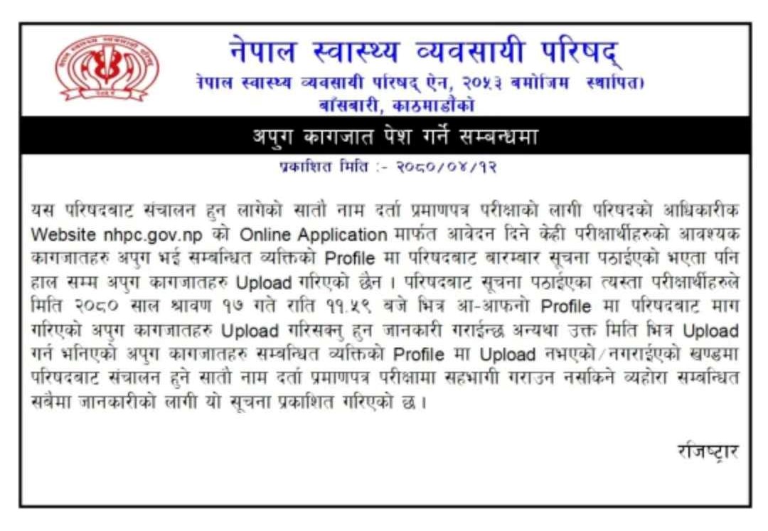
NATIONAL GUIDELINES FOR SNAKEBITE MANAGEMENT IN NEPAL
Snake
Table of Contents(toc)
Introduction
Snakebite is an important occupational hazard affecting farmers, plantation workers, herders and fishermen. Open-style habitation and the practice of sleeping on the floor also expose people to bites from nocturnal snakes, with children being at a particularly high risk. In rural Nepal, snakebite is an important public health problem.
Risk of snakebite in nepal
The agriculturally prosperous terai region with hot climate, high seasonal rainfall, lush natural vegetation, high density of rodents, rich reptiles and amphibian flora make an ideal habitat for snake to live and increase availability of marshy land to undergo hibernation. The abundance of snake and human activities, mainly agriculture, increases the man– snake encounter leading to snakebite. Nocturnal snakebite is also common in Nepal due to the sleeping habit.
NATIONAL GUIDELINES FOR SNAKEBITE MANAGEMENT IN NEPAL PDF
Deciphering the Complexity of ICD-10: A Comprehensive Guide to the
Cornerstone of Modern Healthcare
Table of Contents (toc)
ICD-10
Introduction:
In the intricate web of modern healthcare, where every patient encounter,
diagnosis, and treatment is meticulously documented and analyzed, the
International Classification of Diseases, 10th Revision (ICD-10) stands as a
linchpin. Far more than a simple coding system, ICD-10 is a robust framework
that underpins the entire healthcare data ecosystem, facilitating everything
from billing and reimbursement to public health surveillance and research. In
this comprehensive guide, we embark on a journey to demystify the complexity
of ICD-10, exploring its origins, structure, applications, and future
implications for the healthcare landscape.
I. Understanding ICD-10: A Historical and Conceptual Overview
A. What is ICD-10?
1. Definition and purpose.
The International Classification of Diseases, 10th Revision (ICD-10), is a
globally recognized and standardized system used for classifying diseases,
health conditions, and related phenomena. Developed by the World Health
Organization (WHO), ICD-10 serves as a comprehensive tool for organizing and
categorizing medical diagnoses and procedures. Its primary purpose is to
facilitate the systematic recording, analysis, interpretation, and comparison
of morbidity and mortality data across different populations and settings.
The key components of ICD-10 include alphanumeric codes that represent
specific diseases, conditions, and medical procedures, along with detailed
guidelines for assigning these codes accurately. This classification system
provides a standardized language for healthcare professionals, researchers,
policymakers, and administrators to communicate and exchange health
information effectively.
The primary purposes of ICD-10 are as follows:
1. Disease Classification:
ICD-10 provides a structured framework for
classifying diseases, disorders, injuries, and other health-related conditions
based on etiology, anatomy, and clinical manifestations. By organizing medical
conditions into distinct categories and subcategories, ICD-10 facilitates the
systematic documentation and analysis of health data.
2. Statistical Tracking:
ICD-10 enables the collection, aggregation, and
analysis of health statistics at local, national, and global levels. Health
organizations and government agencies use ICD-10-coded data to monitor disease
trends, track epidemiological patterns, and evaluate public health
interventions. This information is invaluable for informing health policy
decisions, resource allocation, and disease prevention efforts.
3. Reimbursement and Billing:
In many healthcare systems, ICD-10 codes play a
crucial role in reimbursement and billing processes. Health insurers,
government payers, and healthcare providers use these codes to accurately
document and bill for medical services rendered. Accurate coding is essential
for ensuring proper reimbursement, preventing fraud and abuse, and maintaining
financial integrity within the healthcare system.
4. Clinical Research and Epidemiology:
Researchers and epidemiologists rely on
ICD-10 codes to conduct studies on disease prevalence, incidence, risk
factors, and outcomes. By using standardized coding terminology, researchers
can compare data from different studies and populations, identify patterns and
associations, and generate evidence-based insights into disease etiology,
treatment effectiveness, and healthcare delivery.
In summary, the International Classification of Diseases, 10th Revision
(ICD-10), serves as a foundational tool for organizing, categorizing, and
analyzing health-related data. Its widespread adoption and use facilitate
effective communication, decision-making, and quality improvement initiatives
across the global healthcare landscape.
2. Evolution from previous revisions.
B. Historical evolution and significance.
1. Development by the World Health Organization (WHO).
2. Importance of standardization in global healthcare.
C. Global adoption and standardization.
1. Implementation across countries and healthcare
systems.
2. Harmonization efforts for consistency and
interoperability.
II. The Structure of ICD-10: Navigating the Complexity
A. Alphanumeric coding system.
1. Breakdown of the coding format.
2. Meaning and significance of each component.
B. Chapters, sections, and categories.
1. Organization of diseases and conditions.
2. Hierarchical structure for specificity and
granularity.
C. Detailed coding conventions and guidelines.
1. Instructions for code selection and assignment.
2. Tabular and index-based navigation.
III. Purpose and Applications of ICD-10: Beyond Coding and Billing
A. Disease classification and statistical tracking.
1. Role in epidemiology and disease surveillance.
2. Use in monitoring health trends and outcomes.
B. Reimbursement and billing.
1. Linkage to healthcare financing and reimbursement
systems.
2. Importance of accurate coding for revenue cycle
management.
C. Public health surveillance and research.
1. Contribution to health policy and decision-making.
2. Facilitation of clinical and population-based
research.
IV. Transition to ICD-10: Challenges, Lessons, and Benefits
A. Challenges faced during the transition.
1. Training and education requirements.
2. Technical and logistical hurdles.
B. Lessons learned from implementation.
1. Best practices and strategies for successful adoption.
2. Impact on healthcare workflows and productivity.
C. Benefits realized post-implementation.
1. Improved accuracy and specificity in coding.
2. Enhanced data quality and interoperability.
V. Advantages of ICD-10: Driving Healthcare Quality and Innovation
A. Enhanced accuracy and specificity in coding.
1. Ability to capture detailed clinical information.
2. Reduction of coding errors and inaccuracies.
B. Improved data interoperability and analytics.
1. Integration with electronic health records (EHRs) and
health information exchanges (HIEs).
2. Facilitation of data-driven decision-making and
analytics.
C. Better understanding of disease patterns and trends.
1. Identification of emerging health threats and
disparities.
2. Monitoring of healthcare interventions and outcomes.
VI. ICD-10 Coding Guidelines and Updates: Staying Current and Compliant
A. Documentation requirements for code assignment.
1. Importance of thorough clinical documentation.
2. Linkage between coding and documentation integrity.
B. Regular revisions and updates.
1. Role of WHO and national coding authorities.
2. Impact of updates on healthcare practice and policy.
C. Importance of staying informed and compliant.
1. Continuing education and professional development.
2. Compliance with coding standards and regulations.
VII. Future Trends and Innovations in Coding: Anticipating the Next Frontier
A. Integration with electronic health records (EHRs) and
artificial intelligence (AI).
1. Automation of coding processes and decision support.
2. Integration with clinical decision support systems
(CDSS).
B. Expansion of telemedicine and remote coding.
1. Implications for coding workflows and practices.
2. Challenges and opportunities in remote healthcare
delivery.
C. Potential impact of ICD-11 on the healthcare landscape.
1. Anticipated changes and enhancements.
2. Preparation and readiness for future revisions.
Conclusion:
In the ever-evolving landscape of healthcare, the role of ICD-10 cannot be
overstated. As a foundational element of healthcare data management, ICD-10
shapes the way we document, classify, and analyze diseases and health
conditions. By understanding its structure, purpose, and applications,
healthcare professionals can harness the power of ICD-10 to drive quality
improvement, innovation, and better patient outcomes. As we look to the
future, continued advancements in coding technology, coupled with the eventual
transition to ICD-11, promise to further enhance the role of coding in shaping
the healthcare landscape of tomorrow.
Clinical Findings in Chronic Aortic Regurgitation(AR)
Clinical Findings in Chronic Aortic Regurgitation (AR) for medical students, cardiologists, physicians and internists 2023
Clinical Findings in Chronic Aortic Regurgitation
Table of Contents (toc)
Introduction
Aortic regurgitation, a valvular heart disease, is a condition characterized by the backflow of blood from the aorta into the left ventricle of the heart during diastole, the relaxation phase of the cardiac cycle.
Causes of aortic regurgitation
This condition can be attributed to several causes, such as congenital valve abnormalities, rheumatic fever, or aortic root dilatation.
The severity of aortic regurgitation varies and is often categorized as mild, moderate, or severe, depending on the extent of blood leakage.
Diagnosis of aortic regurgitation
To diagnose aortic regurgitation, clinicians typically employ a combination of techniques, including echocardiography, Doppler ultrasound, and magnetic resonance imaging (MRI), to assess the valve function and determine the appropriate treatment approach.
Management of aortic regurgitation
Managing aortic regurgitation may involve medical management, such as medication to reduce symptoms and slow the progression of the disease, or surgical intervention, such as valve repair or replacement, depending on the severity of the condition and the patient’s overall health.
Importance of timely diagnosis
Timely diagnosis and intervention are crucial in preventing the progression of aortic regurgitation, as it can lead to serious complications if left untreated, including heart failure and irreversible damage to the heart muscle..
Signs of aortic regurgitation
Here we have listed multiple symptom and signs of chronic aortic regurgitation a clinician hould be looking for when suspected.
Light House Sign – Blanching & Flushing of forehead
Landolfi’s sign, alternating constriction & dilatation of pupils
Becker’s Sign – visible pulsation of retinal arterioles
De Musset Sign – Bobbing of head
Muller’s Sign – To & fro movement of Uvula
Gerhardt /Sailer Sign– Pulsation of Spleen in Splenomegaly
Rosenbach’s Sign– Hepatic Pulsation
Shelley’s Sign– Pulsation of Cervix
Corrigan’s Sign– Forceful dilatation and quick collapse of Arterial pulse
Water-hammer Pulse
Quincke’s Sign-Nail bed Capillary Pulsation
Wide Pulse Pressure
Hill’s Sign– exaggerated difference in systolic arterial pressure between upper & lower limbs
Traube’s Sign– Pistol Shot sounds over femoral artery
Duroziez’s Sign– Pressure over femoral artery to & fro murmur
1. Which fracture is commonly known as a “dinner fork” deformity due to the characteristic angulation seen on X-rays?
Answer: a) Colles’ fracture
Explanation: Colles’ fracture is a distal radius fracture with dorsal (posterior) angulation of the wrist, resembling a dinner fork deformity. It is typically caused by a fall on an outstretched hand.Continue reading Answer of orthopedics and trauma MCQs 2024
Interim guidance on clinical management of Mpox (Monkeypox) 2079 (2022)
Table of Contents (toc)
Mpox interim guideline NEpal
Monkeypox is a viral zoonotic disease caused by the monkeypox virus, which belongs to the Orthopoxvirus genus, the same group as smallpox. It is primarily found in Central and West Africa but has been increasingly reported in other regions, highlighting its potential to spread beyond endemic areas. The virus is transmitted to humans through close contact with an infected animal, human, or contaminated materials.
Symptoms
Symptoms typically include fever, rash, and swollen lymph nodes, often resembling smallpox but generally less severe. While most cases are mild and self-limiting, severe cases can occur, particularly in immunocompromised individuals or those with comorbidities.
Vaccination
Vaccination against smallpox offers some protection against monkeypox, and public health efforts are focused on surveillance, early diagnosis, and containment to prevent outbreaks.
What are the 10 top health sites of nepal in 2024?
Table of Contenta(toc)
Background of Top 10 health sites of Nepal
Online health seeking has been a new trends now. After acess of internet in the whole nation many of the tradionation methods of service and sales have shifted into online modality.
In the same context, seeking medical help is also slowly transitioning into online modality in the recent years in nepal as well.
In this article we are trying write some review of the top health realted websites of Nepal that provide health information, online counseling and consultation on paid or free form.
This list is entirely a personal opinion and is neither sponsred nor endorsed by anyone. This review neither reflects anyinformation from third party. Continue reading 10 Top health sites of Nepal 2024
Introduction of dyspenea: what is dyspena or shortness of breath
What is dyspnea?
Shortness of breath causes and ddx?
Table of contents(toc)
Introduction of dyspnoea
Dyspena is a condition in which a person feels difficulty breathing.
It is a subjective experience expressed by the patient. The casuse or origin of this may come formvarying reasons including physiological, psychological, social, and environemntal factors.
Thus produced discomfort now is tackled by our physiological response as change in physiological parameters or behavioral modofications including shortnes of breathing.
Dyspnoea can be classified as acute or chronic.
Acute dyspnoea develops withing hours to days while chronic develops over weeks to lnger duration. The causes of acute and chronic dyspnoea might be different depending upon the duration.
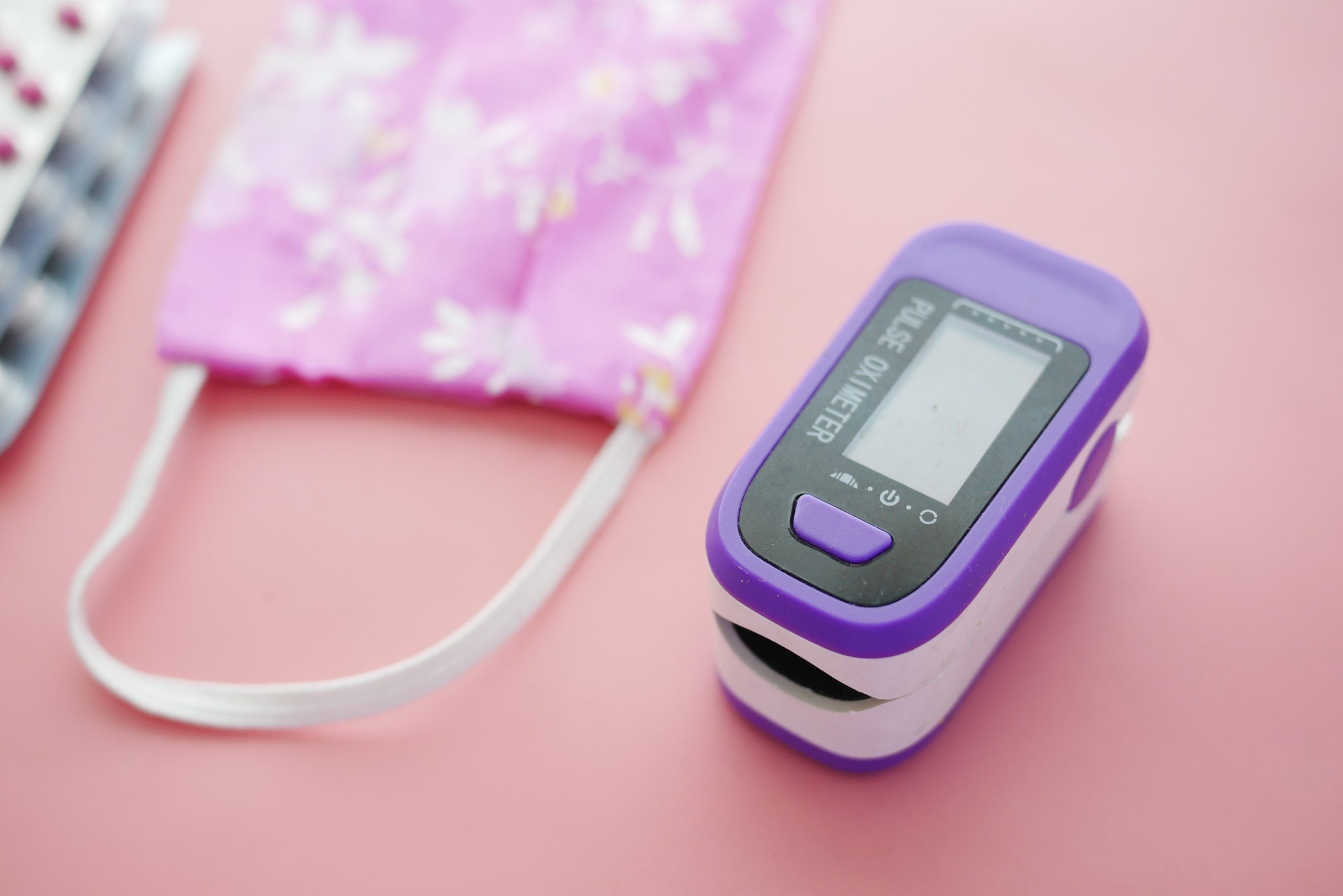
Pulse oxymeter
After careful history taking and examination of the patient having dyspnoea we should make a good list of differential disagnoses. The various differential diagnoses of dyspnoea are summerized in the following list.
The source of causes dyspnea, shortness of breath can be listed as:
Cardiovascular and realted to use of oxygen
Resporatory and chest wall pathology
Psychogenic
Differential diagnoses of dyspnoea/shortness of breath:
1. Causes of acute SOB
Cardiovascular
Cardiogenic
Impaired oxygen delivery
Impaired oxygen use
Respiratory causes
Upper airway
Lower aiway causes
Psychogenic
panic disorder
conversion disorder
drug withdrawal
2. Causes of Chronic SOB
Cardiovascular cause
CHF
Pericarditis
Anemia
Respiratory causes
Pathologies of chest wall, bronchoalveolar system, airways and lung parenchyma
The diseases can be summarised as below:
1. Cardiovascular
Acute MI
CHF/ LH failure
Aortic/Mitral stenosis
Aortic/Mitral Regurgitation
Arrhythmia
Cardiac tamponade
Constrictive pericarditis
Left sided obstructive lesions (atrial myxoma)
Elevated pulmonary venous pressure
2. Respiratory
i) Airway
Asthma
COPD
Upper airway obstruction like foreign body, anaphylaxis, mucus plugging
नेपाल को खोप तालिका बारे छोटो जानकारी (Rastriya Khop talika Nepal)
नेपालमा बच्चा जन्मे देखि दुई वर्षको उमेर सम्म विभिन्न १३ रोग विरुद्धको खोप निःशुल्क लगाइन्छ।
यी खोपहरु सरकारी स्वास्थ्य संस्था हरु मा निःशुल्क पाइन्छ।
गर्भवती महिला लाई दुई डोज टिटानस विरुद्ध को खोप निःशुल्क लगाइन्छ।
कुकुर र रेबिज सार्न सक्ने जनावरले तोकेको व्यक्तिलाई समेत रेबिज विरुद्धको खोप विभिन्न संस्थाहरुमा निःशुल्क लगाइन्छ।
BCG vaccinating a child; SC subcuticular
खोप बारे विशेष जानकारी vaccine information
अहिले नेपाल सरकारले माथि उल्लेखित अवधिमा खोप लगाउन छुटेका बालबालिकाहरूलाई समेत खोप लगाएर रोगहरू बाट जोगाउन पांच वर्षको उमेरका बच्चा हरु लाई समेत खोप लगाउने निर्णय गरेको छ।
यो खोप वैशाख १५ र ३१ गते नेपाल सरकार अन्तर्गत का स्वास्थ्य संस्था हरु मा निःशुल्क लगाइन्छ।
दुई वर्ष को उमेर सम्म खोप लगाइसकेका बच्चा हरु लाई यो अवधिमा थप खोप लगाउन आवश्यक छैन।
खोप लगाउन किन जरुरी छ? Why is vaccine important in Nepal
खोपले बालबालिका हरु लाई १३ थरी रोगहरूबाट जोगाउँछ। यो रोगहरू निम्नानुसार छन्।


")