
What is intermittent fasting?

Table of Contents(toc)

healthy salad

Introduction (What is intermittent fasting?)

Intermittent fasting (IF) has become popular for its potential health
benefits. However, whether it is actually useful depends on various factors
such as individual goals, lifestyle, and health conditions. Here are some key
points on its usefulness:

Benefits of intermittent fasting

Here are some benefits of Intermittent fasting (IF).
  • Weight Loss
  • Improved Insulin Sensitivity
  • Cellular Repair and Autophagy
  • Mental Clarity and Cognitive Function
  • Hormonal Benefits
  • Heart Health
  • Simplicity and Flexibility
  • Potential Drawbacks
  • Sustainability
  • Scientific Support
Now lets discuss each in details:

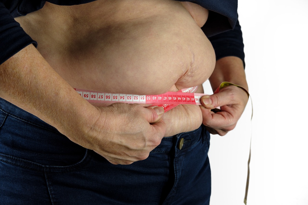
1. Weight Loss

  • Supports Calorie Control:
    By restricting the eating window, many people naturally consume fewer
    calories, which can lead to weight loss.
  • Increases Fat Burning:
    Fasting periods trigger hormonal changes that increase fat breakdown and
    use it for energy.
2.
Improved Insulin Sensitivity

  • Helps Manage Blood Sugar:
    Intermittent fasting can improve insulin sensitivity, making it beneficial
    for managing blood sugar levels, particularly for people with Type 2
    diabetes or prediabetes.
  • Reduces Insulin Resistance: It may help reduce the risk of developing insulin resistance, which is
    a key factor in obesity and type 2 diabetes.

3.
Cellular Repair and Autophagy

  • Cellular Maintenance:
    During fasting, the body enters a state of autophagy, where it breaks down
    and removes dysfunctional proteins and cells, promoting cellular repair.
  • Boosts Longevity: Some
    studies suggest that intermittent fasting may contribute to longevity by
    stimulating autophagy and reducing oxidative stress.

4.
Mental Clarity and Cognitive Function

  • Improved Brain Function: Fasting may support brain health by boosting the production of
    brain-derived neurotrophic factor (BDNF), which plays a role in cognitive
    function and mood regulation.
  • Reduced Inflammation:
    Intermittent fasting may help reduce inflammation, which is linked to
    neurological diseases like Alzheimer’s and Parkinson’s.

5. Hormonal Benefits

  • Increases Growth Hormone: Fasting increases the secretion of human growth hormone (HGH), which
    plays a role in fat loss and muscle preservation.
  • Improved Fat Metabolism: It enhances the breakdown of stored fat for energy, making it easier to
    burn fat rather than carbohydrates.

6. Heart Health

  • Reduces Blood Pressure:
    Some studies suggest intermittent fasting can help lower blood pressure
    and reduce the risk of heart disease.
  • Improves Lipid Profiles: IF may reduce LDL cholesterol, triglycerides, and other markers of
    heart disease, contributing to overall cardiovascular health.

7.
Simplicity and Flexibility

  • Easy to Follow: Many
    people find intermittent fasting simple because it doesn’t require complex
    meal plans or calorie counting.
  • Fits Various Lifestyles: It offers flexibility in terms of when to eat, making it easier to
    adopt into different routines.

    8. Sustainability

    • Long-Term Commitment:
      For IF to be effective, it needs to be practiced consistently over time,
      and some individuals may find it difficult to maintain.
    • Possible Plateaus: Some
      people experience weight loss plateaus after extended periods of fasting,
      requiring adjustments in the routine.

    9. Scientific Support

    • Positive Research:
      Numerous studies support the benefits of intermittent fasting for weight
      loss, metabolic health, and disease prevention.
    • More Research Needed:
      While there is promising evidence, more long-term research is needed to
      fully understand the long-term effects of intermittent fasting.

    Potential Drawbacks of Intermittent fasting (IF)

    • Hunger and Cravings:
      Initially, people may struggle with hunger and cravings, which could
      lead to overeating during eating windows.
    • Disrupts Social Life:
      IF may interfere with social gatherings or family meals, as it requires
      strict eating windows.
    • Not Suitable for Everyone: It may not be appropriate for people with certain health conditions
      like eating disorders, low blood pressure, or pregnant and breastfeeding
      women.

    Types of Intermittent Fasting (How to do intermittent fasting?)

    Intermittent fasting includes different strategies, with alternate-day
    fasting and time-restricted feeding (TRF) being two of the most popular
    approaches. Both have been shown to be effective for weight loss, but
    they do not appear to offer significant benefits over other
    calorie-restricting diets.

    Alternate-Day Fasting

    Alternate-day fasting involves alternating between fasting days and
    eating days. On fasting days, a person consumes about 25% of their daily
    caloric needs, while on feast days, they can eat more freely, typically
    around 125% of their caloric needs.

    • Effectiveness for Weight Loss: Studies show that alternate-day fasting is an effective strategy
      for weight reduction. A network meta-analysis of 24 randomized trials
      found that alternate-day fasting was comparable to regular caloric
      energy restriction diets in terms of weight loss.
    • Example Study: In a
      trial involving 100 individuals with obesity, those following
      alternate-day fasting (25% of total energy needs on fast days and 125%
      on feast days) experienced a weight loss of 6.8% of their body weight
      over six months. This was comparable to those following a regular
      calorie restriction diet (75% of energy needs daily), who lost 6.0% of
      their body weight.

    Time-Restricted Feeding (TRF)

    Time-restricted feeding is a type of intermittent fasting where eating
    is limited to a specific window of time, usually between 8 to 10 hours a
    day (e.g., eating between 12 PM to 8 PM). The extended fasting period
    between meals aligns with natural circadian rhythms and has been
    associated with various health benefits.

    • Simplicity and Benefits: TRF offers a simplified meal-planning approach as it doesn’t
      require calorie counting. The focus is on limiting the eating window,
      which naturally leads to prolonged periods of fasting.
    • Weight Loss and Metabolic Benefits: Short-term studies have suggested that TRF, when aligned with
      circadian rhythms, can help with weight loss and improve metabolic
      parameters, such as insulin sensitivity and fat metabolism.
    • Effectiveness Compared to Calorie Restriction: Some trials have shown that TRF may be beneficial, but its efficacy
      compared to regular calorie restriction remains uncertain. For
      instance, in a randomized trial with 139 adults with obesity,
      participants who followed calorie restriction with TRF (eight
      hours/day) lost 8 kg, while those who followed calorie restriction
      without TRF lost 6.4 kg. However, the difference in weight loss
      between the two groups was not statistically significant at 12 months.

    Mechanisms of Action

    The mechanisms by which intermittent fasting, including TRF, influences
    health are still not completely understood. However, some key factors
    include:

    • Caloric Restriction: Both alternate-day fasting and TRF lead to reduced overall caloric
      intake, contributing to weight loss.
    • Improved Insulin Sensitivity: Intermittent fasting may enhance insulin sensitivity, which can
      lead to better metabolic control and a reduced risk of Type 2
      diabetes.
    • Anti-inflammatory Effects: Both fasting methods may exert anti-inflammatory effects,
      potentially lowering the risk of chronic diseases such as
      cardiovascular disease.

    Know HAPE and HACE if Travelling to Nepal or Travelling to Himalayas

    Table of Contents(toc)

    High Altitude Pulmonary Edema (HAPE) in Nepal: A Silent Killer in the
    Himalayas

    Nepal, home to eight of the world’s fourteen highest peaks, including Mount
    Everest, is a paradise for trekkers and mountaineers. However, the
    breathtaking landscapes also pose serious health risks, one of the most
    dangerous being High Altitude Pulmonary Edema (HAPE). This
    life-threatening condition affects individuals who ascend to high altitudes
    too quickly without proper acclimatization, leading to fluid accumulation in
    the lungs and potentially fatal respiratory failure.

    What is HAPE?

    HAPE is a severe form of altitude sickness caused by exposure to low oxygen
    levels at high elevations, typically above
    2,500 meters (8,200 feet).

    It is characterized by fluid
    leakage from pulmonary capillaries into the lungs due to hypoxic pulmonary
    vasoconstriction, which increases pulmonary arterial pressure. 

    Unlike other
    forms of altitude sickness, HAPE can develop even in healthy individuals with
    no prior history of altitude illness.

    How does HAPE occur?

    Essentially, elevated mean pulmonary artery pressure (>35-40 mmHg) plays a
    crucial role in initiating HAPE, but it is not sufficient by itself. The
    second key factor is uneven vasoconstriction in the pulmonary circulation.

    The process can be explained as follows:

    1. Elevated Pulmonary Artery Pressure:
      The increase in pulmonary artery pressure is triggered by the lower oxygen
      levels at high altitudes, which causes hypoxic pulmonary vasoconstriction.
      This elevated pressure is a significant factor in HAPE but is not the only
      cause.

    2. Uneven Vasoconstriction:
      In the lungs, hypoxia induces vasoconstriction, but this response is not
      uniform across the pulmonary vasculature. Certain capillary beds in the
      lungs constrict less than others, and those areas are exposed to higher
      microvascular pressures (>20 mmHg).

    3. Overperfusion and Capillary Stress:
      These areas of uneven vasoconstriction receive disproportionately more
      blood flow, leading to overperfusion. This increases the stress on the
      alveolar-capillary barrier, which eventually fails under the pressure.

    4. Alveolar-Capillary Barrier Failure and Pulmonary Edema:
      The failure of the alveolar-capillary barrier results in leakage of fluid
      into the alveoli, leading to pulmonary edema. This edema tends to be
      patchy, which is characteristic of HAPE.

    Risk Factors for HAPE

    Several factors contribute to the development of HAPE, including:

    • Rapid Ascent: Climbing too quickly without proper
      acclimatization.
    • Individual Susceptibility: Genetic predisposition can make
      some individuals more prone.
    • Cold Temperatures: Cold exposure can exacerbate pulmonary
      hypertension.
    • Strenuous Physical Activity: Excessive exertion at high
      altitudes increases oxygen demand and stress on the lungs.
    • History of HAPE: Those who have had HAPE before are at
      higher risk.

    Symptoms of HAPE

    HAPE symptoms usually appear within 1-4 days of ascent and worsen if ignored.
    Early signs include:

    • Shortness of breath at rest
    • Persistent dry cough or frothy sputum
    • Rapid heart rate and breathing
    • Cyanosis (bluish skin or lips)
    • Fatigue, confusion, or difficulty walking
    • Crackling sounds in the lungs on auscultation

    Without prompt intervention, HAPE can rapidly progress to
    respiratory failure and death.

    HAPE in Nepal: A Major Concern

    Nepal’s trekking routes, such as
    Everest Base Camp (5,364m), Annapurna Circuit (5,416m), and Manaslu Circuit
    (5,106m)
    , attract thousands of adventurers yearly. However, many suffer from
    altitude-related illnesses due to poor acclimatization and underestimating the
    risks. HAPE cases are frequently reported in places like
    Lukla, Namche Bazaar, and Gorak Shep, where rapid altitude
    gain is common.

    Prevention: The Key to Safety

    Preventing HAPE is crucial, as it is easier to avoid than to treat in remote
    areas. Follow these guidelines:

    • Gradual Ascent: Follow the “300-500 meters per day” rule above 3,000m.
    • Acclimatization Days: Spend an extra night at intervals to
      allow your body to adjust.
    • Hydration and Nutrition: Drink plenty of fluids and consume
      high-energy foods.
    • Avoid Alcohol and Sedatives: These can depress breathing
      and worsen symptoms.
    • Recognize Symptoms Early: Immediate descent is the best
      treatment.
    • Medications: Acetazolamide (Diamox) can aid
      acclimatization, and nifedipine may help prevent HAPE in susceptible
      individuals.

    Treatment and Emergency Response

    If HAPE develops, immediate action is critical:

    • Descend Immediately: The single most effective treatment.
    • Oxygen Therapy: Supplemental oxygen can relieve symptoms.
    • Portable Hyperbaric Chambers: These simulate lower altitude
      conditions and are used in remote trekking areas.
    • Medications: Nifedipine, a calcium channel blocker, reduces
      pulmonary artery pressure.

    Conclusion

    HAPE remains a significant yet preventable hazard for
    trekkers and climbers in Nepal. Proper acclimatization, awareness, and timely
    intervention can save lives. Whether you are trekking to
    Everest Base Camp or exploring the Annapurna Circuit,
    respecting the altitude and listening to your body can ensure a safe and
    memorable journey in the majestic Himalayas.

    Stay informed, climb responsibly, and enjoy Nepal’s mountains
    safely!

    How to wear stethoscope: Mastering the Art of Wearing a Stethoscope – A Complete Guide

    Table of Contents(toc)

    (Dr Chaitanya Joshi, MBBS)

    How to wear a stethoscope: how to use a stethsocope

    doctors showing stethoscope

    Introduction

    The stethoscope is an iconic symbol of healthcare professionals and plays a
    vital role in diagnosing and monitoring patients’ conditions. 

    While it may seem like a straightforward accessory, properly wearing a
    stethoscope is crucial to ensure accurate sound transmission and optimal
    functionality. 

    In this guide, we will walk you through the steps of wearing a stethoscope,
    with a particular focus on how to wear it in your ears for maximum
    effectiveness.

    How to wear a stethoscope or store it (and how not to)

    Proper Ways to Wear a Stethoscope

    1. Around the Neck (Correctly)
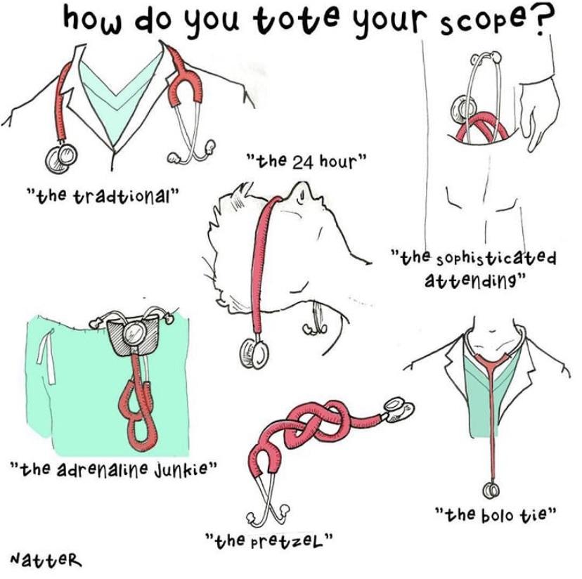
      – Place the tubing behind your neck and let the chest piece hang in front.
      This prevents unnecessary kinking of the tubing.
    2. Over the Shoulders –
      Drape it over your shoulders if you need quick access, but avoid excessive
      stretching.
    3. Use a Dedicated Holster or Pouch
      – Some healthcare professionals prefer clip-on stethoscope holders to
      prevent neck strain.
    4. Keep Earpieces Facing Forward
      – When inserting the earpieces, angle them forward to match the natural
      anatomy of your ear canals.
    5. Adjust the Fit – Ensure
      the headset tension is comfortable by gently squeezing or pulling apart
      the ear tubes.
    6. Clean It Regularly – Wipe
      the diaphragm and tubing with an alcohol swab after use to prevent
      contamination.
    doctor listening to  chest

    How NOT to Wear a Stethoscope

    1. Around the Neck for Long Periods
      – Prolonged hanging around the neck can cause oil buildup and degrade the
      tubing.
    2. Dangling from One Shoulder
      – This can cause it to slip and fall, leading to damage.
    3. Stuffing into a Tight Pocket
      – Bending the tubing too much can cause cracks or deformation.

    Proper Ways to Store a Stethoscope

    1. Flat in a Drawer or Case
      – Lay it flat in a clean drawer or a dedicated case when not in use.
    2. Hanging on a Hook –
      Hang it in a relaxed position to avoid kinking the tubing.
    3. Using a Stethoscope Case
      – A hard or soft case can protect it from dust and physical damage.
    4. Room-Temperature Storage
      – Store in a cool, dry place to prevent tubing degradation.

    How NOT to Store a Stethoscope

    1. Leaving it in a Hot Car
      – Heat exposure can make the tubing brittle and shorten its lifespan.
    2. Coiling Too Tightly –
      Over-bending can cause cracks in the tubing.
    3. Placing Heavy Objects on It
      – Pressure can damage the diaphragm and tubing.
    4. Hanging Near Sharp Edges
      – Avoid hooks or surfaces that could damage the tubing.

    Uses of Stethoscope

    Here is list of Common stethoscope use:

    Cardiac Auscultation

    • Identifying normal heart sounds (S1, S2)
    • Detecting abnormal heart sounds (S3, S4)
    • Recognizing heart murmurs (systolic, diastolic, continuous)
    • Identifying pericardial friction rubs (pericarditis)
    • Evaluating prosthetic heart valve function

    Pulmonary Auscultation

    • Assessing normal breath sounds (vesicular, bronchial, bronchovesicular)
    • Detecting adventitious lung sounds (crackles in pulmonary edema, wheezes
      in asthma, stridor in upper airway obstruction)
    • Identifying pleural rubs (pleuritis)
    • Monitoring post-intubation lung sounds for tube displacement

    Vascular Auscultation

    • Detecting carotid bruits (carotid artery stenosis)
    • Assessing abdominal aortic bruits (abdominal aortic aneurysm)
    • Identifying renal artery bruits (renal artery stenosis)
    • Evaluating femoral bruits (peripheral artery disease)

    Gastrointestinal Auscultation

    • Assessing bowel sounds (normal peristalsis)
    • Detecting hyperactive bowel sounds (gastroenteritis, early bowel
      obstruction)
    • Identifying absent bowel sounds (paralytic ileus, late bowel
      obstruction)

    Obstetric Auscultation

    • Monitoring fetal heart rate using a Doppler stethoscope
    • Assessing fetal well-being during pregnancy

    Blood Pressure Measurement

    • Auscultating Korotkoff sounds for accurate sphygmomanometry
    Blood Pressure Measurement using stethoscope (auscultatory method)

    Critical Care and Emergency Medicine

    • Verifying endotracheal tube placement (equal bilateral breath sounds)
    • Identifying pneumothorax (absent breath sounds on affected side)
    • Assessing pulmonary edema in heart failure (bibasilar crackles)
    • Detecting shock-related bruits in vascular collapse

    Neonatal and Pediatric Assessment

    • Evaluating congenital heart defects (e.g., patent ductus arteriosus,
      ventricular septal defect)
    • Monitoring neonatal lung conditions (e.g., transient tachypnea of
      newborn, respiratory distress syndrome)

    Choosing the Right Stethoscope:

    Before we delve into the proper way to wear a stethoscope, it’s important to
    select the right instrument for your needs. 

    Consider factors such as your area of expertise, comfort, and sound quality
    when purchasing a stethoscope. 

    Opt for a high-quality model from reputable brands to ensure accurate
    auscultation.

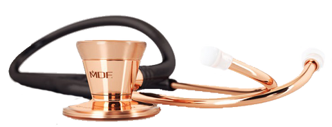
    Image : Two pioneers of stethoscope industry viz littman nad MDF

    Familiarizing Yourself with the Parts:

    A stethoscope typically consists of three main parts: the chestpiece, tubing,
    and earpieces. The chestpiece contains the diaphragm and the bell, which are
    used to listen to different types of sounds. The tubing connects the
    chestpiece to the earpieces, and the earpieces are inserted into the ears for
    sound transmission.

    stethoscope parts diagram

    Adjusting the Earpieces of stethoscope:

    To wear a stethoscope properly, begin by adjusting the earpieces. 

    Each earpiece should fit comfortably in your ears without exerting excessive
    pressure. 

    Gently squeeze or pull the earpieces to adjust the tension, ensuring a snug
    fit while avoiding discomfort or pain. 

    Improperly adjusted earpieces can hinder sound transmission and lead to
    inaccurate auscultation.

    Incorrect Position

    Correct Position

    (Images credit: 3M littman)

    Positioning the Earpieces:

    Insert the earpieces into your ears at the appropriate angle. The earpieces
    should be positioned pointing forward, aligning with the natural angle of your
    ear canal. 

    Ensure that they are not twisted or facing backward, as this can impede sound
    conduction and cause distortion.

    Positioning the Earpieces (credit wikihow)

    Checking Tubing Length:

    Next, check the length of the tubing. Ideally, the tubing should be long
    enough to allow you to auscultate different areas of the patient’s body
    comfortably. 

    However, excessively long tubing can result in sound loss or interference.
    Adjust the length according to your height and arm length, ensuring that it
    doesn’t tangle or drag on the floor.

    Securing the Chestpiece:

    Once the earpieces are in place, secure the chestpiece onto the patient’s
    body. Ensure that the diaphragm or bell is correctly positioned over the area
    of interest.

    For example, use the diaphragm for high-frequency sounds such as heart and
    lung sounds, and the bell for low-frequency sounds like murmurs or bowel
    sounds. 

    Press the chestpiece lightly against the patient’s skin for optimal sound
    transmission.

    Testing Sound Transmission:

    To verify that the stethoscope is correctly positioned and functioning well,
    perform a quick sound check. 

    Listen for the desired sounds and adjust the pressure, angle, or position if
    necessary. Familiarize yourself with the specific sounds produced by your
    hearts and lungs. 

    Listening and interpreting the sounds in stethoscope:

    Then listen to the desired organ of the patient and interpret the sound
    accordingly. Once you have your stethoscope in place, it’s time to start
    listening. 
    To get the best results, you should listen carefully and focus on the sounds
    you hear. Make sure that you are in a quiet environment and that there are no
    distractions that could affect your reading. 
    It’s also important to use the correct technique when listening, such as using
    the diaphragm to listen to high-frequency sounds and the bell for
    low-frequency sounds.

    Basic Sounds of Auscultation

    Crackles Audio   

    Friction Rub  Audio   

    Bowel Sounds Audio 

    Abnormal (increased) Bowel Sounds Audio 

    Normal Breath Sounds Audio 

    Normal Bronchial Breath Sounds Audio 

    Stridor Audio 

    Wheeze Audio 

    Conclusion

    In conclusion, wearing a stethoscope correctly is essential for medical
    professionals. By following these steps, you can ensure that your stethoscope
    is properly fitted and that you can get accurate readings.
    Remember to clean your stethoscope regularly and to listen carefully to the
    sounds you hear. 
     With the right technique, you can make the most of this valuable tool
    and provide the best care for your patients.

    What is the Parotid Gland Swelling One Side ICD 10 and Differentials?

    (toc)Table of Contents

    What is the Parotid gland swelling one side ICD 10 Code?

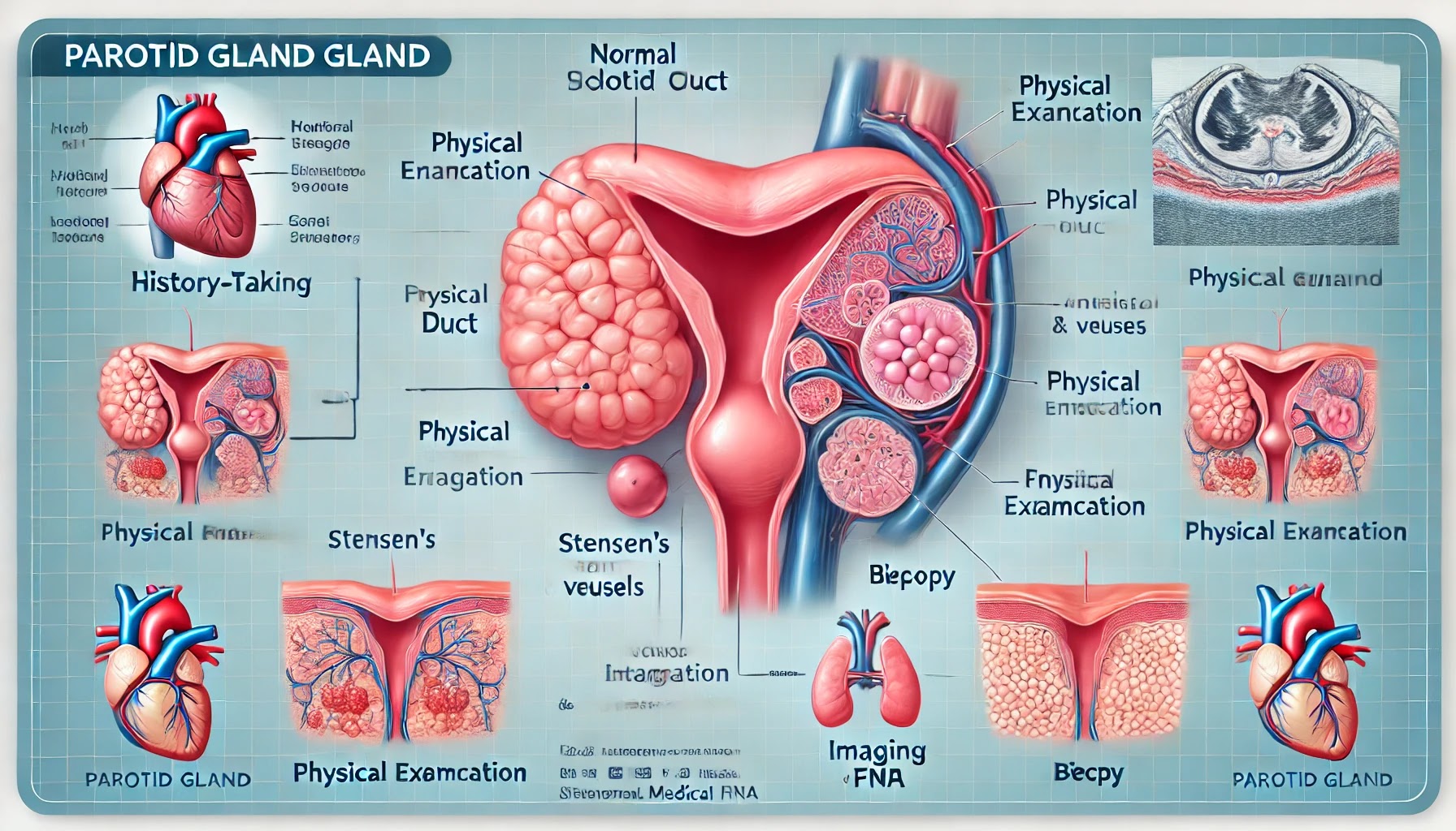
    What is parotid gland?

    The parotid is a large salivary gland located near the jaw that helps produce saliva.
    The parotid glands are the largest of the salivary glands, situated just in front of and below each ear. They secrete saliva into the mouth through ducts, aiding in digestion and oral health by moistening food and helping to break it down.

    Symptoms of Parotid gland Swelling

    Symptoms of parotid gland swelling may include:
    • Pain or Tenderness: Discomfort or pain in the area near the jaw or ear.
    • Swelling: Noticeable enlargement of the gland, causing visible or palpable lumps.
    • Redness or Warmth: Skin over the swollen gland may appear red or feel warm to the touch.
    • Dry Mouth: Reduced saliva production can lead to dryness in the mouth.
    • Difficulty Swallowing: Swelling can make swallowing food or liquids uncomfortable.
    • Fever: An increase in body temperature, often indicating infection or inflammation.
    • Bad Breath: Resulting from reduced saliva flow and potential infection.
    • Difficulty Opening Mouth: Limited movement due to pain or swelling.
    • Taste Changes: Altered sense of taste or unusual taste in the mouth.

    Differential diagnosis of Parotid gland swelling

    Parotid swelling can have various differential diagnoses (DDx), and they can be categorized based on whether the swelling is unilateral (one-sided) or bilateral (both sides).

    Unilateral Parotid Swelling:

    Benign Tumors:

    Pleomorphic adenoma: Most common benign tumor of the parotid gland.
    Warthin’s tumor: Another benign tumor, more common in older males and smokers.

    Malignant Tumors:

    Mucoepidermoid carcinoma: The most common malignant tumor of the parotid gland.
    Adenoid cystic carcinoma: Slow-growing but potentially aggressive tumor.

    Infections:

    Bacterial sialadenitis: Usually due to Staphylococcus aureus or Streptococcus species.
    Viral sialadenitis: Most commonly mumps, especially in unvaccinated individuals.

    Obstructive Causes:

    Sialolithiasis (salivary gland stones): Leads to obstruction of the salivary duct, causing painful swelling.

    Inflammatory Conditions:

    Sarcoidosis: Can present with parotid gland involvement.
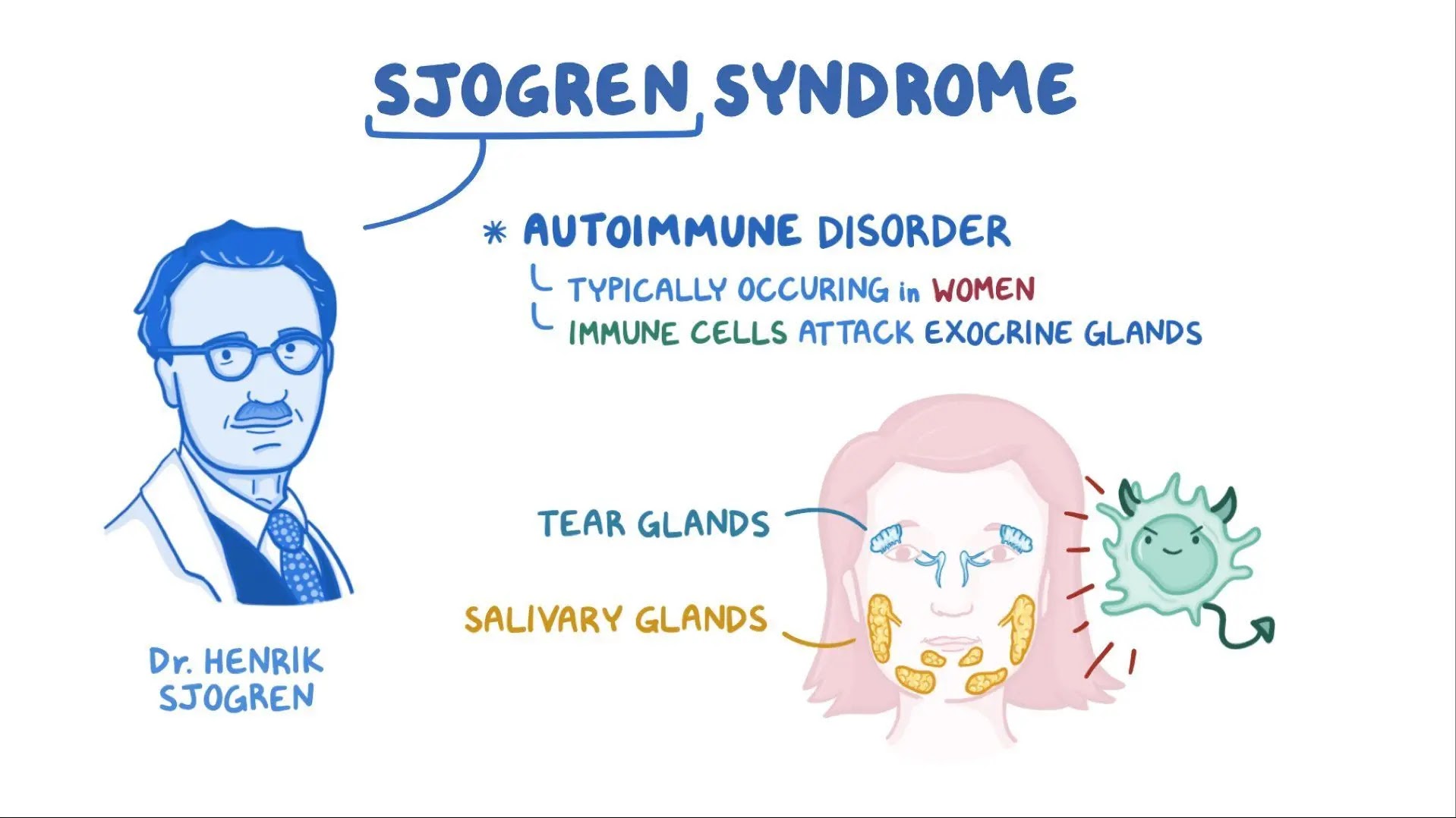
    Sjögren’s syndrome: Autoimmune disease that affects salivary glands, though typically bilateral, it can sometimes present unilaterally.

    Trauma:

    Post-traumatic swelling: Due to direct injury to the parotid gland.

    Bilateral Parotid Swelling:

    Infections:

    Mumps: The most common viral cause, often accompanied by fever and malaise.
    HIV-associated salivary gland disease: May cause bilateral enlargement.

    Autoimmune and Inflammatory Conditions:

    Sjögren’s syndrome: Chronic autoimmune disorder affecting the salivary and lacrimal glands.
    Sarcoidosis: Systemic granulomatous disease that may involve the parotid glands bilaterally.
    sjogren syndrome (from odmosis)

    Metabolic Conditions:

    Alcoholic parotitis: Chronic alcohol abuse can lead to bilateral parotid swelling.
    Diabetes mellitus: Can sometimes be associated with bilateral parotid enlargement.

    Medications:

    Drug-induced parotid enlargement: Certain medications like antihypertensives (e.g., clonidine) can cause bilateral gland enlargement.

    Idiopathic:

    Idiopathic sialadenosis: Non-inflammatory, non-neoplastic bilateral swelling of the parotid glands, often associated with metabolic conditions or malnutrition.
    Identifying the underlying cause of parotid swelling requires a careful clinical evaluation, including history, physical examination, imaging studies (such as ultrasound or MRI), and sometimes biopsy or fine-needle aspiration (FNA).

    लु लाग्नु भनेको के हो? Lu lagnu-lu disease-loo disease meaning- heat stroke -prevention, diagnosis and treatment

    Table of Contents(toc)

    Lu lagnu- heat stroke -prevention,  diagnosis and treatment लु लाग्नु also loo disease nepali

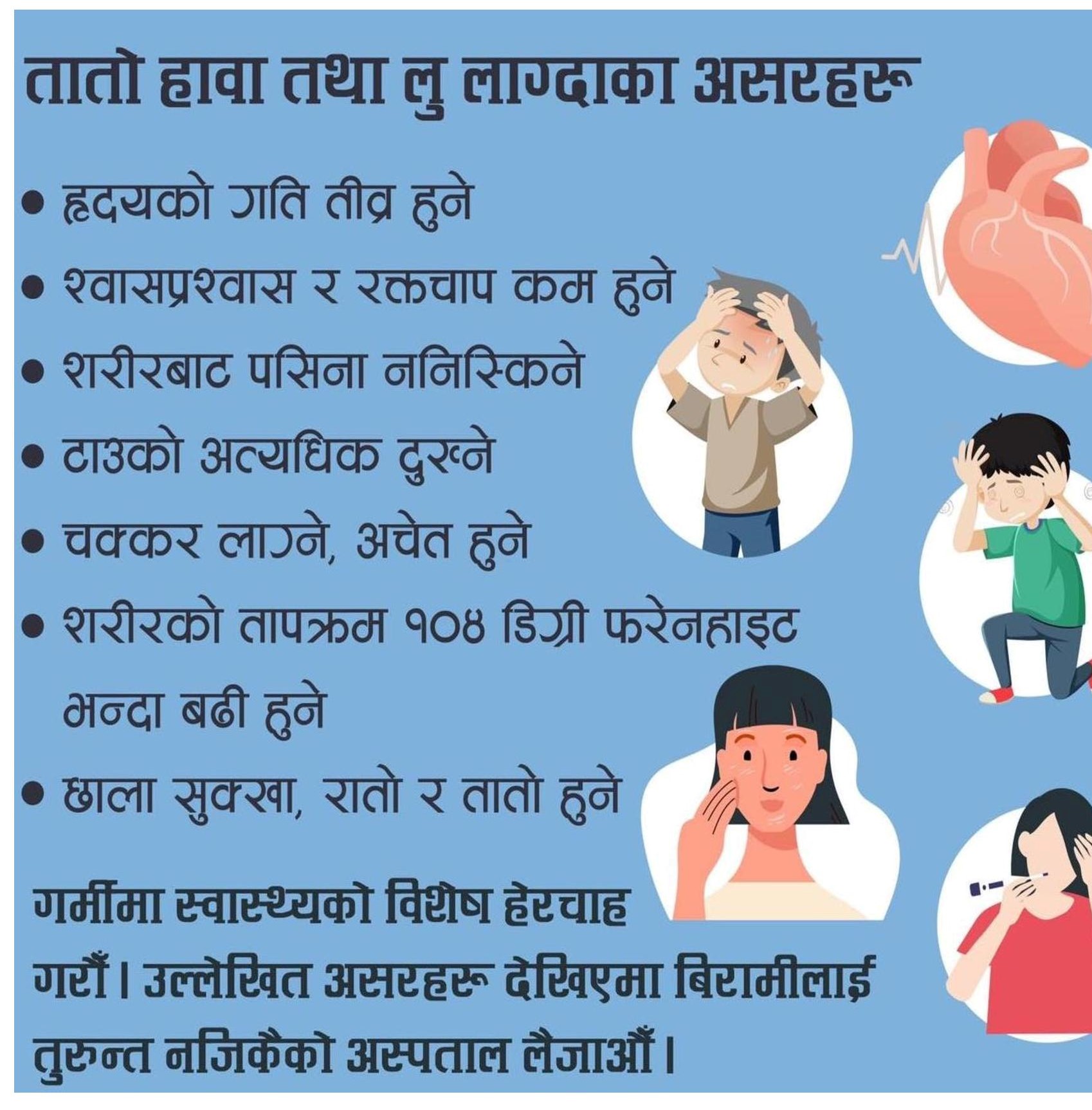
    Recently, Lu, commonly known as heat stroke has been seen very commonly in Nepal and India during summertime. In this article we will discuss regarding pathophysiology, symptoms, and treatment of lu.

    What is lu lagnu? लु लाग्नु meaning of lu in nepali, loo meaning लु लाग्नु भनेको के हो?

    Heat stroke is a range of diseases in which our body is not able to cope with heat and temperature of the environment especially occurring in summertime in the hot, humid climates, like the region of Nepal and India.
    It may cause edema, cramps, rashes, spasm and tetany to syncope, neurological disturbance and death. It is associated with increasedbody temperature more than 40° centigrade.
    लु लाग्नु भनेको अत्यधिक गर्मीका कारणले शरीरले सहन नसकेर देखिने लक्षण हो जसमा ज्यान सुनिने, दुख्ने, मासु बाउँडिने, बेहोस हुने र मानसिक समस्या आउने सम्म हुन सक्छ। यो प्राय शरीरको तापक्रम ४०° भन्दा बढी भएर हुन्छ।

    Causes of loo disease or lu disease: Hyperthermia लु लाग्नुका कारण

    लु लाग्नुको कारण वातावरणको तापक्रम लामो समय सम्म अत्यधिक भएर हो। प्राय तराई तथा गर्मी हुने ठाउँ हरुमा यो हुने गर्दछ। 
    अर्कोथरी हिट स्ट्रोक अत्यधिक कडा शारीरिक काम गर्नाले हुने गर्दछ। 
    यो दुवै थरीले प्राय बच्चा र वृद्धाहरुलाई बढी असार गर्दछ। यसले ज्यान जाने खतरा हुन्छ।
    Both exertional and non exertional heat stroke affect young and old individuals more. Both are associated with high morbidity and mortality rates.
    A hot humid sunmy day

    Pathophysiology of lu disease, loo diasease

    Heat stroke is defined as temperature more tha. 40°C or 104°F associated with neurological dysfunction. Anhidrosis, that is lack of sweating is key feature of geat stroke. 
    लु लाग्नु लाई शरीरको तापक्रम ४०° से वा १०४° फ भन्दा बढी भएर मानसिक समस्या भएको अवस्थालाई बुझिन्छ। यसमा पसिना नआउनु मुख्य चिन्ह हो।
    अत्यधिक गर्मी मौसममा शरीर चिस्याउन पसिना आउने गर्दछ।तर शरीरको कशमाट भन्दा बढी गर्मी भयो भने शरीरले तापक्रम नियन्त्रण गर्न सक्दैन र ।वातावरण सँगै शरीर पनि तातिदैं जान्छ सो को कारणले लक्षणहरू देखिन्छ।
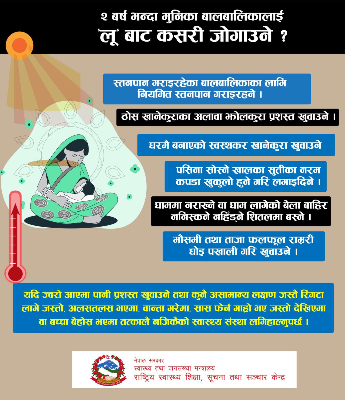
    How to prevent under 2 years age children from lu

    Meaning of lu lagnu, lu disease or loo disease

    लु लाग्नु भनेको अत्यधिक गर्मीले शरीरको तापक्रम सामान्य भन्दा बढी हुनु हो। यो ज्वरो हैन। किन भने यो बाह्य तापक्रमले गर्दा शरीरको तापक्रम बढेको हो र शरीरको तापक्रम नियन्त्रण गर्ने क्षमता असफल भएको अवस्था हो।
    Lu is due to excessive external heat and is not fever. In lu body has lost ability to control temperature. 

    What are symptoms of loo, lu symptoms, heatstroke symptoms

    It can be seen in warm climates and more commonly in people who have excessive hard ore exercises. गर्मी मौसम या अत्यधिक व्यायाम, शारीरिक काम गरेर तलका लक्षण देखिन सक्छ:
    1. मासु देख्ने Myelgia
    2. पेट दुख्ने abdominal pain 
    3. वाकवाकी nausea
    4. वान्ता हुने vomiting
    5. पखाला diarrhea
    6. टाउको दुख्ने headache
    7. रिंगटा लाग्ने diazziness
    8. श्वास फेर्न गाह्रो हुने, गति बढ्ने difficulty breathing, increased respiratory rate
    9. कमजोरी महसुस हुने weakness, tiredness
    10. बेहोस हुने fainting, loss of consciousness
    11. ढल्ने falling
    12. होस् नहुने, दिमागले राम्ररी काम नगर्ने memory losss, disorientation, inability to think properly
    13. मुटुको गति बढ्ने increased heart rate
    14. थकान feeling of tiredness
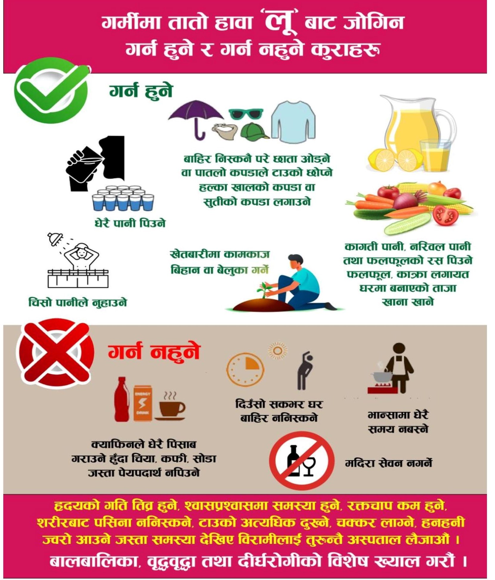
    To do and not to do of lu

    EHS प्राय खेलकुद, व्यायाम गर्ने स्वस्थ वयस्कहरु मा देखिन्छ भने classical NEHS बच्चा र वृद्धालाई बढी देखिन्छ।
    EHS is commonly seen in atheletes while classical NEHS is seen in young and old individuals.
    पुरानो रोग, पानीको कम सेवन, संक्रमण, मोटोपना, निद्राको कमी जस्ता कारणले लु लाग्ने सम्भावना बढाउँछ।
    Chronic illnesses, dehydration, infection, obesity, sleep oack etc rise  hance of lu or heat stroke.

    How to prevent lu, loo prevention

    • Staying in cold environment as far as possible.
    • Drinking plenty of water
    • Taking bathe time to time
    • Eat fruits containing water like cucumber, watermelon etc
    • Do not go outside during daytime
    • Do not drink alcohol
    • Do not drink caffeine containing drinks

    लु लाग्नु भनेको अत्यधिक गर्मीले शरीरको तापक्रम सामान्य भन्दा बढी हुनु हो। यो ज्वरो हैन। किन भने यो बाह्य तापक्रमले गर्दा शरीरको तापक्रम बढेको हो र शरीरको तापक्रम नियन्त्रण गर्ने क्षमता असफल भएको अवस्था हो।

    Read top lecture class: MI class

    Treatment of Lu, heat stroke treatment

    Desert

    लु लागेको संका लागेमा तलका जाँच हरु हर्न सकिन्छ:

    1. ए बि जि ABG
    2. सुगर sugar
    3. सोडियम sodium
    4. पोटासियम potassium
    5. क्यल्सियम calcium
    6. म्याग्नेसियम magnessium
    7. कलेजोको जाँच Liver function tests
    8. सि के Creatinine kinase CK
    9. युरिया urea
    10. क्रिएटिनिन creatinine
    11. मुत्र परीक्षण urine analysis
    Effects of lu

    Complications of heat stroke, loo complications

    There i actual medical treatment except cooling the patient. Some medications used are benzodiazepine, and IV fluids. Diuretics like mannitol may also be needed with large volume infusion to prevent volume overload. Antipyretics and analgesics have no role in heat stroke. The complication include 
    • Short term morbidity
    • Hospitalization
    • Infection
    • Renal failure
    • Neurological deficit
    • Permanent disability
    • Growth retardation
    • Death
    BMI calculator here

    The organs mainly affected by heat are

    • Brain
    • Liver
    • Kidney
    • Lungs
    • Muscle
    • Heart
    • Blood vessels
    • Skin 
    • Sensory organs

    Heat stroke death in nepal: epidemiology

    Every year a sigificant number of children and old age peole die in teria region of nepal due to heat stroke or loo. Many adults are also impacted. 
    sweating boy due to heat

    MOHP: MOHP
    Dr Health RX: Dr Health Rx

    Some images have been taken from MOHP website for awareness purpose.  

    Heatstroke in medscape

    NNC PCL nursing result 2081

    Nursing License Registration for 35rd PCL Nursing

    Check PCL Nursing licensing examination result 2081

    Check below

    Check PDF here pcl nursing result

    download pdf

    click download button to download: 

    click down arrow

    NNC PCL result

    If you have query please ask us. 


    NHPC seventh License examination Notice

    Anesthetic Agents of Choice in Various Conditions and Their Contraindications

    Anesthetic Agents of Choice in Various Conditions and Their Contraindications

    Table of Contents(toc)


    1. Ketamine: The Ideal Agent for Bronchial Asthma

    Ketamine is a potent bronchodilator, making it the anesthetic agent of choice in bronchial asthma. Its ability to maintain airway reflexes and provide both analgesia and sedation makes it a preferred option in asthmatic patients undergoing surgery.

    Contraindications:

    Ketamine increases heart rate (HR), blood pressure (BP), cardiac output (CO), intraocular pressure (IOP), and intracranial tension (ICT), so it should be avoided in conditions such as:

    • Uncontrolled hypertension
    • Ischemic heart disease
    • Glaucoma
    • Elevated intracranial pressure (e.g., traumatic brain injury, space-occupying lesions)

    2. Neuro and Cardiac Anesthesia: Isoflurane

    Isoflurane is the preferred anesthetic for neurosurgery and cardiac surgery due to its ability to maintain cerebral perfusion and cause minimal myocardial depression. It also provides good muscle relaxation and reduces brain metabolic demand.

    Contraindications:

    • Patients with severe hypotension, as Isoflurane can cause vasodilation and drop BP further.
    • Patients with coronary artery disease (CAD) should be monitored closely due to the risk of coronary steal phenomenon.

    3. Hepatic Anesthesia: Isoflurane > Desflurane

    Isoflurane is preferred over desflurane in patients with hepatic dysfunction because it has a more favorable hepatic metabolism profile. While both agents have minimal liver metabolism, Isoflurane is less likely to cause hepatotoxicity compared to halothane.

    Contraindications:

    • Patients with liver failure, where all volatile anesthetics should be used with caution.
    • Desflurane should be avoided in hepatic insufficiency due to its potential to increase liver enzyme levels in susceptible individuals.

    4. Pediatric Anesthesia: Sevoflurane

    Sevoflurane is the anesthetic agent of choice in pediatric anesthesia due to its pleasant odor, rapid induction, and minimal airway irritation. It allows for smooth induction, which is especially useful in uncooperative children.

    Contraindications:

    • Patients at risk of malignant hyperthermia (like those with a genetic predisposition).
    • Patients with severe renal impairment, as sevoflurane metabolism produces fluoride ions, which could potentially affect kidney function.

    5. Daycare Anesthesia: Desflurane & Propofol

    • Desflurane is the volatile agent of choice for daycare surgeries due to its low blood-gas solubility, allowing for rapid emergence from anesthesia.
      • Mnemonic: D for D (Desflurane for Daycare)
    • Propofol is the IV agent of choice for daycare surgery due to its rapid onset, short duration of action, and antiemetic properties.

    Contraindications:

    • Desflurane is not suitable for induction due to its pungency, which can cause airway irritation and coughing.
    • Propofol should be avoided in patients with egg or soy allergies (although true allergies are rare).
    • Not ideal for hemodynamically unstable patients due to its hypotensive effects.

    Conclusion

    Selecting the right anesthetic agent depends on patient-specific conditions and surgical requirements. Understanding the contraindications of each anesthetic is essential for optimizing patient safety and surgical outcomes.

    Errors in Morphogenesis

    Errors in Morphogenesis


    Table of Contents(toc)

    Types of Errors in Morphogenesis

    A. Malformations

    1. Definition – Disturbances in the morphogenesis (development) of an organ.
    2. Occurrence – Mainly during embryogenesis (first 9 weeks of pregnancy).
      • Most occur between the third and ninth weeks of embryogenesis.
      • The most susceptible period is the fourth and fifth weeks when organs form from the germ layers (ectoderm, endoderm, mesoderm).

    B. Deformations

    1. Definition – Caused by extrinsic factors that physically impinge on fetal development in utero.
    2. Occurrence – Between the ninth week and term after fetal organs have developed.
    3. Causes – Most often due to restricted movement in the uterine cavity (uterine restraint). Examples include:
      • Maternal factors – Malformed uterus, large leiomyomas (smooth muscle tumors) in the uterine wall.
      • Placental factors – Oligohydramnios (low amniotic fluid), twin pregnancies.

    C. Disruptions

    1. Definition – A type of deformation that results from the destruction of irreplaceable normal fetal tissue.
    2. Causes – May be due to vascular insufficiency (e.g., thrombosis of placental vessels), trauma, or teratogens.
    3. Example – Amniotic bands:
      • Rupture of the amnion leads to the formation of fibrous bands.
      • These bands encircle fetal parts, causing partial limb amputation or constriction rings around digits.

    D. Agenesis

    1. Definition – Complete absence of an organ due to the absence of the anlage (primordial tissue).
    2. Example – Renal agenesis (absence of one or both kidneys).

    E. Aplasia

    1. Definition – The anlage (primordial tissue) is present but does not develop into a fully formed organ.
    2. Example – Lung aplasia (lung tissue with rudimentary ducts and connective tissue).

    F. Hypoplasia

    1. Definition – The primordial tissue develops incompletely but is histologically normal.
    2. Example – Microcephaly (small brain), hypoplastic left heart syndrome.

    Summary of Errors in Morphogenesis

    1. Malformations

    • Disturbances in the development of an organ.
    • Occur mainly during embryogenesis (first 9 weeks of pregnancy).
      • Most occur between the 3rd and 9th weeks.
      • Highest susceptibility during the 4th and 5th weeks, when organs are forming from germ layers.

    2. Deformations

    • Caused by extrinsic factors that physically affect fetal development in utero.
    • Occur after the 9th week when fetal organs have developed.
    • Most often due to restricted movement in the uterine cavity (uterine restraint).
      • Maternal factors: Malformed uterus, large leiomyomas.
      • Placental factors: Oligohydramnios, twin pregnancies.

    3. Disruptions

    • A type of deformation resulting from destruction of irreplaceable normal fetal tissue.
    • Causes: Vascular insufficiency (e.g., placental thrombosis), trauma, teratogens.
    • Example: Amniotic bands – fibrous bands constrict fetal parts, leading to limb amputation or constriction rings around digits.

    4. Agenesis

    • Complete absence of an organ due to lack of primordial tissue (anlage).
    • Example: Renal agenesis.

    5. Aplasia

    • Primordial tissue (anlage) is present but fails to develop into a functioning organ.
    • Example: Lung aplasia – tissue contains rudimentary ducts and connective tissue but no proper lung formation.

    6. Hypoplasia

    • Incomplete development of an organ; tissue is histologically normal but underdeveloped.
    • Examples:
      • Microcephaly – small brain.
      • Hypoplastic left heart syndrome – incomplete development of the left heart structures.

    Multiple Choice Questions

    1. The primary structural defect of an organ is termed:
      a) Disruption
      b) Malformation
      c) Deformation
      d) Association

    2. Which of the following is an example of deformation?
      a) Renal agenesis
      b) Oligohydramnios-induced limb contracture
      c) Amniotic band syndrome
      d) Microcephaly

    3. At what stage of pregnancy do most malformations occur?
      a) First trimester
      b) Second trimester
      c) Third trimester
      d) After birth

    4. Which condition is an example of a disruption?
      a) Hypoplastic left heart
      b) Amniotic band syndrome
      c) Pulmonary hypoplasia
      d) Diaphragmatic hernia

    5. What is the most susceptible period for malformations during embryogenesis?
      a) First and second weeks
      b) Third to ninth weeks
      c) Ninth to twelfth weeks
      d) After birth

    6. Which of the following best describes agenesis?
      a) Complete absence of an organ due to lack of primordial tissue
      b) Incomplete formation of an organ with normal tissue structure
      c) Absence of an organ despite the presence of rudimentary tissue
      d) Organ damage due to external factors

    7. Which maternal condition can lead to deformation?
      a) Diabetes
      b) Large uterine leiomyomas
      c) Gestational hypertension
      d) Hyperthyroidism

    8. Which of the following best describes hypoplasia?
      a) Complete absence of an organ
      b) Normal tissue with reduced growth
      c) Malformation due to teratogens
      d) A disruption caused by amniotic bands

    9. What is the difference between disruption and deformation?
      a) Disruption occurs due to genetic mutations, while deformation occurs due to external forces.
      b) Deformation occurs after organ formation, while disruption destroys normal fetal tissue.
      c) Deformation is irreversible, while disruption is usually corrected with surgery.
      d) Disruption happens in the second trimester, while deformation happens in the first trimester.

    10. Which of the following is an example of aplasia?
      a) Renal agenesis
      b) Microcephaly
      c) Lung aplasia
      d) Clubfoot


    Answer Key

    1. b) Malformation
    2. b) Oligohydramnios-induced limb contracture
    3. a) First trimester
    4. b) Amniotic band syndrome
    5. b) Third to ninth weeks
    6. a) Complete absence of an organ due to lack of primordial tissue
    7. b) Large uterine leiomyomas
    8. b) Normal tissue with reduced growth
    9. b) Deformation occurs after organ formation, while disruption destroys normal fetal tissue.
    10. c) Lung aplasia

    This structured format is optimized for exam preparation with clear explanations, additional key points, and relevant multiple-choice questions. Let me kno

    Types of Bullets and Forensic Value

    Types of Bullets and Forensic Value

    Table of Contents(toc)

    In forensic ballistics, bullets can be categorized based on their design, material, and purpose. Special types of bullets include:

    1. Hollow-Point Bullets

    • Designed to expand upon impact, increasing stopping power.
    • Commonly used in law enforcement and self-defense.
    • Leaves distinct wound channels, aiding forensic analysis.

    2. Full Metal Jacket (FMJ) Bullets

    • A soft core (usually lead) encased in a harder metal shell (e.g., copper).
    • Less deformation on impact, often passing through the target.
    • Common in military use, leaving clean entry/exit wounds.

    3. Soft-Point (SP) Bullets

    • Partially jacketed with an exposed lead tip.
    • Expands more than FMJ but less than hollow-point bullets.
    • Used in hunting and law enforcement.

    4. Armor-Piercing (AP) Bullets

    • Made with hardened steel, tungsten, or depleted uranium cores.
    • Designed to penetrate hard targets (e.g., body armor, vehicles).
    • Often leaves distinctive markings on surfaces and bodies.

    5. Frangible Bullets

    • Composed of compressed metal powders (e.g., copper/tin).
    • Breaks apart upon impact, minimizing ricochet risks.
    • Often used in training and situations where over-penetration is a concern.

    6. Tracer Bullets

    • Contains a pyrotechnic charge that ignites when fired.
    • Leaves a visible trail, aiding in aiming and tracking.
    • Used in military applications.

    7. Incendiary Bullets

    • Contains chemical compounds (e.g., phosphorus) that ignite upon impact.
    • Used against fuel tanks, aircraft, or for signaling purposes.

    8. Explosive Bullets

    • Designed to detonate on impact.
    • Rare and often restricted due to their destructive capability.

    9. Rubber and Plastic Bullets

    • Non-lethal alternatives used for riot control and crowd dispersal.
    • Can cause blunt force trauma but generally do not penetrate the body.

    10. Wad Cutter & Semi-Wad Cutter Bullets

    • Flat or slightly conical tips designed for clean, circular holes in paper targets.
    • Often used in competitive shooting.

    11. Glaser Safety Slug

    • A frangible bullet with a thin copper jacket and lead shot inside, designed to break apart upon impact.
    • Used to minimize over-penetration in home defense.
    • Forensically, it leaves multiple small wound channels and lacks deep penetration.

    12. Boat-Tail Bullets

    • Has a tapered base to reduce air resistance and improve long-range accuracy.
    • Used in sniper and precision shooting.
    • Ballistic analysis can track trajectory and impact characteristics.

    13. Dum-Dum Bullets

    • Expanding bullets with a modified soft or hollow tip for extreme tissue damage.
    • Initially banned under the Hague Convention due to their lethality.
    • Identified forensically by irregular wound patterns and expanded fragments.

    14. Match-Grade Bullets

    • Precision-made bullets with tight tolerances for competitive shooting.
    • Usually FMJ or hollow-point, but with higher consistency.
    • Rifling marks on these bullets can provide key forensic evidence.

    15. Saboted Bullets (SABOT Rounds)

    • A small-caliber projectile enclosed in a discarding plastic sabot, fired from a larger-caliber barrel.
    • Used in high-velocity rifle ammunition and armor-piercing rounds.
    • The sabot leaves forensic traces, such as polymer residues.

    16. Duplex and Triplex Bullets

    • Consist of two or three projectiles in a single cartridge.
    • Used in experimental military applications.
    • Can create multiple wound tracks, confusing forensic pathologists.

    17. Teflon-Coated Bullets (“Cop Killers”)

    • Originally designed to reduce barrel wear and improve penetration through soft barriers.
    • Incorrectly believed to pierce body armor (actual armor-piercing capability comes from bullet core material).
    • Coating residue can sometimes be detected in forensic analysis.

    18. Caseless Ammunition

    • The bullet is embedded in a solid propellant block, eliminating the need for a brass casing.
    • Rare but used in some advanced military firearms.
    • Forensically, it lacks shell casings, making firearm matching difficult.

    19. Microstamped Bullets

    • Feature laser-etched or stamped identifiers on the base of the bullet.
    • Helps track bullets back to specific manufacturers or purchasers.
    • Still a developing forensic technology.

    20. Electroshock Bullets

    • Experimental rounds that deliver an electric shock upon impact.
    • Designed for non-lethal incapacitation.
    • Forensic examination involves electrical burns and impact analysis.

    Forensic Methods for Bullet Examination

    1. Ballistic Comparison

      • Examines striations left by a gun’s barrel using a comparison microscope.
      • Matches bullets to specific firearms.
    2. Gunshot Residue (GSR) Analysis

      • Identifies lead, antimony, and barium particles left after firing.
      • Can determine shooting distance and whether a suspect fired a gun.
    3. Wound Ballistics Analysis

      • Examines entry/exit wounds, fragmentation, and tissue damage.
      • Helps determine bullet type, trajectory, and velocity.
    4. Trajectory Reconstruction

      • Uses mathematical models to track a bullet’s path and point of origin.
    5. Chemical Analysis

    • Spectroscopy can determine bullet composition, revealing its manufacturer or unique properties.

    Forensic Methods for Bullet Examination

    1. Ballistic Comparison

      • Examines striations left by a gun’s barrel using a comparison microscope.
      • Matches bullets to specific firearms.
    2. Gunshot Residue (GSR) Analysis

      • Identifies lead, antimony, and barium particles left after firing.
      • Can determine shooting distance and whether a suspect fired a gun.
    3. Wound Ballistics Analysis

      • Examines entry/exit wounds, fragmentation, and tissue damage.
      • Helps determine bullet type, trajectory, and velocity.
    4. Trajectory Reconstruction

      • Uses mathematical models to track a bullet’s path and point of origin.
    5. Chemical Analysis

      • Spectroscopy can determine bullet composition, revealing its manufacturer or unique properties.
    6. Striation marks (from barrel rifling).
    7. Deformation patterns (to identify bullet type).
    8. Residue analysis (to determine if a bullet was fired).
    9. Entry/exit wound characteristics (to infer bullet type and velocity).

    Scientists Develop Gene-Edited Lettuce to Combat Micronutrient Deficiencies

    Scientists Develop Gene-Edited Lettuce to Combat Micronutrient Deficiencies

    Table of Contents(toc)


    Jerusalem, March 7, 2025 – A team of researchers from the Hebrew University of Jerusalem has successfully developed a gene-edited lettuce variety with significantly enhanced nutritional value. Using CRISPR gene-editing technology, the scientists increased levels of essential micronutrients, including β-carotene (provitamin A), zeaxanthin, and ascorbic acid (vitamin C), making this modified lettuce a potential tool in the fight against global micronutrient deficiencies.

    The study, published in Plant Biotechnology Journal, was led by Prof. Alexander Vainstein from the university’s Robert H. Smith Faculty of Agriculture, Food, and Environment. The researchers demonstrated that targeted genetic modifications could significantly boost the nutritional content of lettuce without negatively affecting its growth, appearance, or yield.

    Key findings from the study include:

    • A 2.7-fold increase in β-carotene, which is essential for vision, immune function, and skin health.
    • A significant boost in zeaxanthin, an antioxidant that protects against age-related macular degeneration.
    • A 6.9-fold increase in vitamin C, which strengthens the immune system and aids iron absorption.

    The breakthrough highlights the potential of CRISPR technology in enhancing food nutrition without introducing foreign DNA, unlike traditional GMO methods. “This study is an important step toward developing healthier food options that can help address widespread nutrient deficiencies in modern diets,” said Prof. Vainstein.

    Micronutrient deficiencies, often referred to as “hidden hunger,” affect millions worldwide, particularly in developing regions where access to diverse diets is limited. The development of nutrient-rich crops through gene editing could play a crucial role in tackling this global health issue.

    As researchers continue refining gene-editing techniques, this innovative approach could pave the way for future biofortified crops, offering a sustainable and efficient solution to malnutrition.

    The implications of this breakthrough extend beyond just improving individual health. Nutrient-rich crops like gene-edited lettuce could help reduce dependence on dietary supplements and fortified foods, making essential vitamins more naturally available through daily consumption.

    Moreover, the ability to enhance multiple nutrients simultaneously without affecting crop yield is a major advancement in agricultural biotechnology. Traditional breeding methods often require years to achieve similar results, whereas CRISPR allows for precise and efficient modifications in a much shorter time frame.

    Potential for Wider Agricultural Applications

    The success of this study could inspire further applications of gene-editing technology in other staple crops, such as rice, wheat, and maize, which are primary food sources for millions. Scientists are already exploring similar approaches to enhance the nutritional profiles of various vegetables and grains to combat global malnutrition.

    Additionally, gene-edited crops could be developed to have increased resistance to environmental stressors such as drought, pests, and soil deficiencies, making them more sustainable and suitable for cultivation in regions facing harsh agricultural conditions.

    Regulatory and Public Perception Challenges

    Despite the promising benefits of CRISPR-edited foods, regulatory hurdles and public perception remain key challenges. While gene-edited crops do not contain foreign DNA like traditional genetically modified organisms (GMOs), some countries still regulate them similarly. Public education on the safety and advantages of gene editing will be crucial for widespread acceptance.

    Prof. Vainstein and his team hope that their research will contribute to changing the narrative around gene-edited foods by demonstrating their potential to improve public health without compromising food safety or environmental sustainability.

    Future Prospects

    Looking ahead, the research team plans to further optimize the gene-editing process to fine-tune nutrient levels and explore potential commercial applications. If regulatory approvals are granted, consumers could see nutrient-enhanced lettuce and other biofortified vegetables in supermarkets within the next few years.

    As global food security challenges continue to grow, innovations like this gene-edited lettuce offer a promising step toward a future where healthier, more nutritious food is accessible to all.

    Global Health Impact

    The development of gene-edited lettuce could play a pivotal role in addressing micronutrient deficiencies, particularly in regions where malnutrition is a significant public health challenge. According to the World Health Organization (WHO), deficiencies in vitamin A, vitamin C, and other essential micronutrients affect over 2 billion people globally, contributing to weakened immune systems, impaired vision, and increased mortality rates.

    By offering a naturally nutrient-rich alternative, gene-edited crops could help vulnerable populations gain better access to essential vitamins without relying heavily on expensive supplements or fortified foods. This innovation aligns with global efforts like the United Nations Sustainable Development Goals (SDGs), which aim to end hunger and improve nutrition by 2030.

    Scientific Milestone in Precision Agriculture

    The study highlights how CRISPR technology is revolutionizing agriculture by making precise genetic changes without altering the plant’s natural characteristics. This advancement opens the door for “precision agriculture”, where crops are tailored not only for higher yields but also for improved health benefits.

    Prof. Vainstein emphasized the broader implications of this technology, stating:

    “Our approach demonstrates that gene editing can improve food quality without sacrificing agricultural performance. This could pave the way for a new generation of crops that directly address the nutritional needs of populations worldwide.”

    Next Steps and Commercialization

    The research team is now collaborating with agricultural partners to conduct field trials and assess the performance of gene-edited lettuce under real-world farming conditions. If successful, they plan to seek regulatory approvals in various countries to introduce this biofortified lettuce to the market.

    Additionally, the scientists are exploring similar gene-editing techniques in other leafy vegetables, such as spinach and kale, which are widely consumed and could benefit from enhanced nutrient profiles.

    Conclusion

    The gene-edited lettuce developed by the Hebrew University of Jerusalem represents a significant step forward in the fight against hidden hunger. By combining cutting-edge biotechnology with a commitment to global health, this breakthrough has the potential to reshape the future of agriculture and nutrition.

    As regulatory frameworks evolve and public awareness grows, gene-edited crops could become a powerful tool in building a more sustainable and equitable food system, bringing healthier diets within reach for millions around the world.

    Credits: New, More Nutritional Lettuce Plant Developed by Hebrew University Researchers Using CRISPR Gene Editing, https://www.afhu.org/2025/03/07/new-more-nutritional-lettuce-plant-developed-by-hebrew-university-researchers-using-crispr-gene-editing/

    Article at: Yarin Livneh, Ehud Leor-Librach, Dor Agmon et al, Combined enhancement of ascorbic acid, β-carotene and zeaxanthin in gene-edited lettuce,  https://onlinelibrary.wiley.com/doi/10.1111/pbi.70018,  doi: https://doi.org/10.1111/pbi.70018

    (please contact me for any copyright infringement action before reporting, i will happily remove it if reported)

    (for educational purpose only)

    Exit mobile version