STANDARD TREATMENT PROTOCOL OF EMERGENCY HEALTH SERVICE PACKAGE 2023

STANDARD TREATMENT PROTOCOL OF EMERGENCY HEALTH SERVICE PACKAGE

CONTENTS

STP BHS

FOREWORD iii

ACKNOWLEDGEMENT V

ABBREVIATIONS X

BACKGROUND 1

Definition of Emergency Health Services 1

Rationale of the STP 2

Development Process of the STP 3

Utilization of the STP 3

Assumptions made for the implementation of the STP 4

How to use the STP 4

ABCDE Approach in the Emergency Room 7

Adult Basic Life Support 28

Paediatric Basic Life Support 29

Paediatric Cardiac Arrest Algorithm 30

Adult Cardiac Arrest Algorithm 31

Adult Tachycardia with a Pulse Algorithm 32

Adult Bradycardia Algorithm 33

Adult Post- Cardiac Arrest Care Algorithm 34

LIST OF EMERGENCY CONDITIONS 35

1. Respiratory Emergencies 37

2. Cardiac Emergencies 62

3. Neurological Emergencies 89

4. Gastrointestinal Emergencies 101

5. Genitourinary Emergencies 128

6. Gynaecology and Obstetrical Emergencies 135

7. Orthopedics and Trauma 152

8. Metabolic Emergencies 168

9. Ocular Emergencies 179

10. ENT Emergencies 182

11. Burns 185

12. Mental Health Emergencies 191

13. Toxicological Emergencies 204

14. Snake bite, Animal bite, Insect bite 225

15. Pediatric Emergencies 234

16. Miscellaneous 244

STANDARD TREATMENT PROTOCOL OF EMERGENCY HEALTH SERVICE PACKAGE

viii

ANNEXES 253

ANNEX I: Schedule 2: Emergency Health Services 254

ANNEX II: List of Essential Medicines 257

Annex III: Patient Referral Form 270

ANNEX IV: Participants of Pre Planning/ Preliminary Consultative

Meeting with key government officials on STP of EHS 271

ANNEX V: Participants of Consultative TWG Meeting on STP of EHS 272

ANNEX VI: Participants of Consultative Meeting with Subjects/

Emergency Experts on STP of EHS 273

ANNEX VII: Participants of Consultative Meeting with Professional

Council and Association Members on STP of EHS 274

ANNEX VIII: Participants of High Level Consultative Meeting on STP of EHS 275

BIBLIOGRAPHY 277

STANDARD TREATMENT PROTOCOL OF EMERGENCY HEALTH SERVICE PACKAGE

ix

LIST OF FIGURES

Figure 1 Head tilt and chin lift 10

Figure 2 Head tilt and chin lift 10

Figure 3 Jaw thrust 11

Figure 4 Jaw thrust 11

Figure 5 Oropharyngeal airway (Guedel airway) 12

Figure 6 Nasopharyngeal airway 12

Figure 7 Bag and mask ventilation (One person technique) 13

Figure 8 Bag and mask ventilation (Two person technique) 13

Figure 9 LMA (Laryngeal mask airway) 14

Figure 10 Endotracheal tube 15

Figure 11 Cervical collar application 25

Figure 12 Cervical collar application 25

Figure 13 Log roll 27

Figure 14 Log roll 27

Figure 15 Acute anterior wall myocardial infarction 68

Figure 16 Acute inferior wall myocardial infarction 68

Figure 17 Atrial fibrillation with fast ventricular rate 76

Figure 18 Supraventricular tachycardia 78

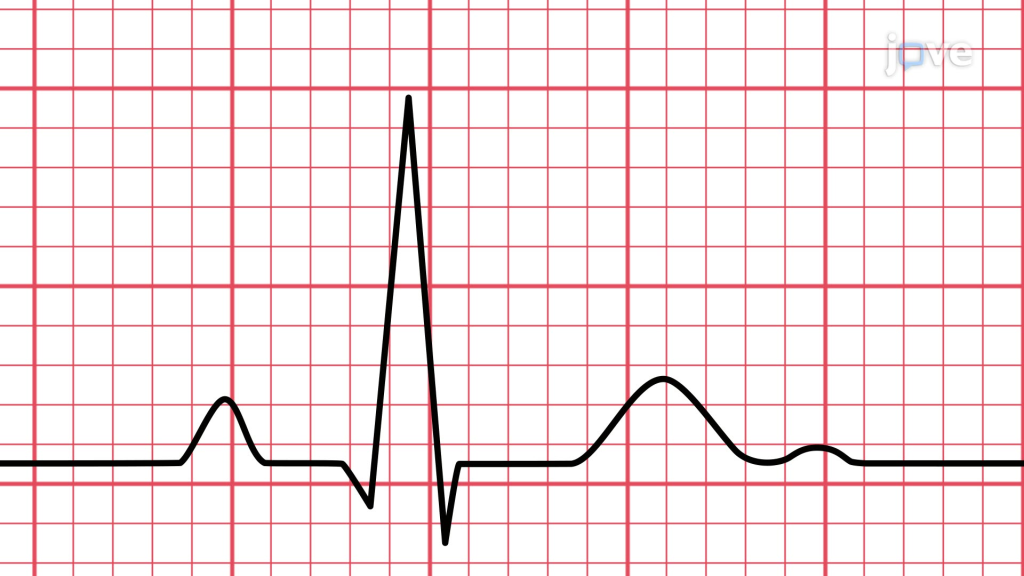
Figure 19 Complete heart block

download

Tetanus prophylaxis guideline nepal 2023

Tetanus prophylaxis guideline nepal 2023

Please go through the pdf for tetanus prophylaxis guideline nepal 2023 below.
vaccine

Tetanus guideline

Appropriate tetanus prophylaxis should be administered as soon as possible following a wound but should be given even to patients who present late for medical attention. This is because the incubation period is quite variable; most cases occur within 8 days, but the incubation period can be as short as 3 days or as long as 21 days. For patients who have been vaccinated against tetanus previously but who are not up to date, there is likely to be little benefit in administering human tetanus immune globulin more than 1 week or so after the injury. However, for patients thought to be completely unvaccinated, human tetanus immune globulin should be given up to 21 days following the injury; Td or Tdap should be given concurrently to such patients

Package of Essential Non-communicable Diseases (PEN)

 Package of Essential Non-communicable Diseases (PEN)

PEN module book

what is pen package who nepal

 Package of Essential Non-communicable Diseases (PEN) was training developed for trainignn doctors and paramedics regardaing non communicalve diseasae.
 Package of Essential Non-communicable Diseases (PEN) is a trainign program for health professionals to increase if NCD treatment in rural communities

How to perform ABG and interpretation ABG analysis results

 How to perform and interpret ABG analysis

Introduction to ABG:

ABG stands for arterial blood gas. ABG is a common test done in case of critical care and severely ill patient to know the arterial bloog gas status of the patient along with various other parameters.
Sample for ABG analysis

Normal ABG value ranges:

ABG practice questions:

How to read and ECG: Mnemonic for ECG reading fast

Introduction

A structured approach makes ECG interpretation systematic and reliable. A commonly used mnemonic is:

RRAHI

  • R = Rhythm
  • R = Rate
  • A = Axis
  • H = Hypertrophy
  • I = Ischemia

How to Calculate Heart Rate in Regular Rhythm ECG

For a regular rhythm, use either of the following methods:

Method 1 (Big boxes)

  • Take any two R waves
  • Count the number of big boxes between them
  • Divide 300 by that number
  • The result is the heart rate

Method 2 (Small boxes)

  • Take any two R waves
  • Count the number of small boxes between them
  • Divide 1500 by that number
  • The result is the heart rate

How to Calculate Heart Rate in Irregular Rhythm ECG

For irregular rhythms, use a longer rhythm strip:

  • Take 10 big boxes in the rhythm strip
  • Count the number of QRS complexes (R waves) within those 10 big boxes
  • Multiply that number by 30

👉 This gives the heart rate in beats per minute (bpm)

Completed Formula

Heart rate = (Number of QRS complexes in 10 big boxes) × 30

Why this works:

  • 1 big box = 0.2 seconds
  • 10 big boxes = 2 seconds
  • 60 seconds ÷ 2 seconds = 30

So you are converting a 2-second sample into a 1-minute rate.


How to Check Rhythm of an ECG

To determine the rhythm:

  • Check if R–R intervals are regular or irregular
  • Look for P waves before every QRS complex
  • Ensure P waves have a consistent shape
  • Confirm that each P wave is followed by a QRS

Normal (Sinus Rhythm) Features:

  • Regular rhythm
  • One P wave before each QRS
  • Normal PR interval
  • Heart rate between 60–100 bpm

How to Check Axis of an ECG

Axis refers to the overall direction of electrical activity in the heart.

Basic Method (using limb leads):

  • Look at Lead I and aVF
Lead IaVFAxis
++Normal axis
+Left axis deviation
+Right axis deviation
Extreme axis deviation

Normal Axis:

  • Between -30° to +90°

The electrical impulse normally:

  • Starts from the right atrium
  • Travels toward the left ventricle
  • Occurs 60–100 times per minute

How to Identify Myocardial Ischemia on ECG

Look for these three key changes:

  • ST segment changes
    • Elevation → possible acute injury
    • Depression → ischemia
  • T wave changes
    • Inversion
    • Flattening
  • Q waves
    • May indicate previous myocardial infarction

How to Identify Hypertrophy on ECG

Hypertrophy leads to increased voltage (amplitude) on ECG.

Left Ventricular Hypertrophy (LVH)

A commonly used method:

  • S wave in V1 + R wave in V5 or V6 ≥ 35 mm

Additional clues:

  • Deep S waves in V1
  • Tall R waves in V5/V6
  • Possible left axis deviation

This completes the full ECG reading framework using the RRAHI approach, covering all essential steps: rate, rhythm, axis, hypertrophy, and ischemia.

Thyroid function test: TFT how to interpret TFT and know these dangerous disease at home

Thyroid gland


Core Tests

  • TSH (Thyroid Stimulating Hormone) → most sensitive
  • Free T4 (FT4) → active hormone
  • Free T3 (FT3) → useful in hyperthyroidism
  • ± Antibodies:
    • Anti-TPO
    • TRAb (TSH receptor antibody)

Step 1: Look at TSH first

          TSH
        /  |   \
     Low Normal High

IF TSH LOW → Think Hyperthyroid OR Central Hypothyroid

TSH ↓
  |
  ├── FT4 ↑ / FT3 ↑ → PRIMARY HYPERTHYROIDISM
  │        (e.g. Graves', toxic nodules)
  │
  ├── FT3 ↑ only → T3 TOXICOSIS
  │
  ├── FT4 normal, FT3 normal → SUBCLINICAL HYPERTHYROIDISM
  │
  └── FT4 ↓ → CENTRAL (SECONDARY) HYPOTHYROIDISM
           (pituitary/hypothalamic disease)

Diagnoses

  • Graves’ disease
  • Toxic multinodular goiter
  • T3 toxicosis
  • Central hypothyroidism

IF TSH HIGH → Think Hypothyroidism

TSH ↑
  |
  ├── FT4 ↓ → PRIMARY HYPOTHYROIDISM
  │        (thyroid gland failure)
  │
  ├── FT4 normal → SUBCLINICAL HYPOTHYROIDISM
  │
  └── FT4 ↑ → RARE:
           - TSH-secreting adenoma
           - Thyroid hormone resistance

Diagnoses

  • Hashimoto thyroiditis
  • Iodine deficiency
  • Post-thyroidectomy
  • TSH-secreting pituitary adenoma

IF TSH NORMAL → Check FT4/FT3 carefully

TSH normal
  |
  ├── FT4 ↓ → CENTRAL HYPOTHYROIDISM
  │
  ├── FT4 ↑ → TSHOMA / RESISTANCE
  │
  └── FT4 normal → EUTHYROID

SUMMARY FLOWCHART (HIGH-YIELD)

                TSH
         ┌──────┼──────┐
        ↓       N       ↑
        |       |       |
     Check     Check    Check
     FT4/FT3   FT4      FT4
        |       |        |
   ┌────┼───┐   |    ┌───┼────┐
  ↑     N   ↓   |   ↓    N     ↑
Hyper Subclin Central   Primary Subclin Rare
thyroid hyper  hypo     hypo    hypo   causes

SPECIAL PATTERNS (EXAM FAVORITES)

1. Euthyroid Sick Syndrome

  • TSH: normal/low
  • FT3: ↓ (early)
  • FT4: ↓ (late)

👉 Seen in severe illness (ICU patients)


2. Subclinical Disease

ConditionTSHFT4
Subclinical hyperthyroidismNormal
Subclinical hypothyroidismNormal

3. Pregnancy Changes

  • TSH ↓ (1st trimester)
  • FT4 slightly ↑ early

4. Drug Effects

  • Amiodarone → hypo OR hyper
  • Steroids → ↓ TSH
  • Lithium → hypothyroidism

MEMORY TRICKS

“TSH runs opposite to thyroid hormones”

  • ↑ TSH = ↓ thyroid function (usually)
  • ↓ TSH = ↑ thyroid function

“Think CENTRAL if TSH doesn’t match FT4”

  • Low TSH + Low FT4 → pituitary problem
  • Normal TSH + Low FT4 → pituitary problem

“Subclinical = TSH abnormal, FT4 normal”


QUICK CLINICAL PROTOCOL

Suspected Hypothyroidism

  1. Check TSH
  2. If ↑ → check FT4
  3. If FT4 ↓ → treat
  4. If FT4 normal → monitor / anti-TPO

Suspected Hyperthyroidism

  1. Check TSH
  2. If ↓ → check FT4, FT3
  3. If elevated → confirm cause:
    • TRAb → Graves’ disease
    • Scan → nodules

Suspected Pituitary Disease

  • Always check:
    • FT4
    • TSH (may be misleading)

COMMON PITFALLS

  • Relying only on TSH in pituitary disease
  • Ignoring FT3 in hyperthyroidism
  • Misinterpreting illness-related changes

ULTRA-SHORT REVISION

  • TSH ↑ + FT4 ↓ → Primary hypothyroid
  • TSH ↓ + FT4 ↑ → Hyperthyroid
  • TSH ↑ + FT4 normal → Subclinical hypo
  • TSH ↓ + FT4 normal → Subclinical hyper
  • TSH ↓ + FT4 ↓ → Central hypothyroid

RFT- Renal function test (notes summary)

RFT- Renal function test: Indication, Interpretatin, Normal Range

RFT- Renal function test: Indication, Interpretatin, Normal Range

Table of Contents(toc)

Introduction of Renal Function Test (RFT)

Renal Function Tests (RFTs) are a group of blood and urine tests used to assess the health and efficiency of the kidneys. These tests help in evaluating how well the kidneys are filtering waste products, maintaining electrolyte balance, and regulating fluid levels in the body. 

RFTs are essential tools in diagnosing and monitoring kidney diseases, as well as in managing systemic conditions like hypertension and diabetes that can impact kidney function. 

By analyzing key parameters such as serum creatinine, blood urea nitrogen (BUN), estimated glomerular filtration rate (eGFR), and electrolytes, healthcare providers can detect early signs of renal impairment and initiate appropriate interventions.

Indications for RFT

  • Suspected kidney disease
  • Hypertension
  • Diabetes mellitus (routine monitoring)
  • Urinary tract infections (recurrent or severe)
  • Edema (swelling of legs/face)
  • Hematuria (blood in urine)
  • Proteinuria (protein in urine)
  • Monitoring patients on nephrotoxic drugs (e.g., aminoglycosides, NSAIDs)
  • Preoperative assessment
  • Chronic kidney disease (CKD) follow-up

Components of RFT

  1. Serum Creatinine
  2. Blood Urea Nitrogen (BUN) / Urea
  3. Estimated Glomerular Filtration Rate (eGFR)
  4. Serum Electrolytes (Na⁺, K⁺, Cl⁻, HCO₃⁻)
  5. Urinalysis (protein, glucose, blood, specific gravity)
  6. Creatinine Clearance (if needed)

Normal Ranges

(Note: may vary slightly by lab)

  • Serum Creatinine:
    • Males: 0.7 – 1.3 mg/dL
    • Females: 0.6 – 1.1 mg/dL
  • Blood Urea Nitrogen (BUN): 7 – 20 mg/dL
  • Urea (alternative to BUN): 20 – 40 mg/dL
  • eGFR:
    • Normal: >90 mL/min/1.73 m²
    • Mild decrease: 60–89
    • Moderate: 30–59
    • Severe: 15–29
    • Kidney failure: <15
  • Creatinine Clearance: 90 – 140 mL/min
  • Serum Sodium (Na⁺): 135 – 145 mEq/L
  • Serum Potassium (K⁺): 3.5 – 5.0 mEq/L
  • Serum Bicarbonate (HCO₃⁻): 22 – 29 mEq/L

Interpretation

  • ↑ Creatinine & BUN: Indicates impaired renal function or acute/chronic kidney disease
  • ↓ eGFR: Suggests declining kidney function
  • ↑ Potassium (Hyperkalemia): Common in advanced renal failure
  • ↓ Sodium (Hyponatremia): Seen in fluid overload or renal salt wasting
  • Urinalysis:
    • Proteinuria: Indicates glomerular damage
    • Hematuria: Suggests infection, stones, or glomerulonephritis
    • Low Specific Gravity: May suggest tubular dysfunction

Please learn to read a Lipid profile report

Lipid profile- Know the danger of cholesterol in a single test

Table of contents(toc)

Introduction Lipid panel

Lipid simply means fat or cholesterol in general. 
But lipid profile contains multiple parameters viz. Total cholesterol, triglycerides, high density lipoprotein, low density lipoprotein and very low density lipoprotein.

These all parameters have their own reference range and own function in our body. 
Now we will discuss each of the above aprameter individually.

Parameters that are measuted in lipid profile

Here are the parameters measured in Lipid profile
  1. Total cholesterol
  2. triglycerides
  3. high density lipoprotein
  4. low density lipoprotein
  5. very low density lipoprotein

Total Cholesterol

Reference ranges:


 < 200 mg/dL (Desirable)

• 200–239 mg/dL: Borderline high

• ≥ 240 mg/dL: High

Triglycerides

Reference ranges:


< 150 mg/dL (Normal)

• 150–199 mg/dL: Borderline high

• 200–499 mg/dL: High

• ≥ 500 mg/dL: Very high

High density lipoprotein

Reference ranges:


Men: > 40 mg/dL (Optimal)

• Women: > 50 mg/dL (Optimal)

• ≥ 60 mg/dL is considered protective against heart disease

Low density lipoprotein

Reference ranges:

Optimal: < 100 mg/dL

• 100–129 mg/dL: Near optimal

• 130–159 mg/dL: Borderline high

• 160–189 mg/dL: High

• ≥ 190 mg/dL: Very high


Very low density lipoprotein

Reference ranges: 5–30 mg/dL (Normal range)
  • Atherosclerosis risk of deranged lipid profile
  • Heart disease risk of deranged lipid profile
  • Arterial plaque buildup of deranged lipid profile
  • Coronary artery disease and deranged lipid profile
  • Cardiovascular health and deranged lipid profile
  • Cholesterol testing
  • Lipid profile interpretation
  • Cholesterol management
  • Dietary cholesterol impact
  • Lifestyle modifications
  • Statins and cholesterol control
  • Exercise and cholesterol levels
  • Genetic predisposition to high cholesterol
  • Importance of regular lipid screening
Exit mobile version