Causes of Anemia in Infants

Causes of Anemia in Infants (List Only):

Table of Contents(toc)

1. Nutritional causes

  • Iron deficiency
  • Vitamin B12 deficiency
  • Folate deficiency
  • Protein-energy malnutrition

2. Blood loss

  • Gastrointestinal bleeding (e.g., anal fissure, Meckel’s diverticulum
  • Birth trauma or internal hemorrhage
  • Iatrogenic blood loss (frequent phlebotomy in NICU)
  • Occult bleeding (cow’s milk protein-induced colitis)

3. Hemolytic causes

  • Hemolytic disease of the newborn (Rh or ABO incompatibility)
  • G6PD deficiency
  • Hereditary spherocytosis
  • Thalassemia
  • Sickle cell disease
  • Infections causing hemolysis (e.g., malaria, sepsis)

4. Bone marrow failure / decreased production

  • Aplastic anemia
  • Congenital pure red cell aplasia (Diamond–Blackfan anemia)
  • Transient erythroblastopenia of childhood
  • Bone marrow infiltration (leukemia, storage diseases)

5. Chronic disease / inflammation

  • Anemia of chronic infection or inflammation
  • Renal failure (↓ erythropoietin)

6. Physiologic / other causes

  • Physiologic anemia of infancy
  • Prematurity (low iron stores, immature erythropoiesis)
  • Hypothyroidism
  • Chronic blood loss from parasites (hookworm in older infants)

How to store breast milk for future use?

1. Purpose

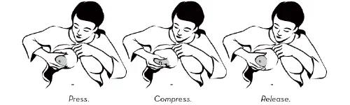
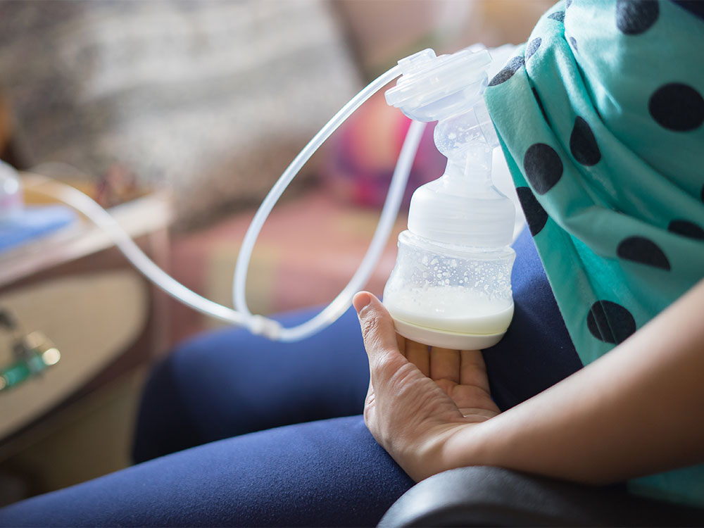
Proper storage of expressed breast milk (EBM) helps preserve its nutritional and immunological properties for later feeding when the mother is away.

2. Containers for Storage

  • Use clean, sterilized glass or BPA-free plastic containers with tight-fitting lids.
  • Breast milk storage bags (food-grade, pre-sterilized) are convenient for freezing.
  • Avoid ordinary plastic bottles or disposable liners.

3. Labeling

  • Each container should be labeled with:
  • Date and time of expression
  • Baby’s name (if used in hospital or daycare)

4. Storage Guidelines

LocationTemperatureDurationRemarks
Room temperatureUp to 25°C (77°F)4–6 hoursKeep in a cool, clean area away from direct sunlight
Refrigerator (back portion)2–4°C (35–40°F)Up to 72 hours (3 days)Do not store in refrigerator door due to temperature fluctuation
Freezer (separate door)–18°C or lowerUp to 3–6 monthsKeep in small portions; leave space for expansion
Deep freezer–20°CUp to 6–12 monthsBest for long-term storage
Insulated cooler box with ice packs~15°CUp to 24 hoursFor temporary transport or travel

5. Thawing and Warming

  • Thaw in refrigerator overnight or by placing the container in warm water (<37°C).
  • Do not microwave or boil — destroys antibodies and nutrients.
  • Swirl gently (do not shake) to mix separated fat layers.
  • Once thawed, use within 24 hours and do not refreeze.

6. Hygiene

  • Wash hands before expressing or handling milk.
  • Use clean pump parts and containers for each session.
  • Avoid touching the inside of lids or bottles.

7. Key Points

  • Always use oldest milk first (“first in, first out”).
  • Discard leftover milk from feeding bottle after use.
  • Observe for odor or curdling — discard if spoiled.

Summary:

Safe breast milk storage requires hygienic handling, correct temperature, and appropriate duration. Properly stored milk retains most of its nutritional, immunological, and protective properties, ensuring safe feeding for infants when direct breastfeeding isn’t possible.

What causes Large and Thick placenta (placentomegaly)?

Placentomegaly — Causes

Table of Contents(toc)

Definition:

Placentomegaly refers to an abnormally thick or enlarged placenta, typically defined as:

  • >4 cm thick at 20 weeks gestation, or

  • >6 cm thick at term.

plaenta normal anterior image

I. Maternal Causes

  1. Diabetes mellitus (especially poorly controlled)

    → due to villous edema and increased fetal size.

  2. Maternal anemia (especially severe)

    → compensatory placental hypertrophy to improve oxygen transfer.

  3. Maternal infection

    • TORCH infections (Toxoplasmosis, Rubella, CMV, Herpes)

    • Syphilis, Malaria, Hepatitis, HIV

  4. Hypertension with superimposed infection or diabetes

  5. Rh isoimmunization (leading to fetal hydrops and placental edema)


II. Fetal Causes

  1. Fetal hydrops (immune or non-immune)

    • Most common fetal cause.

    • Due to excessive fluid accumulation → placental edema.

  2. Chromosomal abnormalities

    • Trisomy 13, 18, 21, Triploidy, Turner syndrome

  3. Fetal anemia (any cause, e.g., parvovirus B19 infection)

  4. Twin-to-twin transfusion syndrome (recipient twin side)

  5. Large-for-gestational-age (LGA) fetus

  • Often secondary to maternal diabetes.

a large placenta

III. Placental / Cord Causes

  1. Chorioangioma (benign vascular tumor of placenta)

  2. Molar pregnancy (partial mole)

  3. Chronic villitis or placentitis

  4. Placental edema due to venous obstruction (cord anomalies)


IV. Other / Miscellaneous Causes

  1. Congenital infections (CMV, syphilis, toxoplasmosis, parvovirus)

  2. Placental transfusion syndromes

  3. Maternal-fetal hemorrhage

  4. High altitude pregnancies (chronic hypoxia)


Mnemonic (for quick recall):

“BIG PLACENTA”

  • B – Beta-thalassemia / fetal anemia

  • I – Infections (TORCH, malaria, syphilis)

  • G – Gestational diabetes

  • P – Parvovirus / Polyhydramnios

  • L – Large baby (LGA)

  • A – Aneuploidy (Trisomy 13/18/21)

  • C – Chorioangioma

  • E – Erythroblastosis fetalis (Rh isoimmunization)

  • N – Nonimmune hydrops

  • T – Twin-to-twin transfusion

  • A – Anemia (maternal or fetal)

Key Takeaways:
  • An enlarged placenta isn’t usually a reason to panic. The word is big (placentomegaly), but most of the time it doesn’t cause problems.
  • Some conditions play a role. A larger placenta may be linked to hypertension, anemia, or diabetes — but your doctor will monitor these and the health of your baby.
  • Your baby’s growth matters most. Even if your placenta measures big, steady fetal development is the important goal, so keep up your regular appointments to be sure everything’s right on track.

Rett Syndrome Notes for MD and DM level

Rett Syndrome

Definition

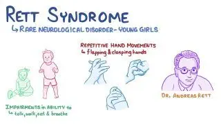
Rett syndrome is a neurodevelopmental disorder that primarily affects girls, characterized by normal early development followed by regression of acquired skills, especially speech and purposeful hand movements, with onset typically between 6–18 months of age.


Etiology

  • Genetic cause: Mutation in MECP2 gene (methyl-CpG-binding protein 2) on the X chromosome (Xq28)

  • Inheritance: Usually sporadic (de novo); rarely familial

  • Pathophysiology: Dysfunction of MECP2 protein → abnormal brain maturation and synaptic development


Epidemiology

  • Affects 1 in 10,000–15,000 female births

  • Lethal in males (most do not survive infancy unless mosaic or XXY)


Clinical Features

Phases of Disease

  1. Early Onset (6–18 months)

    • Normal development initially

    • Gradual loss of interest in surroundings

    • Loss of purposeful hand skills

    • Deceleration of head growth (acquired microcephaly)

  2. Rapid Destructive Phase (1–4 years)

    • Loss of speech and purposeful hand use

    • Stereotyped hand movements: hand-wringing, washing, clapping, or mouthing

    • Gait ataxia, truncal apraxia

    • Autistic-like behavior

  3. Plateau Phase (2–10 years)

    • Some improvement in social interaction and eye contact

    • Persistent motor problems and seizures

  4. Late Motor Deterioration (>10 years)

    • Progressive scoliosis, muscle wasting, rigidity, dystonia

    • Loss of ambulation in many cases


Other Features

  • Breathing abnormalities: hyperventilation, apnea during wakefulness

  • Seizures: common (up to 90%)

  • Bruxism, cold/purple extremities (autonomic dysfunction)

  • Sleep disturbances

  • Growth retardation


Investigations

  • Genetic testing: MECP2 mutation analysis (diagnostic)

  • EEG: slowing with epileptiform activity

  • MRI brain: may show nonspecific atrophy

  • Metabolic tests: normal (to rule out other causes)


Diagnosis

  • Clinical + confirmed MECP2 mutation

  • Diagnostic criteria include:

    • Regression after normal early development

    • Loss of purposeful hand skills and spoken language

    • Gait abnormalities

    • Stereotypic hand movements


Differential Diagnosis

  • Autism spectrum disorder

  • Angelman syndrome

  • Cerebral palsy (especially ataxic type)

  • Childhood disintegrative disorder


Management

  • No cure – supportive and multidisciplinary care

    • Physiotherapy & occupational therapy: maintain mobility

    • Speech therapy: communication support (eye-tracking devices)

    • Antiepileptics: for seizures

    • Nutritional support: adequate calories, manage feeding difficulties

    • Behavioral therapy: improve interaction

    • Orthopedic care: for scoliosis, contractures


Prognosis

  • Progressive but non-degenerative

  • Life expectancy: many survive into adulthood (40–50 years)

  • Main causes of death: sudden unexplained death, pneumonia, cardiac arrhythmias


Mnemonic (Key features)“RETT”

  • R = Regression (speech, hand skills)

  • E = Episodic breathing abnormalities

  • T = Typical hand movements (wringing, washing)

  • T = Tiny head (acquired microcephaly)

Hypothyroidism in Neonates and Children

Table of Contents(toc)

1. Definition

Hypothyroidism is a clinical state resulting from deficiency of thyroid hormone production or action, leading to a generalized slowing of metabolic processes.

It may be:

  • Congenital (Neonatal) – present at birth.

  • Acquired (Childhood) – develops later due to autoimmune, iatrogenic, or other causes.


2. Classification

A. Based on Level of Defect

Type Site of Defect TSH T4/T3
Primary Thyroid gland
Secondary Pituitary ↓/N
Tertiary Hypothalamus ↓/N
Peripheral (Resistance) Target tissue N/↑ N/↑

B. Based on Onset

  • Congenital hypothyroidism (CH)

  • Acquired hypothyroidism


3. Epidemiology

  • CH: ~1 in 2,000–4,000 live births.

  • More common in females.

  • Acquired form common in older children/adolescents, often autoimmune (Hashimoto’s).

thyroid gland


4. Etiology

A. Congenital Hypothyroidism

  1. Thyroid dysgenesis (80–85%)

    • Agenesis, ectopy, or hypoplasia.

    • Usually sporadic.

  2. Dyshormonogenesis (10–15%)

    • Inborn errors of thyroid hormone synthesis (autosomal recessive).

    • E.g. TPO, TG, Pendrin, NIS mutations.

  3. Central hypothyroidism (rare)

    • Pituitary/hypothalamic malformation, midline defects.

  4. Transient CH

    • Iodine excess/deficiency, maternal antibodies or antithyroid drugs.

  5. Thyroid hormone resistance – extremely rare.

B. Acquired Hypothyroidism

  • Autoimmune thyroiditis (Hashimoto’s) – most common.

  • Iodine deficiency/excess.

  • Post-irradiation or post-surgical.

  • Drugs: amiodarone, lithium, interferon-α.

  • Secondary causes: pituitary tumors, craniopharyngioma.


5. Pathophysiology

↓ Thyroid hormone → ↓ metabolic activity → impaired CNS myelination, growth retardation, delayed bone maturation.

  • In neonates: irreversible neurodevelopmental impairment if untreated.

  • In older children: growth failure and delayed puberty predominate.


6. Clinical Features

A. Neonatal / Congenital

Often asymptomatic at birth due to transplacental maternal T4.

Typical features (develop over weeks):

  • Prolonged jaundice

  • Lethargy, hypotonia

  • Feeding difficulty, constipation

  • Large fontanelles

  • Macroglossia

  • Umbilical hernia

  • Cold, dry skin

  • Hoarse cry

  • Poor growth

  • Delayed bone age

  • Delayed milestones (later)

B. Childhood / Acquired

  • Growth retardation, short stature

  • Weight gain with poor height velocity

  • Fatigue, cold intolerance

  • Constipation

  • Dry skin, coarse hair

  • Bradycardia

  • Delayed puberty / menstrual irregularities

  • Pseudoprecocious puberty (rare, due to high TRH → prolactin ↑)

  • Goiter (especially in Hashimoto’s)


7. Investigations

A. Screening

  • Neonatal screening: heel-prick sample at 48–72 hr.

    • Primary TSH (most programs).

    • Elevated TSH → confirm with serum free T4.

B. Diagnostic Tests

Test Interpretation
Serum TSH, Free T4 ↓T4 with ↑TSH → primary hypothyroidism
T3 less reliable in neonates
Thyroglobulin Low in agenesis, high in dyshormonogenesis
Imaging Thyroid scan (99mTc or I-123) – ectopy, agenesis, uptake defects
Ultrasound Gland location and size
Antibodies (TPO, Tg) Positive in autoimmune
Bone age X-ray Delayed
Additional: Pituitary MRI if central hypothyroidism suspected

8. Complications (if untreated)

  • Neurologic: irreversible intellectual disability, deaf-mutism, spasticity.

  • Growth: severe stunting, delayed bone age.

  • Metabolic: dyslipidemia.

  • Cardiac: bradycardia, pericardial effusion.


9. Management

A. Principles

  • Early, adequate, lifelong replacement.

  • Monitor and titrate carefully to maintain euthyroid state.

B. Drug

  • Levothyroxine (L-T4) – drug of choice.

    • Dose:

      • Neonates: 10–15 µg/kg/day.

      • Infants: 8–10 µg/kg/day.

      • Children: 4–6 µg/kg/day.

      • Adolescents: 2–4 µg/kg/day.

    • Given on empty stomach (preferably crushed with water or milk).

C. Monitoring

Age Frequency Parameters
0–6 mo Every 2 wk till T4 normal, then q1–2 mo T4, TSH
6–12 mo q2–3 mo
1–3 yr q3–4 mo
>3 yr q6–12 mo

Target: Free T4 in upper half of normal range, TSH normal.

D. Developmental Follow-up

  • Neurodevelopmental assessment

  • Hearing evaluation

  • Growth chart monitoring


10. Prognosis

  • Normal IQ if therapy started within first 2 weeks of life.

  • Delay in treatment → irreversible intellectual deficit.

  • Acquired forms usually fully reversible with treatment.


11. Key Differentials

  • Pituitary insufficiency

  • Hypothyroxinemia of prematurity

  • Chronic systemic illness (euthyroid sick syndrome)

  • Constitutional growth delay


12. Summary Table

Feature Congenital Acquired
Onset Birth Childhood/adolescence
Cause Dysgenesis > dyshormonogenesis Hashimoto’s most common
Presentation Lethargy, constipation, macroglossia Growth failure, delayed puberty
TSH High High
T4 Low Low
Rx Levothyroxine Levothyroxine
Prognosis Excellent if early Excellent

References

  1. Nelson Textbook of Pediatrics, 22nd ed.

  2. Indian Academy of Pediatrics Guidelines (2021) — Screening and management of congenital hypothyroidism.

  3. Endocrine Society Clinical Practice Guideline (2020) – Congenital Hypothyroidism.

  4. Sperling MA, Pediatric Endocrinology, 5th ed.

Leishmaniasis (Complete Notes)

Leishmaniasis — MD Pediatrics Note (Based on Nelson Textbook of Pediatrics)

Table of Contents(toc)

Introduction

  • Leishmaniasis is a spectrum of protozoal diseases caused by Leishmania species, transmitted by the bite of infected female phlebotomine sandflies.

    cutaneous leishmaniasis

  • Disease manifestations depend on the species involved and the host immune response.

  • Major clinical forms:

    1. Visceral leishmaniasis (VL / kala-azar)

    2. Cutaneous leishmaniasis (CL)

    3. Mucocutaneous leishmaniasis (MCL)


Etiology and Classification

Form Causative Species Geographic Distribution
Visceral L. donovani, L. infantum (chagasi) South Asia, East Africa, Latin America
Cutaneous L. tropica, L. major, L. mexicana, L. braziliensis Middle East, Africa, Americas
Mucocutaneous L. braziliensis complex Central & South America

Epidemiology

  • Endemic in >80 countries; affects poor, rural populations.

  • Vectors: Phlebotomus (Old World), Lutzomyia (New World).

  • Reservoirs: Humans (L. donovani), dogs, rodents.

  • Transmission: Sandfly bite, rarely congenital or via transfusion.

Phlebotomus

Pathogenesis

  • Inoculation of promastigotes → engulfed by macrophages → transform into amastigotes → intracellular multiplication → spread to RES (liver, spleen, bone marrow).

  • Disease severity depends on cell-mediated immunity (CMI).

    • Strong CMI → localized CL.

    • Poor CMI → disseminated VL.

lifecycle of L donovani

Clinical Features

A. Visceral Leishmaniasis (Kala-azar)

  • Incubation: 2 weeks–18 months.

    Visceral Leishmaniasis (Kala-azar)
  • Onset: Insidious.

  • Major triad:

    1. Fever: Remittent, double-quotidian, or irregular.

    2. Hepatosplenomegaly: Marked splenomegaly, moderate hepatomegaly.

    3. Pancytopenia: due to hypersplenism and marrow infiltration.

  • Other features:

    • Weight loss, wasting, darkening of skin (“kala-azar” = black fever)

    • Lymphadenopathy (esp. African form)

    • Anemia, bleeding, infections (esp. bacterial superinfection)

    • Growth retardation and cachexia in chronic disease.

Post-kala-azar dermal leishmaniasis (PKDL):
  • Occurs months–years after VL cure.

  • Hypopigmented macules, papules, nodules (face, arms, trunk).

  • Serves as a reservoir in endemic areas (notably India).


B. Cutaneous Leishmaniasis

  • Lesion: Painless papule → ulcer with indurated margin (“oriental sore”).

  • Usually heals spontaneously in 3–6 months but leaves scar.

  • Chronic forms may resemble lupus vulgaris.


C. Mucocutaneous Leishmaniasis

  • Extension from cutaneous lesion (nasal/oral mucosa).

  • Causes destructive ulcerations → severe disfigurement.

mucocutaneous leishmaniasis

Laboratory Diagnosis

1. Direct demonstration

  • Amastigotes (Leishman–Donovan bodies) in:

    • Splenic aspirate (most sensitive, but risky)

    • Bone marrow aspirate (safe, moderately sensitive)

    • Lymph node or buffy coat smear

  • Giemsa-stained smears show:

    • Oval amastigotes (2–5 μm) with nucleus and kinetoplast inside macrophages.

2. Culture

  • Novy–MacNeal–Nicolle (NNN) medium → promastigote growth.

3. Serologic tests

  • rK39 dipstick test: Highly sensitive & specific for VL (field use).

  • Direct agglutination test (DAT), IFA, ELISA also available.

4. Molecular tests

  • PCR: Highly sensitive, species-specific; used in reference labs.

5. Hematology

  • Pancytopenia, hypergammaglobulinemia, elevated ESR.


Treatment

First-line (Visceral Leishmaniasis)

  • Liposomal Amphotericin B (preferred):

    • Total dose 10–21 mg/kg (varies by region/protocol)

    • Short-course regimens effective.

  • Alternative:

    • Amphotericin B deoxycholate: 1 mg/kg/day × 15–20 doses (toxic, nephrotoxic)

    • Miltefosine: 2.5 mg/kg/day (max 150 mg/day) × 28 days (oral)

    • Paromomycin (IM): 11 mg/kg/day × 21 days

    • Combination regimens (to prevent resistance):

      • Single-dose liposomal amphotericin B + short-course miltefosine or paromomycin.

Cutaneous Leishmaniasis

  • Local therapy (cryotherapy, intralesional antimony) for small lesions.

  • Systemic therapy for multiple, mucosal, or immunocompromised cases:

    • Miltefosine, liposomal amphotericin B, or pentavalent antimonials.

Mucocutaneous Leishmaniasis

  • Liposomal amphotericin B or pentavalent antimonials for ≥28 days.


Prevention and Control

  • Vector control: Insecticide spraying, bed nets.

  • Reservoir control: Treat dogs, cull infected reservoirs.

  • Personal protection: Repellents, protective clothing.

  • Early diagnosis and treatment reduce transmission.

  • Vaccine: None yet in routine use; trials ongoing.


Complications

  • Secondary bacterial infections

  • Severe anemia, hemorrhage

  • Disseminated infection in HIV patients

  • PKDL (in endemic regions like India/Nepal)


Prognosis

  • Excellent with prompt diagnosis and treatment.

  • Mortality >90% if untreated (mainly from secondary infections, cachexia).


Key Points from Nelson

  • Visceral leishmaniasis should be suspected in any febrile child with splenomegaly and pancytopenia in an endemic area.

  • rK39 test is the diagnostic test of choice in field settings.

  • Liposomal amphotericin B is the preferred therapy in both immunocompetent and immunocompromised children.

  • PKDL represents an important reservoir for ongoing transmission.

  • HIV co-infection complicates disease course and increases relapse risk.

Fanconi Anemia Notes for Doctors and PG Aspirants

Fanconi Anemia (FA)

Table of Contents(toc)

Category: Inherited bone marrow failure syndrome (IBMFS)
Inheritance: Autosomal recessive (rarely X-linked)
Gene defects: >22 genes identified (FANCA, FANCC, FANCG most common) → defective DNA interstrand crosslink repair.

fanconi anemia notes

1. Pathophysiology

  • Defect in DNA repair (Fanconi/BRCA pathway) → chromosomal breakage and hypersensitivity to DNA cross-linking agents (e.g., mitomycin C, diepoxybutane).

  • Progressive bone marrow failure (due to stem cell depletion) and genomic instability → predisposition to malignancies.

  • Multisystem developmental abnormalities due to impaired cell proliferation during embryogenesis.


2. Epidemiology

  • Incidence: ~1 in 100,000–250,000 live births.

  • Carrier frequency: ~1 in 200.

  • Median age of diagnosis: 7–9 years.

  • ~90% develop marrow failure by age 40.


3. Clinical Features

A. Hematologic

  • Pancytopenia (usually first manifests with thrombocytopenia or macrocytic anemia).

  • Progressive bone marrow hypoplasia.

  • Increased fetal hemoglobin (HbF).

  • Myelodysplastic syndrome (MDS) or acute myeloid leukemia (AML) risk ↑ markedly.

B. Physical anomalies (present in ~75%)

  • Growth: Short stature, low birth weight.

  • Skeletal: Radial ray defects—absent/hypoplastic thumb, radius anomalies.

  • Skin: Café-au-lait spots, hypopigmentation, hyperpigmentation.

  • Head/Face: Microcephaly, triangular face, microphthalmia.

  • Genitourinary: Renal agenesis, horseshoe kidney, hypoplastic gonads, undescended testes, infertility.

  • Cardiac: Structural heart defects.

  • ENT: Hearing loss.

  • GI: Duodenal atresia, anal anomalies (occasionally).

C. Endocrine/Metabolic

  • Hypothyroidism, glucose intolerance, gonadal failure, low IGF-1.

D. Malignancy risk

  • AML, MDS, and solid tumors (esp. head & neck SCC, gynecologic SCC, liver tumors) due to chromosomal instability.


4. Investigations

Test Finding/Use
CBC Pancytopenia, macrocytosis, increased HbF
Bone marrow biopsy Hypocellular marrow with fatty replacement
Chromosomal breakage test Diagnostic — increased breaks after exposure to diepoxybutane (DEB) or mitomycin C
Molecular genetic testing Confirms FANCA–FANC gene mutations
Flow cytometry for CD34 Decreased hematopoietic stem cells
Ultrasound abdomen Renal anomalies
Endocrine profile Hypothyroidism, gonadal failure screening

5. Differential Diagnosis

  • Acquired aplastic anemia

  • Dyskeratosis congenita

  • Shwachman-Diamond syndrome

  • Diamond–Blackfan anemia


6. Management

Supportive

  • Regular CBC monitoring.

  • Transfusion support (RBCs, platelets) — minimize iron overload.

  • Iron chelation therapy if ferritin ↑.

  • Androgens (e.g., oxymetholone, danazol) → stimulate erythropoiesis (transient benefit).

  • G-CSF for neutropenia (short-term).

  • Avoid DNA-damaging agents (chemotherapy, radiation).

Curative

  • Allogeneic hematopoietic stem cell transplantation (HSCT)only curative therapy for marrow failure.

    • Ideal: HLA-matched sibling donor.

    • Conditioning regimens: low-intensity to minimize toxicity (avoid alkylators, irradiation).

Malignancy surveillance

  • Annual oral, gynecologic, and dermatologic exams.

  • CBC every 3–6 months.

  • Avoid smoking, alcohol, and UV exposure.

Endocrine and developmental care

  • Hormonal replacement as indicated (thyroid, sex steroids, GH).

  • Orthopedic/surgical correction for congenital anomalies.


7. Prognosis

  • Median survival (without HSCT): ~20–30 years.

  • With HSCT: markedly improved, though risk of secondary malignancy persists.

  • Lifelong surveillance for cancer and organ dysfunction required.


8. Key Points for Exams

  • Classic triad: Bone marrow failure + congenital anomalies + cancer predisposition.

  • Diagnostic hallmark: Chromosomal breakage test positive with DEB/Mitomycin C.

  • Curative therapy: HSCT.

  • Common mutation: FANCA.

  • AML/MDS risk: markedly increased.

  • Androgens improve counts transiently but cause virilization/hepatotoxicity.

नाडीबाट रगत किन निकालिन्छ (मुख्य कारण)?

 नाडीबाट रगत किन निकालिन्छ ?

Table of Contents(toc)

हामी प्रायः रगत परीक्षणका लागि हातको नसाबाट (vein) रगत निकालिन्छ भन्ने कुरा जान्दछौं। तर कहिलेकाहीँ स्वास्थ्यकर्मीले नाडीबाट (artery) पनि रगत निकाल्छन्। यो सामान्य रगत परीक्षणभन्दा फरक र विशिष्ट उद्देश्यका लागि गरिन्छ।

ABG sampling technique why and when

 नाडीबाट रगत निकाल्नुको मुख्य कारण — “Arterial Blood Gas (ABG)” परीक्षण

नाडीबाट रगत निकाल्ने मुख्य उद्देश्य Arterial Blood Gas (ABG) test हो।
यो परीक्षणले शरीरमा रहेका अक्सिजन (O₂), कार्बन डाइअक्साइड (CO₂)रगतको अम्ल–क्षार (pH) सन्तुलन कस्तो छ भन्ने देखाउँछ।

यो जानकारी फोक्सो र मुटुको कार्य कस्तो छ भन्ने बुझ्न अत्यन्त जरुरी हुन्छ।


यो परीक्षण कहिले गरिन्छ ?

  1. जब बिरामीलाई अक्सिजन कमी (hypoxia) को शंका हुन्छ।

  2. सास फेर्न गाह्रो भएको अवस्थामा (जस्तै– दमा, COPD, pneumonia, ARDS)।

  3. भेन्टिलेटरमा राखिएका बिरामीहरूमा, अक्सिजनको मात्रा ठिक छ कि छैन भनेर हेर्न।

  4. गम्भीर रोगीहरूमा, अम्ल–क्षार सन्तुलन (acid–base balance) पत्ता लगाउन।

  5. सर्जरीपछि वा गम्भीर संक्रमण (sepsis) भएका बिरामीहरूमा।


कुन नाडीबाट निकालिन्छ ?

सबैभन्दा धेरै प्रयोग हुने नाडीहरू:

  • Radial artery (कलाईको नाडी) – सबैभन्दा सामान्य र सुरक्षित।

  • Femoral artery (जाँघको नाडी) – आपतकालमा प्रयोग।

  • Brachial artery (काँधतर्फको नाडी) – कहिलेकाहीँ प्रयोग।

ABG गर्नुअघि प्रायः Allen’s test गरिन्छ, जसले हातको रक्तप्रवाह सुरक्षित छ कि छैन भन्ने पक्का गर्छ।


कसरी निकालिन्छ ?

  1. बिरामीलाई आराम दिन्छ।

  2. छालालाई सफा गरिन्छ (antiseptic)।

  3. नाडीको धड्कन भेटाएर सुई प्रयोग गरी सिधै नाडीभित्र सुई प्रवेश गरिन्छ।

  4. रगत सिधै syringe मा स्वचालित रूपमा भरिन्छ, किनकि नाडीको दबाब (pressure) बढी हुन्छ।

  5. त्यसपछि तुरुन्तै syringe लाई बर्फमा राखी ल्याबमा पठाइन्छ ताकि ग्यासहरू नबदलिऊन्।


नसाबाट होइन, नाडीबाट किन ?

नसाको रगतले शरीरको अक्सिजन र कार्बन डाइअक्साइडको सन्तुलन सही रूपमा देखाउँदैन, किनभने त्यो पहिले नै ऊतकहरूबाट फर्किएको हुन्छ।
तर नाडीको रगत भने फोक्सोबाट निस्किएको ताजा अक्सिजनयुक्त रगत हो, जसले शरीरको साँच्चिकै ग्यास स्थिति जनाउँछ।

त्यसैले फोक्सो, सासफेर्ने प्रणाली वा अक्सिजन थेरापी मूल्याङ्कन गर्न नाडीबाट रगत आवश्यक पर्छ।


के जोखिम हुन्छ ?

सामान्यतया सुरक्षित भए पनि केही साइड इफेक्ट हुन सक्छन् —

  • नाडीमा दबाबको कारण दुखाइ वा निलो दाग (bruise)

  • कहिलेकाहीँ रगत बग्ने वा clot बन्ने समस्या

  • धेरै पटक सुई लगाउँदा नाडीको क्षति वा हात सुन्निनु

त्यसैले यो परीक्षण प्रशिक्षित स्वास्थ्यकर्मी (जस्तै चिकित्सक वा नर्स) ले मात्र गर्नुपर्छ।


सारांशमा

नाडीबाट रगत निकाल्नु साधारण परीक्षण होइन, तर अत्यन्त महत्त्वपूर्ण चिकित्सकीय प्रक्रिया हो जसले शरीरको अक्सिजन, कार्बन डाइअक्साइड र अम्ल–क्षार सन्तुलनबारे सटीक जानकारी दिन्छ।
यसले चिकित्सकलाई बिरामीको सासफेर्ने स्थिति बुझ्न, भेन्टिलेटर मिलाउन, र उपचारको प्रभाव मूल्याङ्कन गर्न मद्दत गर्छ।

Exit mobile version