ESR Vs CRP Video


{
“@context”: “http://schema.org”,
“@type”: “VideoObject”,
“name”: “ESR Vs CRP: What are the differences?”,
“description”: “ESR Vs CRP: What are the differences? Subscribe: https://www.youtube.com/@cjoshi0?sub_confirmation=1 Features of ESR (Erythrocyte Sedimentation Rate) Measures the rate at which red blood cells settle in a tube over one hour. Indirect marker of inflammation. Slow response time (takes days to rise and fall). Less sensitive; affected by factors like age, anemia, and pregnancy. Non-specific; can be elevated in infections, chronic diseases, pregnancy, and anemia. Better for tracking chronic inflammatory diseases (e.g., rheumatoid arthritis). Affected by other factors like age, anemia, and plasma protein levels. Normal range varies by age and gender (e.g., ≤20 mm/hr for young adults). Used in conditions like autoimmune diseases, chronic infections, and malignancies. Features of CRP (C-Reactive Protein) Measures the concentration of CRP, a protein produced by the liver in response to inflammation. Direct marker of inflammation. Fast response time (rises within hours, decreases quickly). More sensitive and specific for inflammation. More specific to acute inflammation and infection. Better for detecting and monitoring acute inflammation (e.g., bacterial infections, sepsis). Less affected by external factors. Normal range is typically less than 10 mg/L (may vary slightly by lab). Used in conditions like bacterial infections, sepsis, and cardiovascular risk assessment. NHPC, NPC, NNC, NMC examinations. CTEVT examinations, MBBS and nursing class. —————————————————————– Subscribe: https://www.youtube.com/@cjoshi0?sub_confirmation=1 @cjoshi0 @MysticHealthcare Social Media Platform Link Facebook https://www.facebook.com/chaitanya.com.np Facebook https://www.facebook.com/MysticHealthNP Facebook https://www.facebook.com/theunavailableusername YouTube https://www.youtube.com/@cjoshi0 YouTube https://www.youtube.com/@MysticHealthcare WhatsApp https://wa.me/+9779848843589 WhatsApp https://wa.me/+9779861481571 Instagram https://www.instagram.com/joshi.chaitanya/ Instagram https://www.instagram.com/mystichealthcare/ Instagram https://www.instagram.com/dr_chaitanya_mbbs TikTok https://www.tiktok.com/chaitanya.com.np Reddit https://www.reddit.com/user/cjoshii/ Snapchat https://snapchat.com/add/thecjoshi Pinterest https://www.pinterest.com/cjoshi0/ LinkedIn https://www.linkedin.com/in/cjoshi0/ ———————————————- Subscribe: https://www.youtube.com/@cjoshi0?sub_confirmation=1 LinkedIn https://www.linkedin.com/company/mystic-healthcare-private-limited/ Twitter https://twitter.com/cJoshi0 Twitter https://twitter.com/mystichealthnp Telegram https://t.me/CJoshi0 Viber viber://add?number=9779848843589 Viber viber://add?number=9779861481571 Website https://www.chaitanya.com.np/ Website http://www.mystichealth.com.np/ Website https://www.doctorhealthrx.com/”,
“thumbnailUrl”: “https://i.ytimg.com/vi/EejPSlBlEqs/default.jpg”,
“uploadDate”: “2025-02-20T17:30:40Z”,
“duration”: “PT8M57S”,
“embedUrl”: “https://www.youtube.com/embed/EejPSlBlEqs”,
“interactionCount”: “27”
}

Most Common Complication of High Myopia

Most Common Complication of High Myopia

Table of Contents(toc)


Most Common Complication of High Myopia


The Most Common Eye Problem Is


function checkAnswer(event, questionId, correctAnswer) {
event.preventDefault();
let selectedOption = document.querySelector(`input[name=”${questionId}”]:checked`);
let resultDiv = document.getElementById(`${questionId}-result`);

if (!selectedOption) {
resultDiv.innerHTML = “Please select an answer!”;
resultDiv.style.backgroundColor = “#ffdddd”;
} else if (selectedOption.value === correctAnswer) {
resultDiv.innerHTML = “Correct!”;
resultDiv.style.backgroundColor = “#d4edda”;
} else {
resultDiv.innerHTML = “Incorrect! Try again.”;
resultDiv.style.backgroundColor = “#ffdddd”;
}

resultDiv.style.display = “block”;
}

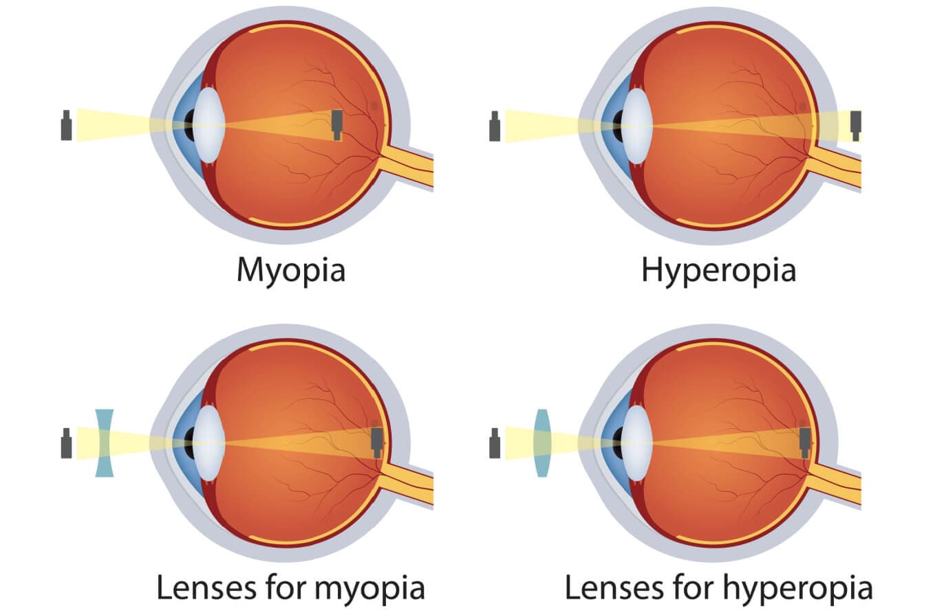
Myopia, or nearsightedness, and hyperopia, or farsightedness, are both common refractive eye conditions that blur vision. 

Complications of Myopia:

  • Myopic crescents (on the temporal side)
  • Posterior staphyloma
  • Patchy choroidal atrophy within the posterior pole
  • Vitreous syneresis
  • Breaks in Bruch’s membrane with accompanying choroidal atrophy (lacquer
    cracks)
  • Subretinal neovascular membrane with overlying retinal pigment epithelial
    hyperplasia (Foster-Fuchs spot)
  • Retinal detachment

Key Concepts on vision:

  • Physiological myopia is the
    most common eye disorder worldwide.
  • In myopia, parallel rays of
    light focus
    in front of the retina.
  • In hypermetropia, parallel
    rays of light focus
    behind the retina.
  • A 1 mm change in axial
    length leads to a
    3D change in refraction.
    • Shortening causes
      hypermetropia
    • Lengthening causes
      myopia
  • A 1 mm change in corneal
    curvature can lead to
    6D hypermetropia.
  • At birth, the eye is normally hypermetropic.
  • Optical treatment:
    • Myopia → Concave lens
    • Hypermetropia →
      Convex lens
  • Presbyopia: Physiological
    insufficiency of accommodation due to loss of elasticity of the lens capsule
    (age-related).
  • Astigmatism: Unequal
    refractive error in two eyes.
  • Aniseikonia: Unequal
    image shape/size in the visual cortex.
  • Aphakia: Absence of the
    crystalline lens.
    Treatment of choice
    Posterior chamber IOL implantation.
  • The most common eye disorder worldwide is physiological myopia, a refractive error.
  • The most common cause of blindness worldwide: Cataract (more than refractive error and aphakia blindness).
  • The most common cause of irreversible blindness worldwide: Glaucoma.

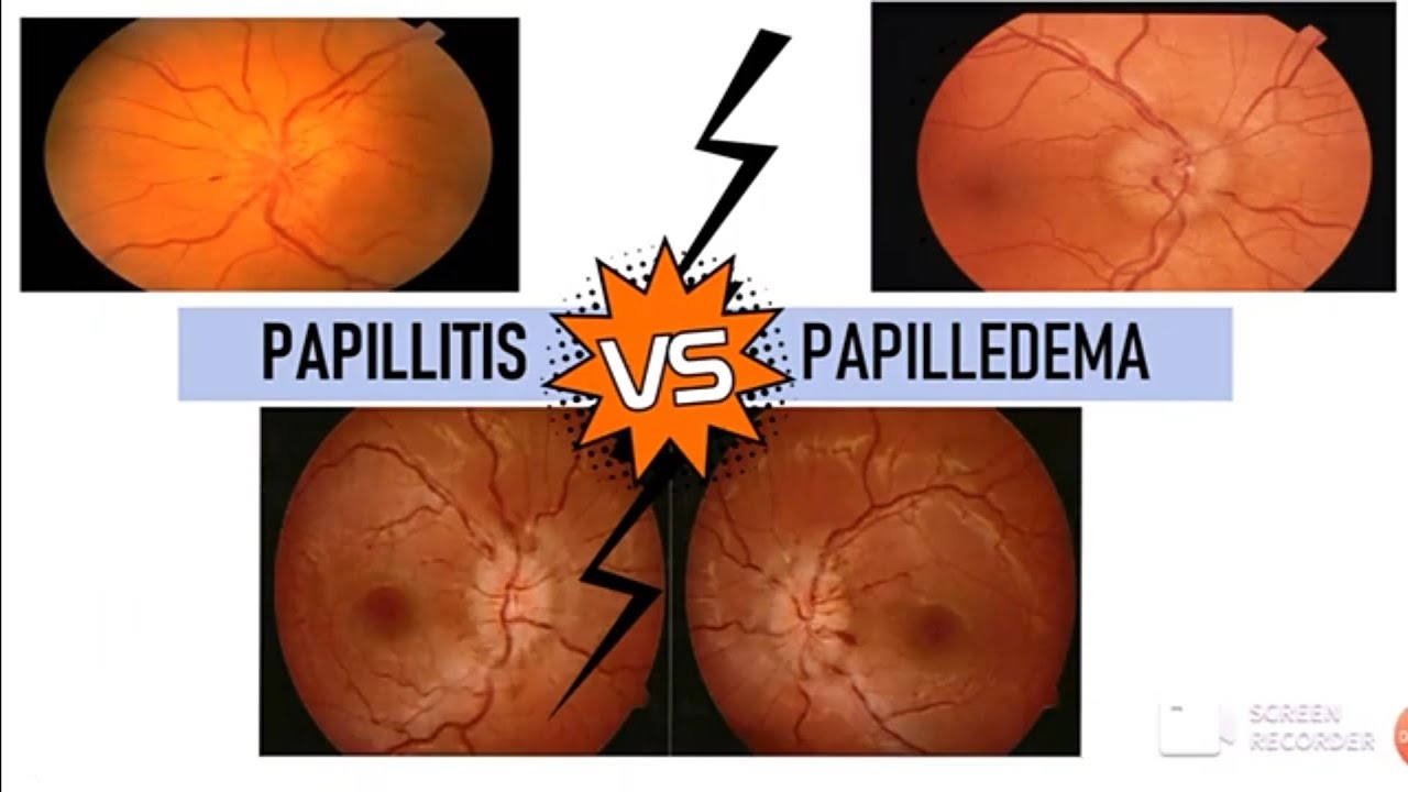
Differences : Papillitis and Papilledema

Papillitis Vs Papilledema

Here is a detailed table comparing
Papillitis (Optic Neuritis) and Papilledema:

FeaturePapillitis (Optic Neuritis)Papilledema
Usually AffectsUnilateral (U/L)Bilateral (B/L)
OnsetSuddenInsidious/Chronic
Loss of VisionMarked, painfulNegligible, gradual
Pain with Eye MovementPresentAbsent
TendernessPresent at the insertion of MR & SRAbsent
Other SymptomsUsually not seenHeadache, nausea, vomiting (↑ ICP)
Swelling (Edema) of Disc2-3D swelling>3D swelling, marked blind spot enlargement
Visual Field DefectCentrocecal scotomaEnlarged blind spot
Color VisionReduced (Dyschromatopsia)Normal
Pupillary Reflex (RAPD)Present (Positive)Absent
Posterior VitreousFine opacities presentClear
CauseOptic neuritis, Multiple sclerosis, infectionsIncreased intracranial pressure (tumor, hydrocephalus, meningitis)
ManagementSteroids (IV Methylprednisolone)Treat underlying cause (reduce ICP)

MCQs on Vision and Optic Pathology

  1. What is the most common cause of papilledema?
    a) Optic neuritis
    b) Increased intracranial pressure
    c) Glaucoma
    d) Retinal detachment
  2. Which of the following is NOT a feature of papilledema?
    a) Bilateral involvement
    b) Enlarged blind spot
    c) Sudden painful vision loss
    d) Absence of RAPD
  3. A patient with optic neuritis is likely to have which of the following?
    a) Unilateral sudden vision loss
    b) Marked papilledema
    c) Gradual vision loss
    d) Normal pupillary reflexes
  4. The presence of a relative afferent pupillary defect (RAPD) suggests:
    a) Glaucoma
    b) Optic neuritis
    c) Retinal detachment
    d) Conjunctivitis
  5. What is the primary treatment for optic neuritis?
    a) Acetazolamide
    b) Intravenous steroids
    c) Antibiotics
    d) Antihypertensives
  6. Which condition is associated with a centrocecal scotoma?
    a) Papilledema
    b) Papillitis
    c) Retinitis pigmentosa
    d) Diabetic retinopathy
  7. Which of the following is NOT a common cause of increased intracranial pressure?
    a) Brain tumor
    b) Hydrocephalus
    c) Optic neuritis
    d) Meningitis
  8. What visual defect is classically seen in papilledema?
    a) Central vision loss
    b) Enlarged blind spot
    c) Peripheral scotoma
    d) Homonymous hemianopia
  9. A 25-year-old female with multiple sclerosis presents with unilateral painful vision loss. What is the most likely diagnosis?
    a) Glaucoma
    b) Papilledema
    c) Papillitis (Optic neuritis)
    d) Retinal detachment
  10. The presence of dyschromatopsia (impaired color vision) is most commonly associated with:
    a) Papillitis
    b) Papilledema
    c) Cataract
    d) Age-related macular degeneration

Answer Key:

  1. b
  2. c
  3. a
  4. b
  5. b
  6. b
  7. c
  8. b
  9. c
  10. a

Trachoma – Key Points

Trachoma – Key Points

Table of Contents(toc)


Introduction

  • Definition: A chronic
    infectious eye disease caused by
    Chlamydia trachomatis.
  • Transmission: Spread
    through direct contact with infected secretions, contaminated objects, and
    flies.
  • Symptoms: Eye redness,
    irritation, discharge, photophobia, and progressive corneal scarring.
  • Complications: Corneal
    opacity, trichiasis (inward-growing eyelashes), and blindness.
  • Epidemiology: Leading cause
    of infectious blindness globally, prevalent in poor hygiene and low-income
    regions.

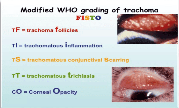
Stages of Trachoma (WHO Grading System)

  1. TF (Trachomatous Inflammation – Follicular): Presence of five or more follicles (>0.5 mm) in the upper tarsal
    conjunctiva.
  2. TI (Trachomatous Inflammation – Intense): Thickened, inflamed upper tarsal conjunctiva with pronounced redness.
  3. TS (Trachomatous Scarring):
    Visible scarring of the tarsal conjunctiva.
  4. TT (Trachomatous Trichiasis): Inturned eyelashes rubbing against the eyeball.
  5. CO (Corneal Opacity):
    Opacity leading to significant visual impairment or blindness.

SAFE Strategy for Trachoma Control
(WHO)

  1. S – Surgery: For
    trichiasis to prevent corneal damage.
  2. A – Antibiotics: To treat
    active infection and reduce transmission.
  3. F – Facial cleanliness:
    Encouraging hygiene to prevent spread.
  4. E – Environmental improvement: Ensuring clean water supply and sanitation.

Antibiotics for Trachoma

  1. Azithromycin (preferred
    drug)

    • Dose:
      • Adults: 1 g orally, single dose
      • Children: 20 mg/kg (maximum 1 g), single dose
    • Duration: Single-dose treatment, repeated annually in endemic areas.
  2. Tetracycline (1%) ophthalmic ointment

    • Dose: Apply to both eyes twice daily
    • Duration: 6 weeks
  3. Erythromycin
    (alternative to azithromycin)

    • Dose:
      • Adults: 500 mg orally twice daily
      • Children: 12.5 mg/kg orally four times daily
    • Duration: 14 days

{
“@context”: “https://schema.org”,
“@type”: “FAQPage”,
“mainEntity”: [{
“@type”: “Question”,
“name”: “What are Stages of Trachoma and mnemonic?”,
“acceptedAnswer”: {
“@type”: “Answer”,
“text”: “TF (Trachomatous Inflammation – Follicular): Presence of five or more follicles (>0.5 mm) in the upper tarsal conjunctiva.
TI (Trachomatous Inflammation – Intense): Thickened, inflamed upper tarsal conjunctiva with pronounced redness.
TS (Trachomatous Scarring): Visible scarring of the tarsal conjunctiva.
TT (Trachomatous Trichiasis): Inturned eyelashes rubbing against the eyeball.
CO (Corneal Opacity): Opacity leading to significant visual impairment or blindness.
Mnemonic is FISTO.”
}
},{
“@type”: “Question”,
“name”: “What is SAFE Strategy for Trachoma Control (WHO)?”,
“acceptedAnswer”: {
“@type”: “Answer”,
“text”: “S – Surgery: For trichiasis to prevent corneal damage.
A – Antibiotics: To treat active infection and reduce transmission.
F – Facial cleanliness: Encouraging hygiene to prevent spread.
E – Environmental improvement: Ensuring clean water supply and sanitation.”
}
},{
“@type”: “Question”,
“name”: “What are treatments of Trachoma?”,
“acceptedAnswer”: {
“@type”: “Answer”,
“text”: “Azithromycin , Tetracycline (1%) ophthalmic ointment or Erythromycin . (pls Confirm dosage and route)”
}
}]
}

Contraindications of Tooth Extraction

Contraindications of Tooth Extraction

Table of Contents(toc)


Introduction

Tooth extraction is a medical procedure that may not be suitable for all patients, especially those with certain health conditions. Below is a categorized list of general, local, absolute, and relative contraindications.

1. General Contraindications

These are conditions where tooth extraction should be avoided due to overall health concerns:

  • Cardiac Diseases:

    • Valvular, Rheumatic, or Ischemic heart disease
    • Congestive heart failure
    • Hypertension
    • Patients on anticoagulation therapy
  • Blood Diseases:

    • Severe anemia
    • Leukemia
    • Hemophilia
    • Agranulocytosis
  • Addison’s Disease

  • Patients on Corticosteroids:

    • Requires dose adjustment
  • Liver Diseases:

    • Jaundice and liver disease
    • Vitamin K deficiency
    • Blood clotting factor deficiencies (Prothrombin, Fibrinogen)
  • Diabetes Mellitus:

    • Poor wound healing due to decayed healing processes
  • Thyrotoxicosis

  • Pregnancy:

    • Especially during the first and third trimesters
    • Risks of abortion, premature delivery
    • Gravid uterus causing discomfort
    • Reduced stamina for opening the mouth
  • Very Old Age:

    • Decreased physiological response
    • Non-healing tissues and increased risk of infection
  • Epileptic Patients

  • Presence of Fever:

    • Infections can complicate healing
  • Debilitating Diseases:

  • Psychosis/Neurosis:

    • Mental health conditions affecting cooperation
  • Allergy to Local Anesthetics (LA)

  • Incorporative Patients:

    • Conditions where patient cooperation is compromised
  • During Menstruation:

    • Hormonal changes may affect healing and tolerance

2. Local Contraindications

These are conditions that affect the immediate area of the tooth that may cause complications:

  • Acute Infections:

    • Presence of infection at the extraction site
  • Malignancy:

    • Growth may spread rapidly after the procedure
  • Irradiated Jaw:

    • Risk of acute osteoradionecrosis due to lack of blood supply (endarteritis obliterans)

3. Absolute Contraindications

Certain conditions make tooth extraction entirely inappropriate:

  • Hemangioma:

    • Increased risk of bleeding
  • Arteriovenous Malformation (AV Malformation):

    • Bleeding that cannot be controlled
  • In the case of such conditions, if extraction is necessary:

    • Place the tooth back in the socket and apply pressure to control bleeding

4. Relative Contraindications

These are conditions where tooth extraction may be performed with caution or special consideration:

  • Diabetes Mellitus:

    • May require special management due to healing concerns
  • Valvular Heart Disease:

    • And other similar conditions where dental management requires careful planning and monitoring

Deep Vein Thrombosis (DVT)

 Deep Vein Thrombosis (DVT) : Note For Doctors

Table of Contents(toc)
 Deep Vein Thrombosis (DVT)


Definition:

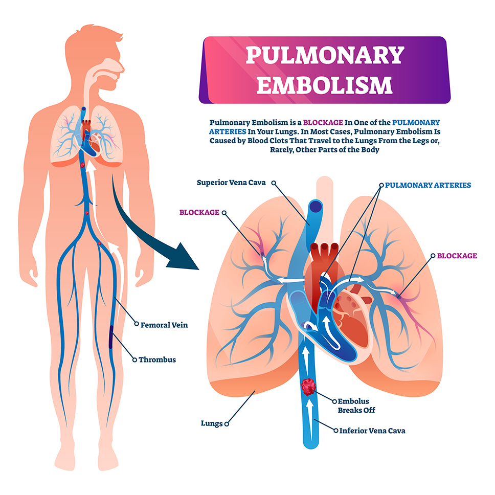
  • Deep Vein Thrombosis (DVT): The formation of a thrombus (blood clot) within the deep veins, most commonly in the lower extremities. If untreated, it can lead to severe complications such as pulmonary embolism (PE).

Pathophysiology:

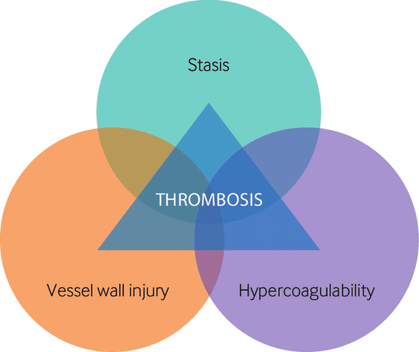
  • Virchow’s Triad: Three key factors contributing to thrombogenesis:
    • Stasis of blood flow (e.g., immobility, prolonged sitting, heart failure)
    • Endothelial injury (e.g., trauma, surgery, catheter insertion)
    • Hypercoagulability (e.g., genetic disorders like Factor V Leiden, malignancy, pregnancy)
  • Clot formation begins when platelets adhere to the endothelial surface, followed by fibrin deposition and aggregation of blood cells. The clot can extend, causing venous obstruction.
virchows triad

Etiology and Risk Factors:

  • Primary Risk Factors:
    • Prolonged immobility (post-surgery, prolonged bed rest, long-duration travel)
    • Surgical procedures (especially orthopedic surgeries: hip, knee)
    • Trauma (fractures, surgery, etc.)
    • Malignancy (increased clotting tendency due to tumor-derived procoagulants)
    • Pregnancy and postpartum (due to increased estrogen levels and venous stasis)
    • Oral contraceptives and hormone replacement therapy (estrogen increases clotting risk)
    • Genetic thrombophilia (e.g., Factor V Leiden mutation, Prothrombin gene mutation)
  • Secondary Risk Factors:
    • Age > 60 years
    • Obesity
    • Family history of DVT or PE
    • Smoking
    • Chronic conditions like heart failure, varicose veins, and inflammatory bowel disease.

Clinical Presentation:

  • Common Symptoms:
    • Unilateral leg swelling: Most common clinical feature, often with a sense of heaviness.
    • Pain: Deep, aching pain in the affected leg, aggravated by standing or walking.
    • Erythema: Redness and warmth over the affected area.
    • Palpable cord: The thrombus may feel like a firm, rope-like structure along the affected vein.
  • Classic Signs:
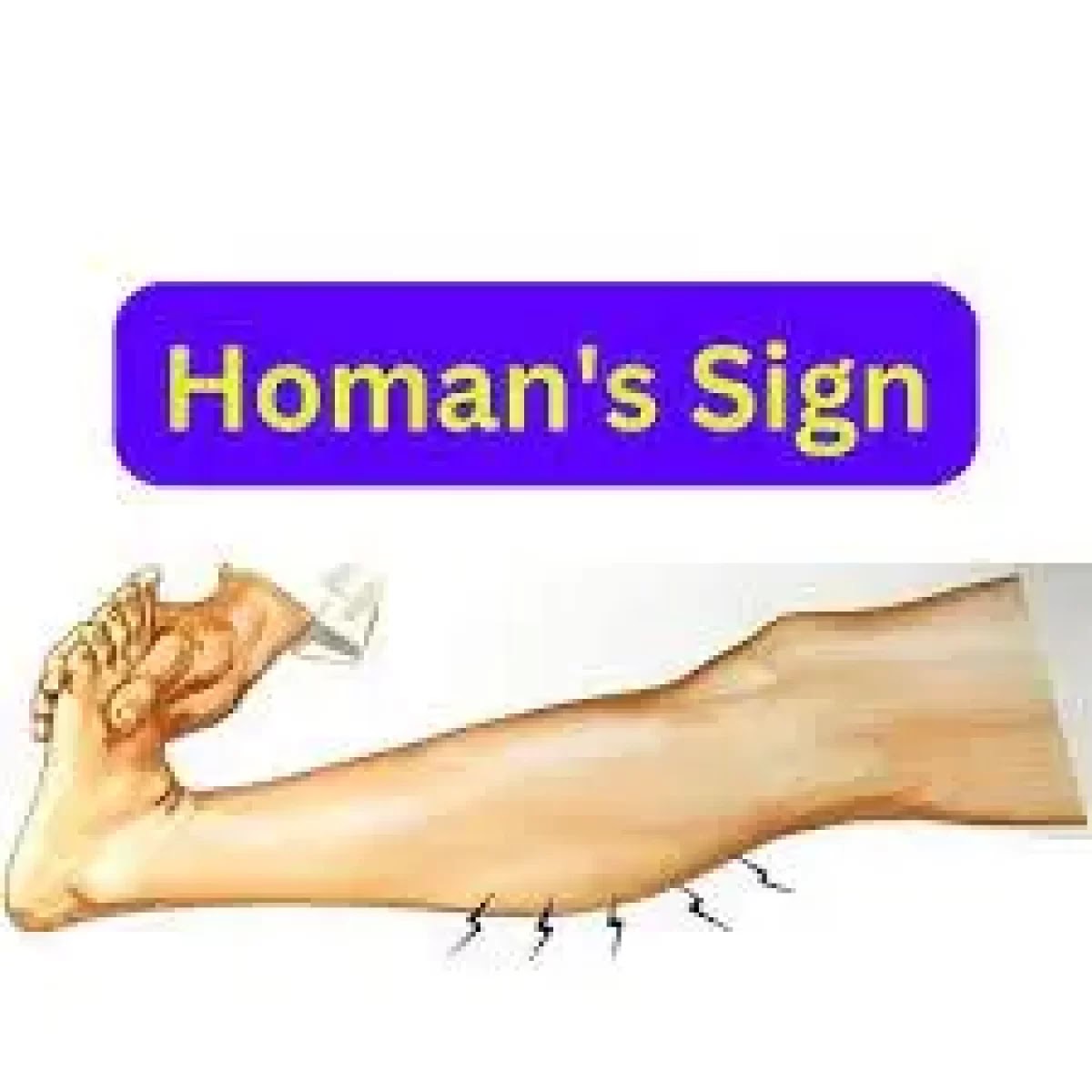
    • Homan’s sign: Pain on dorsiflexion of the foot (not highly sensitive or specific).
    • Positive Homans or Lowenberg test: Pain with calf compression, though less commonly used in modern clinical practice.

Complications:

  • Pulmonary Embolism (PE): The most serious complication. Clots from DVT may dislodge and travel to the pulmonary circulation, causing a blockage.
  • Post-thrombotic Syndrome (PTS): Chronic condition resulting from long-term venous hypertension, causing pain, swelling, and skin changes.
  • Chronic Venous Insufficiency: Due to damage to venous valves, leading to chronic swelling and skin changes.

Diagnosis:

  1. Clinical Assessment:

    • Clinical probability can be assessed using the Wells score (for DVT and PE), which factors in risk factors and clinical presentation.
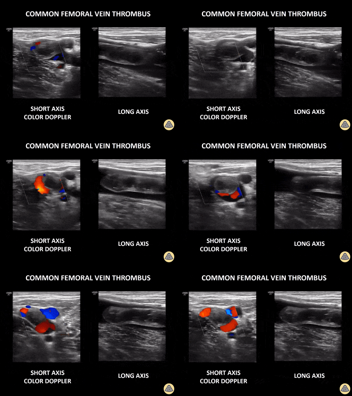
  2. Ultrasound (Doppler):

    • The gold standard for diagnosing DVT. High-frequency ultrasound assesses for the presence of a thrombus, venous compression, and blood flow.
  3. D-dimer:

    • Elevated D-dimer levels indicate fibrin degradation products, suggesting clot formation. However, it lacks specificity, and can be raised in other conditions (e.g., infection, cancer).
    • Sensitivity >95%, but specificity is low, especially in low-risk patients.
  4. CT Venography/Magnetic Resonance Venography (MRV):

    • Used in selected cases when ultrasound is inconclusive or inaccessible.
  5. Contrast Venography:

    • The gold standard historically but is less commonly used today due to its invasiveness and the rise of ultrasound.

Management:

  1. Anticoagulation Therapy:

    • Initial Treatment:
      • Low molecular weight heparin (LMWH) (e.g., enoxaparin) or unfractionated heparin (UH) for immediate anticoagulation.
      • Direct oral anticoagulants (DOACs) (e.g., rivaroxaban, apixaban) as an alternative to LMWH.
    • Long-term Management:
      • Warfarin (Coumadin), INR monitored (goal INR 2.0-3.0), or continued use of DOACs for 3-6 months based on risk factors and the nature of the clot.
  2. Thrombolysis:

    • Recombinant tissue plasminogen activator (rt-PA) or urokinase for large, symptomatic clots or in cases with life-threatening PE. Reserved for severe cases.
  3. Thrombectomy or Catheter-directed Thrombolysis:

    • Surgical intervention or catheter-based removal may be considered in patients with massive DVT or failure of anticoagulation therapy.
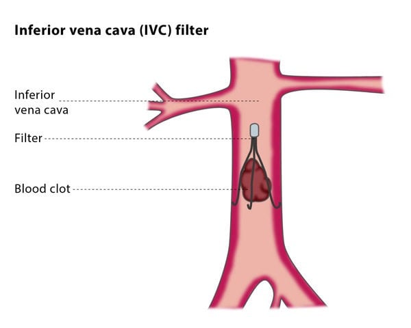
  4. Inferior Vena Cava (IVC) Filter:

  • Used in patients with contraindications to anticoagulation (e.g., active bleeding) or recurrent PE despite anticoagulation therapy.
  • Compression Stockings:

    • Used to reduce swelling, prevent post-thrombotic syndrome, and improve venous return in chronic cases.
  • Prevention:

    • Prophylaxis:
      • Early mobilization and exercises for hospitalized patients.
      • Low-dose heparin or LMWH for high-risk surgical patients.
      • Intermittent pneumatic compression devices for critically ill patients.
      • Compression stockings for those at risk, especially post-operatively.

    Follow-up and Monitoring:

    • Regular monitoring of anticoagulation levels, especially for warfarin (INR monitoring), and for signs of bleeding complications.
    • For patients on DOACs, renal function should be monitored periodically.

    Tip of the day

    Casoni’s Test : Notes (Echinococcus)

    Casoni’s Test : Notes for Doctors

    Table of Contents(toc)
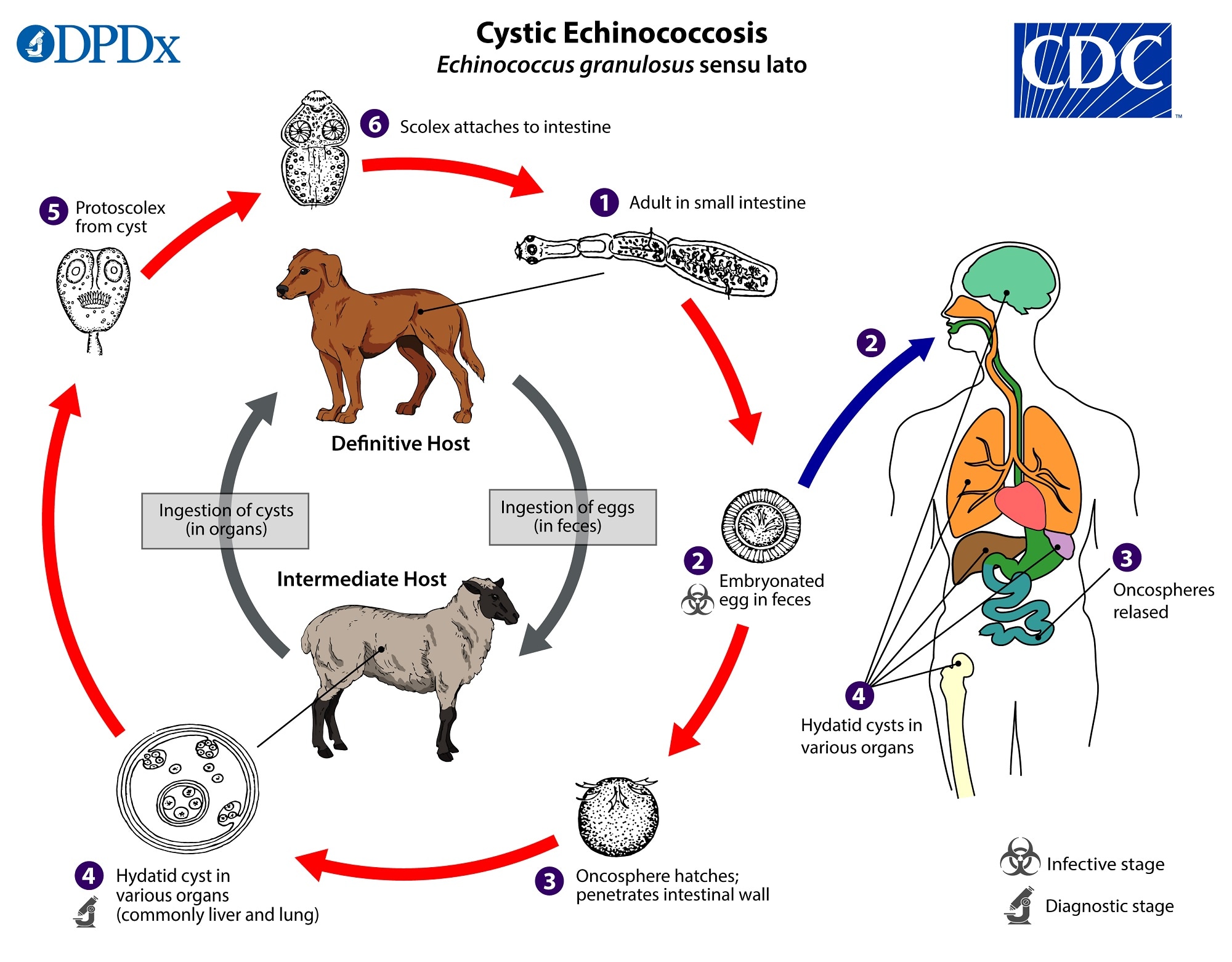
    E granulosus lifecycle Souce and Copyright: CDC


    Lets atart with a questin
     Casoni’s test is done to diagnose …
        a. Fascioliasis
        c. Hookworm
        b. Hydatid cyst
        d. Schistoomiasis

    Solution

    .reveal-box {
    display: none;
    margin-top: 10px;
    padding: 10px;
    border: 1px solid #ccc;
    background-color: #f9f9f9;
    }
    .reveal-btn {
    cursor: pointer;
    display: inline-block;
    padding: 8px 12px;
    background-color: #007bff;
    color: white;
    border: none;
    border-radius: 5px;
    }

    Ans: ‘b’
    Casoni’s test (intradermal skin test) is an immediate hypersensitivity skin
    test used in the diagnosis of hydatid cyst.
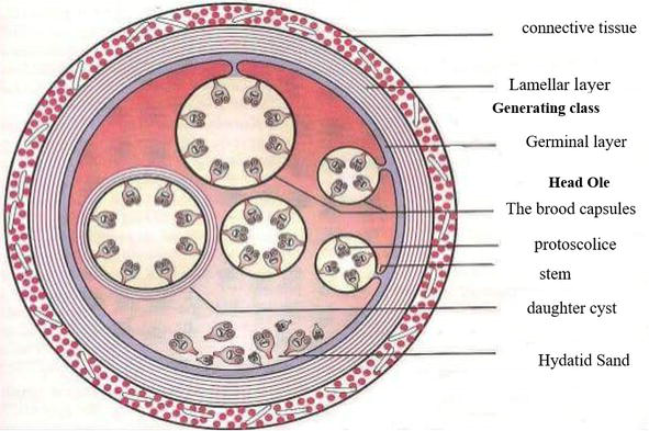
    Echinococcus cyst cross section

    Hydatid cyst

    • Pathogenesis infection is acquired by grass/vegetables contaminated with
      dog feces containing larval cyst of E. granulosus.
    • Man is dead end host & accidental host
    • Dog is definitive host
    • Cyst may occur in liver (m/c site in adults); lung (m/c site in
      children)

    USG Finding in Hydatid Cyst

    • USG is diagnostic (multiloculated cyst)

    PAIR procedure for Hydatid cyst: 

    Percutaneous puncture (P) of cyst under USG guidance, aspiration (A) of its
    content, injection (I) of scolicidal agent & reaspiration (R). (for
    small size < 5-6 cm).

    Scolicidal agents for Hydatid Cyst

    hypertonic 2% saline, sodium hypochloride, cetrimide, chlorhexidine

    Common postoperative complications

    Common postoperative complications

    Table of Contents(toc)


    Lets start with a MCQ

    Question

    All of the following are immediate post operative complications except
    a. Aspiration
    b. Myocardial infarction
    c. Deep vein thrombosis
    d. Neurogenic shock
    Ans: ‘c’ deep vein thrombosis

    Explanation and comments

    General postoperative complications

    Immediate:

    • Primary haemorrhage
    • Basal atelectasis
    • Shock: blood loss, acute MI
    • Low urine output: inadequate fluid replacement
    • Neurogenic shock
    • Aspiration

     Early 

    • Pain
    • Acute confusion
    • Nausea & vomiting: analgesia or related, paralytic
    • ileus
    • Fever
    • Secondary haemorrhage (d/t infection)
    • DVT Pneumonia
    • Acute urinary retention
    • UTI
    • Paralytic ileus
    •  Pressure sore

    Late

    • Bowel obstruction d/t fibrous adhesions
    • Incisional hernia
    • Persistent sinus
    • Recurrence of reason for surgery eg – malignancy
    • Keloid formation
    • Cosmetic appearance

    attrition vs abrasion vs erosion

    Chronic Dental Conditions in the Elderly

    Table of Contents(toc)


    Conditions that develop over a long period, particularly in older individuals, include attrition, abrasion, and erosion.

    Attrition

    Attrition refers to the wearing away of tooth substance due to mastication.

    Causes:

    • Coarse, gritty diet
    • Nervous habits (e.g., grinding teeth during anxiety)
    • Bruxism (grinding teeth at night)
    • Chewing on pipes

    Sites Affected:

    • Anterior teeth: Incisal edges
    • Posterior teeth: Occlusal surfaces

    Clinical Features:

    • Affected surfaces appear smooth and polished.
    • In advanced cases, incisal edges and cusps wear away, appearing peg-like, while the occlusal surface becomes flat or even hollowed.
    • Despite severe attrition, pulp exposure is rare due to compensatory dentine formation.
    • Attrition is incompatible with caries and periodontal disease since these conditions lead to tooth destruction and mobility, preventing attrition from occurring.
    • Attrition may have a protective role against caries by eliminating stagnation areas on occlusal surfaces.

    Abrasion

    Abrasion is the pathological wearing away of teeth by foreign substances.

    Causes:

    • Chewing tobacco
    • Vigorous tooth brushing, especially with abrasive toothpaste
    • Professions involving repetitive biting habits (e.g., cutting thread)
    • Hard tooth brushing with a horizontal sweeping action (most common cause)

    Sites Affected:

    • Cervical region of teeth (near the cementoenamel junction)—most susceptible to abrasion
    • Corner teeth—most severely affected

    Clinical Features:

    • Significant gingival recession is observed, but no gingivitis occurs due to effective plaque removal.
    • Progressive exposure of cementum and dentine, leading to grooved defects.

    Erosion

    Erosion refers to the progressive dissolution of tooth structure, typically caused by acids, though some cases have unknown etiologies.

    Causes:

    • Occupational exposure (e.g., battery or acid factory workers exposed to acid fumes)
    • Dietary factors:
      • Habitual sucking of citrus fruits for prolonged durations
      • Excessive consumption of carbonated soft drinks (high in phosphoric acid), leading to developmental caries
    • Gastroesophageal conditions:
      • Chronic regurgitation of acidic gastric juice, as seen in acid peptic disease (APD), gastroesophageal reflux disease (GERD), and early pregnancy, predominantly affecting the palatal surface
    • Erosion of unknown origin: Manifests as shallow, highly polished lesions on the labial surface

    Treatment:

    • Identification and elimination of the cause (occupational, dietary, medical factors)
    • Application of protective coatings
    • Use of fluoridated toothpaste
    • Iontophoresis with sodium fluoride (NaF) for enamel reinforcement

    GCS Calculator

    GCS Calculator

    .result {
    margin-top: 20px;
    padding: 10px;
    background: #007BFF;
    color: white;
    border-radius: 5px;
    font-size: 20px;
    }

    #severity { font-size: 18px; margin-top: 10px; }

    /* Buttons */
    button {
    margin: 10px;
    padding: 10px;
    border: none;
    border-radius: 5px;
    cursor: pointer;
    }

    Glasgow Coma Scale (GCS) Calculator

    Spontaneously (4)
    To speech (3)
    To pain (2)
    No response (1)

    Oriented (5)
    Confused (4)
    Inappropriate words (3)
    Incomprehensible sounds (2)
    No response (1)

    Obeys command (6)
    Moves to localized pain (5)
    Flex to withdraw from pain (4)
    Abnormal flexion (3)
    Abnormal extension (2)
    No response (1)

    Total GCS Score: 15

    🟢 Mild Brain Injury

    function calculateGCS() {
    let eyeResponse = parseInt(document.getElementById(“eyeResponse”).value);
    let verbalResponse = parseInt(document.getElementById(“verbalResponse”).value);
    let motorResponse = parseInt(document.getElementById(“motorResponse”).value);

    let totalGCS = eyeResponse + verbalResponse + motorResponse;
    document.getElementById(“gcsScore”).innerText = totalGCS;

    let severity = document.getElementById(“severity”);
    if (totalGCS >= 13) {
    severity.innerText = “🟢 Mild Brain Injury”;
    } else if (totalGCS >= 9) {
    severity.innerText = “🟡 Moderate Brain Injury”;
    } else {
    severity.innerText = “🔴 Severe Brain Injury”;
    }
    }

    Exit mobile version