Table of Contents
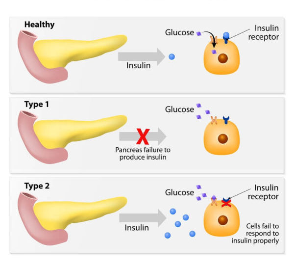
Diabetes is a chronic medical condition characterized by high levels of blood sugar (glucose). This occurs either because the body doesn't produce enough insulin (a hormone that regulates blood sugar) or because the cells don't respond properly to the insulin that is produced. Insulin is necessary for the body to effectively use glucose as a source of energy.

History for diabetes mellitus type 1 and 2:
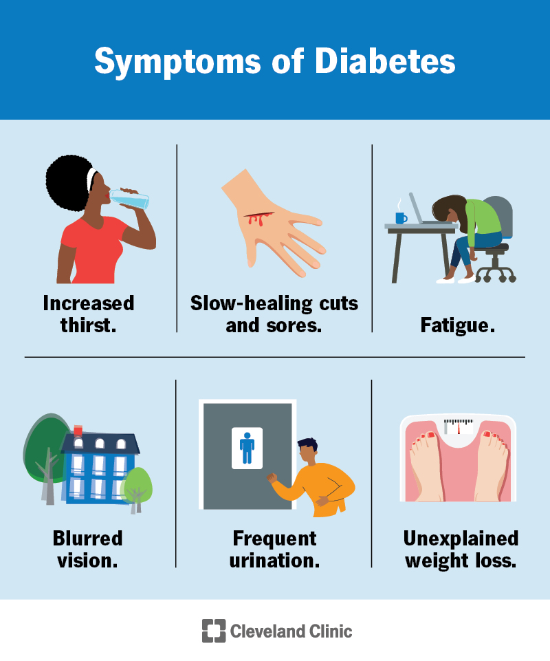
- Symptoms of hyperglycemia
- Thirst, dry mouth
- Polyuria
- Nocturia
- Tiredness, fatigue, lethargy
- Noticeable change in weight (usually weight loss)
- Blurring of vision
- Pruritus vulvae, balanitis (genital candidiasis)
- Nausea; headache
- Hyperphagia; predilection for sweet foods
- Mood change, irritability, difficulty in concentrating, apathy
- Family history

Physical examination for diabetes mellitus type 1 and 2
- BMI
- Retinal examination
- Orthostatic blood pressure
- Foot examination
- Peripheral pulses
- Insulin injection sites
- Peripheral neuropathy
Type 1 Vs Type 2 Diabetes mellitus DM
| Type 1 | Type 2 | |
| Onset | Sudden | Gradual |
| Age at onset | Any (mostly young) | Mostly in adults |
| Body habitus | Thin or normal | Often obese |
| Ketoacidosis | Common | Rare |
| Autoantibodies | Usually + | Absent |
| Endogeneous insulin | Low or absent | Normal, decreased or increased |
| Concordance in identical twins | ~ 50% | ~90% |
| Prevalence | Less prevalent | More prevalent (~90-95% of US diabetics) |
| Biochemical | C-peptide disappears | C-peptide persists |

Increased bloog glucose sugar level definition is called when
Normal Blood Glucose
- FPG <100 mg/dL (5.6 mmol/L)
- Two-hour glucose during OGTT <140 mg/dL (7.8 mmol/L)
Categories of increased risk for diabetes:
- •Impaired fasting glucose(IFG)
- FPG between 100 and 125 mg/dL (5.6 to 6.9 mmol/L).
- •Impaired glucose tolerance(IGT)
- Two-hour - 75 g OGTT between 140 and 199 mg/L (7.8 to 11.0 mmol/L).
- •A1C – Persons with 5.7 to 6.4 percent (39 to 46 mmol/mol
Diagnostic criteria of Diabetes mellitus
OR
2. Fasting Plasma Glucose ≥126 mg/dL (7.0 mmol/L)
(Fasting is defined as no caloric intake for at least eight hours.)
OR
3. Two-hour plasma glucose ≥200 mg/dL (11.1 mmol/L) during an OGTT.
(The test should be performed using a glucose load containing the equivalent of 75-gram anhydrous glucose dissolved in water.)
OR
4. In a patient with classic symptoms of hyperglycemia or hyperglycemic crisis, a random plasma glucose ≥200 mg/dL (11.1 mmol/L).
1. A1C ≥6.5 percent
OR
2. Fasting Plasma Glucose ≥126 mg/dL (7.0 mmol/L)
(Fasting is defined as no caloric intake for at least eight hours.)
OR
3. Two-hour plasma glucose ≥200 mg/dL (11.1 mmol/L) during an OGTT.
(The test should be performed using a glucose load containing the equivalent of 75-gram anhydrous glucose dissolved in water.)
OR
4. In a patient with classic symptoms of hyperglycemia or hyperglycemic crisis, a random plasma glucose ≥200 mg/dL (11.1 mmol/L).

Advice to patients with Impaired glucose tolerance
- Have an increased risk both of progression to type 2 diabetes and of developing macrovascular disease
- Advice lifestyle modification reduces the risk of progression in IGT
- Monitor annually by measurement of fasting blood glucose
- Other cardiovascular risk factors treate aggressively
For Management of Diabetes mellitus Please refer to this link.
Thank you for being here.