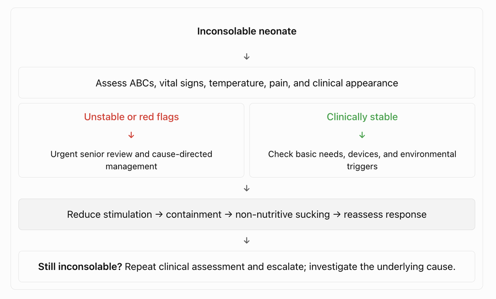
In the NICU, an inconsolable neonate should be approached as a potentially distressed or unwell baby first, and a baby needing soothing second. The key is to identify the cause, reduce stimulation, provide containment, and reassess the response.
neonate with a mother
1. First: exclude an acute clinical problem
Before spending 10–15 minutes trying to settle the baby, perform a rapid assessment.
Check immediately:
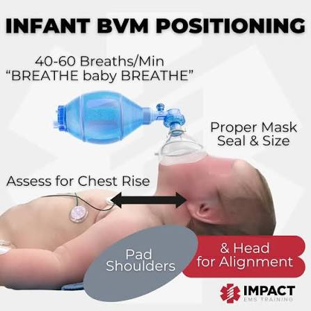
Airway, breathing, work of breathing, and oxygen saturation.
Heart rate, temperature, perfusion, and blood pressure if indicated.
Blood glucose, particularly in preterm, low-birth-weight, or symptomatic neonates.
Abdominal distension, tenderness, bilious vomiting, bloody stool, or feeding intolerance.
IV cannula, infusion lines, CPAP interface, NG/OG tube, urinary catheter, and pressure areas.
Signs of infection, seizures, trauma, or acute neurological deterioration.
Red flags: new high-pitched or weak cry, lethargy, apnea, cyanosis, temperature instability, poor perfusion, bulging fontanelle, seizures, a tense abdomen, or sudden unexplained persistent crying. Escalate promptly to the NICU clinician.
Arrange urgent clinical and neurological assessment.
Withdrawal
Tremors, increased tone, irritability, disturbed sleep, relevant antenatal drug exposure
Review maternal medication and substance exposure and assess using the appropriate neonatal withdrawal protocol.
Skin injury or limb discomfort
Swelling, redness, tenderness, reduced limb movement, pressure marks
Inspect the skin, extremities, digits, skin folds, and pressure points.
Device-related discomfort
Crying after CPAP application, cannulation, adhesive placement, or tube insertion
Check the position and fit of devices, IV sites, lines, and tubing for pressure, infiltration, displacement, or injury.
Overstimulation
Crying after repeated handling, excessive light, noise, or procedures
Reduce sensory input, provide gentle containment, and allow recovery.
4. Additional techniques that work well in the NICU
Skin-to-skin care: Consider kangaroo mother care when the baby is clinically stable and the team considers it safe. Monitor temperature, airway position, and respiratory support.
Gentle rocking: Only if appropriate for the baby’s condition and local protocol. Avoid vigorous rocking or shaking.
Non-nutritive sucking: Often helpful in preterm infants, especially during stressful care.
Facilitated tucking: Maintain flexion by gently supporting the infant’s limbs during procedures.
Pain relief: For procedures, use appropriate measures such as non-nutritive sucking, breastfeeding when feasible, and prescribed sucrose according to local neonatal protocols. Sucrose is not a treatment for unexplained persistent crying.
Review recent interventions: Suctioning, heel pricks, cannulation, CPAP prongs, adhesive dressings, and repeated handling can all trigger distress.
5. What if nothing works?
If the baby remains inconsolable after initial soothing, do not keep repeating the same interventions indefinitely.
Reassess vital signs and the baby’s overall appearance.
Check glucose if indicated and review the feeding history, urine output, stool, and recent procedures.
Examine the abdomen, fontanelle, skin, extremities, and device interfaces.
Review medications and possible withdrawal, infection, or other underlying illness.
Escalate to the NICU senior or neonatologist if the crying is persistent, unusual, unexplained, or accompanied by any abnormal findings.
Do not give sedatives simply to stop crying. Opioids, benzodiazepines, and other sedatives can cause respiratory depression and may obscure clinical deterioration. Use them only for a defined indication under appropriate medical supervision.
Antibiotic choice, pediatric doses, organism-specific duration,
and practical management points based on the Nelson approach.
Nelson-based • School-aged children & adolescents
01. Initial approach
In an older child with suspected acute bacterial meningitis,
treatment must begin urgently after blood cultures and lumbar
puncture, if these can be obtained without delaying antibiotics.
Initial empirical treatment should cover the most likely pathogens,
particularly Streptococcus pneumoniae and
Neisseria meningitidis.
Clinical priority
Do not delay antibiotics for CT or lumbar puncture when LP is
unsafe or would substantially delay treatment. Obtain blood cultures
if feasible, but start antibiotics promptly.
Regimen selection depends on age, immunization status, local
antimicrobial resistance, allergy history, and whether the infection
is community-acquired or healthcare-associated.
02. Empirical antibiotic therapy
For children older than 3 months, including school-aged children
and adolescents, the classic empiric regimen is:
First-line empiric treatment
Ceftriaxone + Vancomycin
OR
Cefotaxime + Vancomycin
Administer intravenously at meningitis doses while awaiting
CSF Gram stain, culture, and susceptibility results.
Why add vancomycin?
The third-generation cephalosporin covers common bacterial meningitis
pathogens. Vancomycin adds coverage against potentially
cephalosporin-resistant pneumococci.
Pediatric meningitis doses
Ceftriaxone
100 mg/kg/day IV
Usually once daily, or 50 mg/kg/dose every 12 hours.
Cefotaxime
200 mg/kg/day IV
Divide into doses every 6 hours in the cited Nelson regimen.
Vancomycin
60 mg/kg/day IV
Divide into doses every 6 hours in the cited Nelson regimen.
Monitor according to institutional protocol.
Dose and safety note
Confirm the current maximum dose, renal adjustment, infusion details,
and vancomycin therapeutic drug monitoring requirements using your
local pediatric antimicrobial protocol.
03. Antibiotic duration by organism
The following durations represent classic durations for uncomplicated
bacterial meningitis. Modify treatment according to culture,
susceptibility, clinical response, CSF sterilization, and complications.
Organism
Preferred treatment and total duration
Streptococcus pneumoniae
Ceftriaxone or cefotaxime if susceptible.
10–14 days.
If resistant to penicillin and third-generation cephalosporins,
continue an active regimen guided by susceptibility,
including vancomycin when indicated.
Neisseria meningitidis
Penicillin G if susceptible, or ceftriaxone/cefotaxime.
5–7 days.
Haemophilus influenzae
Ceftriaxone or cefotaxime, particularly for
beta-lactamase-producing strains.
7–10 days.
Unknown bacterial pathogen
Ceftriaxone or cefotaxime.
7–10 days may be appropriate in an
uncomplicated, clinically improving child when the CSF
profile supports bacterial meningitis but no organism is
identified.
Gram-negative bacilli
Use an active agent such as cefotaxime, ceftazidime,
or another susceptibility-directed antibiotic.
Treatment is generally prolonged, often
at least 3 weeks in the classic regimen.
Other clinically important pathogens
Pathogen
Treatment and duration
Listeria monocytogenes
Ampicillin; usually at least 21 days in high-risk patients.
Group B Streptococcus
Usually 14–21 days.
Pseudomonas aeruginosa
An active antipseudomonal agent, such as ceftazidime or
cefepime, selected according to susceptibility.
Prolonged treatment is often required.
Remember
Listeria and group B streptococcal meningitis are especially relevant
in neonates and selected immunocompromised patients. Gram-negative
and complicated infections require individualized treatment.
04. Culture-directed treatment
Once the CSF Gram stain, culture, PCR, and susceptibility results
become available, narrow therapy to the most appropriate active
antibiotic.
1
Suspected bacterial meningitis
Start ceftriaxone or cefotaxime plus vancomycin IV.
2
Identify the pathogen
Review CSF Gram stain, culture, PCR, and antimicrobial
susceptibility results.
3
Narrow the antibiotic regimen
Stop vancomycin when it is no longer needed and the identified
organism is adequately covered by the selected beta-lactam,
provided susceptibility and clinical circumstances support
de-escalation.
4
Complete the appropriate course
Continue organism-specific treatment, accounting for effective
therapy, prior antibiotics, clinical response, complications,
and CSF sterilization.
When should vancomycin be continued?
Continue vancomycin when there is a meaningful possibility of
cephalosporin-resistant pneumococcal meningitis, until susceptibility
results permit narrowing.
If the pneumococcus is susceptible to ceftriaxone or cefotaxime,
a third-generation cephalosporin can generally be used alone.
Severe immediate beta-lactam allergy
Alternative therapy requires specialist guidance. Chloramphenicol
is discussed as an alternative in some Nelson regimens for children
older than 1 month, but local resistance, availability, toxicity,
and susceptibility must be considered.
05. Practical bedside points
Urgency
Do not delay antibiotics for CT or an unsafe or substantially
delayed lumbar puncture.
CSF sterilization
With appropriate therapy, CSF generally becomes sterile within
24–48 hours in susceptible bacterial meningitis.
Repeat LP
Not routine in an uncomplicated, clinically improving case.
Consider it for resistant pneumococcal disease, gram-negative
meningitis, poor response, or suspected failure of sterilization.
Dexamethasone
Consider the first dose immediately before or concurrently with
the first antibiotic dose when indicated, particularly when
pneumococcal or Hib meningitis is suspected.
Warning: suspected meningitis
This is a medical emergency. Stabilize airway, breathing, and
circulation, manage seizures and shock, and administer empiric
antibiotics promptly. Adapt management to the child’s clinical
condition and local protocol.
06. Exam-oriented recall
A 9-year-old child presents with acute bacterial meningitis.
Before culture results are available, what is the most appropriate
empiric regimen?
Correct answer: B — Ceftriaxone + vancomycin.
This combination provides empiric coverage of common bacterial
meningitis pathogens, including potentially resistant
Streptococcus pneumoniae. Once culture and susceptibility
results are available, therapy should be narrowed appropriately.
07. Rapid revision
Question
High-yield answer
Empiric therapy in older children?
Ceftriaxone or cefotaxime + vancomycin.
Typical pneumococcal duration?
10–14 days.
Typical meningococcal duration?
5–7 days.
Typical Hib duration?
7–10 days.
When can vancomycin be stopped?
When susceptibility and clinical circumstances support narrowing.
Should LP delay antibiotics?
No.
08. References and clinical note
Primary reference:
Nelson Textbook of Pediatrics — bacterial meningitis:
initial antibiotic therapy and duration of treatment.
Important:
Confirm the current edition-specific Nelson recommendations,
maximum doses, local antimicrobial resistance patterns, and
institutional pediatric antimicrobial protocol before applying
treatment to a patient.
Work of breathing is assessed mainly by respiratory effort, chest movement, and signs of respiratory distress.
According to Nelson Textbook of Pediatrics, work of breathing (WOB) refers to the effort required to breathe—i.e., the amount of respiratory muscle activity needed to move air into and out of the lungs.
Clinically, Nelson assesses increased WOB by looking for signs of respiratory distress, particularly:
Work of breathing = the effort required to generate ventilation and move air through the respiratory system.
Increased WOB means the child must generate greater-than-normal respiratory effort to maintain adequate ventilation.
High-yield distinction: WOB ≠ respiratory rate alone. A child can have severe increased WOB even with a relatively normal RR, particularly when respiratory muscle fatigue is developing.
Parameters to assess work of breathing
Parameter
What to look for
Significance
Respiratory rate
Tachypnea for age
Early sign of increased WOB
Nasal flaring
Alae nasi widening during inspiration
↓ upper-airway resistance
Retractions / recession
Suprasternal, intercostal, subcostal, substernal
Increased negative intrathoracic pressure
Grunting
Expiratory grunt
Maintains end-expiratory pressure; important neonatal distress sign
Head bobbing
Especially infants
Severe respiratory effort
Tracheal tug
Downward movement of trachea on inspiration
Increased inspiratory effort
See-saw/paradoxical breathing
Chest moves inward while abdomen moves outward, or vice versa
Severe distress/fatigue
Accessory muscle use
SCM, scalene, abdominal muscles
Increased WOB
Abnormal chest movement
Asymmetry, poor expansion
Airway/lung pathology
Breath sounds
Wheeze, stridor, crackles, diminished/absent air entry
Identifies cause/severity
Expiratory phase
Prolonged expiration
Particularly obstructive disease
Grunting
Especially in neonates
Suggests significant respiratory distress
Posture
Tripod/sniffing position
Compensatory increase in airway patency
Ability to speak/feed
Full sentences vs words; feeding interruption in infants
Functional measure of WOB
Mental status
Irritability → lethargy → exhaustion
Late/severe respiratory compromise
Oxygenation
SpO₂, cyanosis
Consequence rather than direct measure of WOB
Quick severity framework
Mild ↑ WOB
Tachypnea
Mild nasal flaring
Mild intercostal/subcostal recession
Child alert and feeding reasonably well
Moderate ↑ WOB
Marked retractions
Nasal flaring
Accessory muscle use
Grunting
Difficulty feeding/speaking
Persistent tachypnea
Severe ↑ WOB / impending failure
Severe recession
Head bobbing
Paradoxical/see-saw breathing
Poor air entry or “quiet chest”
Exhaustion, altered consciousness
Apnea/bradypnea
Cyanosis despite oxygen
Important: A falling respiratory rate is not necessarily improvement. In a tiring child, tachypnea may progress to a normal or low RR as respiratory muscles fatigue—often a dangerous sign.
For bedside pediatric assessment, the highest-yield things to document are RR + retractions + nasal flaring + grunting + accessory muscle use + air entry + SpO₂ + mental status/feed/speech ability.
For a child with prolonged fever + hepatosplenomegaly + persistent leukocytosis, I would use the following as a bedside differential/workup table, with particular attention to infections relevant to Nepal and hematologic disease.
For persistent leukocytosis + HSM specifically, I would make sure the report gives the absolute neutrophil, lymphocyte, monocyte, eosinophil and basophil counts—not just total WBC. That single detail can substantially narrow this differential.
Q1. According to WHO/AAP recommendations, exclusive breastfeeding should be continued for the first:
A. 3 months
B. 4 months
C. 6 months
D. 12 months
Q2. Deficiency of which vitamin is the classic cause of nutritional rickets in infants?
A. Vitamin A
B. Vitamin C
C. Vitamin D
D. Vitamin K
Q3. What is the recommended daily vitamin D supplementation dose for exclusively and partially breastfed infants?
A. 100 IU/day
B. 200 IU/day
C. 400 IU/day
D. 1000 IU/day
Q4. Vitamin K is administered at birth primarily to prevent which condition?
A. Neonatal jaundice
B. Vitamin K deficiency bleeding (hemorrhagic disease of the newborn)
C. Necrotizing enterocolitis
D. Neonatal sepsis
Q5. A child presents with generalized edema, hypoalbuminemia, a fatty liver, and skin/hair changes, but has been receiving adequate calories with very low protein intake. This is most consistent with:
A. Marasmus
B. Kwashiorkor
C. Vitamin A deficiency
D. Scurvy
Q6. Marasmus differs from kwashiorkor in that marasmus is characterized by:
A. Prominent edema with preserved muscle mass
B. Severe wasting of fat and muscle without edema, due to deficiency of both calories and protein
C. Normal weight-for-height with fatty liver
D. Isolated micronutrient deficiency only
Q7. In a well infant, which laboratory marker is the most sensitive early indicator of depleted iron stores before anemia develops?
A. Serum ferritin
B. Hemoglobin
C. Mean corpuscular volume (MCV)
D. Reticulocyte count
Q8. Exclusively breastfed term infants are generally recommended to start iron supplementation or iron-rich complementary foods by what age?
A. 1 month
B. 4 months
C. 9 months
D. 12 months
Q9. A malnourished child presents with dry conjunctivae, Bitot spots, and corneal xerosis progressing toward keratomalacia. This is due to deficiency of:
A. Vitamin A
B. Vitamin B1 (thiamine)
C. Vitamin D
D. Vitamin E
Q10. A child with poor dietary vitamin C intake presents with gum swelling/bleeding, perifollicular hemorrhages, and corkscrew body hairs. The diagnosis is:
A. Scurvy
B. Rickets
C. Pellagra
D. Beriberi
Q11. Perioral and acral erythematous, scaly/vesiculobullous dermatitis with diarrhea and alopecia in an infant is classically associated with deficiency of:
A. Zinc
B. Copper
C. Selenium
D. Iodine
Q12. An infant on prolonged fat-restricted parenteral nutrition without adequate lipid develops a dry, scaly rash, poor growth, and thrombocytopenia. This is most consistent with deficiency of:
A. Essential fatty acids (linoleic acid)
B. Vitamin E
C. Biotin
D. Vitamin B12
Q13. During nutritional rehabilitation of a severely malnourished child, the electrolyte abnormality most characteristic of refeeding syndrome is:
A. Hyperphosphatemia
B. Hypophosphatemia
C. Hypernatremia
D. Hypercalcemia
Q14. In chronic protein-energy malnutrition, which anthropometric parameter is affected LAST (most preserved)?
A. Weight
B. Height/length
C. Head circumference
D. Mid-upper arm circumference
Q15. Failure to thrive is most commonly identified on growth charts by:
A. A single weight measurement at the 25th percentile
B. Weight persistently below the 3rd–5th percentile or crossing down two major percentile lines over time
C. Head circumference above the 95th percentile
D. Height above the 90th percentile
Q16. Periconceptional supplementation with which micronutrient reduces the risk of neural tube defects?
A. Folate (folic acid)
B. Vitamin B12
C. Iodine
D. Vitamin D
Q17. An exclusively breastfed infant of a strict vegan mother (with no B12 supplementation) presents with pallor, developmental regression, and macrocytic anemia. The most likely deficiency is:
A. Vitamin B12 (cobalamin)
B. Folate
C. Iron
D. Vitamin B6
Q18. WHO growth standards use weight-for-age, length/height-for-age, weight-for-length/height, and BMI-for-age primarily to assess a child’s:
A. Immunization status
B. Nutritional status and growth pattern
C. Developmental milestones
D. Bone age
Q19. The first-line dietary management for an infant with confirmed cow’s milk protein allergy who is not exclusively breastfed is:
A. Soy-based formula
B. Extensively hydrolyzed formula (or amino acid–based formula if severe/refractory)
C. Whole cow’s milk diluted with water
D. Goat’s milk formula
Q20. Childhood obesity is defined as a BMI-for-age and sex at or above which percentile on standard growth charts?
Q1. According to WHO/AAP recommendations, exclusive breastfeeding should be continued for the first:
Correct Answer: C. 6 months
Explanation: Exclusive breastfeeding (no other liquids or solids, only breast milk) is recommended for the first 6 months of life, with continuation of breastfeeding alongside complementary foods up to 1 year of age or beyond.
High-Yield Pearl: Complementary feeding should begin around 6 months — introduced too early, it displaces breast milk and increases infection risk; too late, it risks micronutrient deficiency (especially iron and zinc).
Q2. Deficiency of which vitamin is the classic cause of nutritional rickets in infants?
Correct Answer: C. Vitamin D
Explanation: Vitamin D deficiency impairs intestinal calcium and phosphate absorption, leading to defective mineralization of growing bone (rickets) and, in adults, osteomalacia.
High-Yield Pearl: Exclusively breastfed infants, dark-skinned infants, and those with minimal sun exposure are at highest risk — hence universal vitamin D supplementation from birth.
Q3. What is the recommended daily vitamin D supplementation dose for exclusively and partially breastfed infants?
Correct Answer: C. 400 IU/day
Explanation: All breastfed and partially breastfed infants should receive 400 IU/day of oral vitamin D beginning in the first few days of life, continued until the infant is weaned to at least 1 L/day (about 1 quart) of vitamin D–fortified formula or whole milk.
High-Yield Pearl: Formula-fed infants taking less than 1 L/day of fortified formula also need supplementation — fortified formula alone rarely meets the requirement until intake is substantial.
Q4. Vitamin K is administered at birth primarily to prevent which condition?
Correct Answer: B. Vitamin K deficiency bleeding (hemorrhagic disease of the newborn)
Explanation: Newborns have low vitamin K stores, poor placental transfer, and sterile gut flora (which normally synthesizes vitamin K), predisposing them to bleeding. A single intramuscular dose of vitamin K at birth prevents early, classic, and late forms of vitamin K deficiency bleeding.
High-Yield Pearl: Late VKDB (2–12 weeks) can present with intracranial hemorrhage and is strongly associated with exclusive breastfeeding without vitamin K prophylaxis — oral regimens are less reliable than a single IM dose.
Q5. A child presents with generalized edema, hypoalbuminemia, a fatty liver, and skin/hair changes, but has been receiving adequate calories with very low protein intake. This is most consistent with:
Correct Answer: B. Kwashiorkor
Explanation: Kwashiorkor results from severe protein deficiency in the presence of relatively adequate energy (calorie) intake. It produces edema (from hypoalbuminemia), hepatomegaly with fatty infiltration, skin depigmentation/desquamation, and sparse, discolored (‘flag sign’) hair.
High-Yield Pearl: The edema of kwashiorkor can mask underlying wasting and make the child appear less malnourished than they are — always check for pitting edema and serum albumin when assessing severe malnutrition.
Q6. Marasmus differs from kwashiorkor in that marasmus is characterized by:
Correct Answer: B. Severe wasting of fat and muscle without edema, due to deficiency of both calories and protein
Explanation: Marasmus is a global energy/protein deficiency leading to severe loss of subcutaneous fat and muscle wasting (‘skin and bones’ appearance) without the edema seen in kwashiorkor.
High-Yield Pearl: Marasmic kwashiorkor (mixed form, with both wasting and edema) carries the highest mortality risk among severe acute malnutrition presentations.
Q7. In a well infant, which laboratory marker is the most sensitive early indicator of depleted iron stores before anemia develops?
Correct Answer: A. Serum ferritin
Explanation: Serum ferritin reflects total body iron stores and falls before hemoglobin or MCV change, making it the earliest marker of iron depletion (though it can be falsely elevated as an acute-phase reactant during inflammation).
High-Yield Pearl: Iron deficiency progresses in stages: depleted stores (↓ferritin) → iron-deficient erythropoiesis (↓transferrin saturation) → iron deficiency anemia (↓Hgb, microcytosis) — hemoglobin alone misses the earliest stage.
Q8. Exclusively breastfed term infants are generally recommended to start iron supplementation or iron-rich complementary foods by what age?
Correct Answer: B. 4 months
Explanation: Breast milk iron content, while well absorbed, is low, and fetal iron stores typically last about 4–6 months. The AAP recommends iron supplementation (1 mg/kg/day) for exclusively breastfed infants starting at 4 months until iron-rich complementary foods (such as iron-fortified cereal or pureed meats) are reliably introduced.
High-Yield Pearl: Preterm infants have lower iron stores at birth and need earlier, higher-dose iron supplementation (typically starting by 2 weeks to 1 month of age).
Q9. A malnourished child presents with dry conjunctivae, Bitot spots, and corneal xerosis progressing toward keratomalacia. This is due to deficiency of:
Correct Answer: A. Vitamin A
Explanation: Vitamin A deficiency causes xerophthalmia — a spectrum from night blindness and conjunctival xerosis to Bitot spots (foamy, keratinized conjunctival patches) and, if untreated, corneal ulceration/keratomalacia leading to irreversible blindness.
High-Yield Pearl: Vitamin A deficiency also impairs immune function and increases mortality from measles and diarrheal disease — WHO recommends vitamin A supplementation with measles treatment in deficient populations.
Q10. A child with poor dietary vitamin C intake presents with gum swelling/bleeding, perifollicular hemorrhages, and corkscrew body hairs. The diagnosis is:
Correct Answer: A. Scurvy
Explanation: Vitamin C is a cofactor for collagen hydroxylation; its deficiency (scurvy) impairs collagen synthesis, producing gingival bleeding, perifollicular hemorrhages, corkscrew hairs, and subperiosteal hemorrhage causing bone pain and pseudoparalysis in infants.
High-Yield Pearl: Infantile scurvy classically presents with irritability and pseudoparalysis of the legs from painful subperiosteal hemorrhage, often mistaken for non-accidental trauma.
Q11. Perioral and acral erythematous, scaly/vesiculobullous dermatitis with diarrhea and alopecia in an infant is classically associated with deficiency of:
Correct Answer: A. Zinc
Explanation: Zinc deficiency (acquired or the inherited form, acrodermatitis enteropathica) causes a characteristic triad of periorificial/acral dermatitis, diarrhea, and alopecia, along with growth failure and impaired immune function.
High-Yield Pearl: Zinc deficiency should be considered in infants with unexplained perioral dermatitis plus poor growth — treatment with oral zinc produces dramatic skin improvement within days.
Q12. An infant on prolonged fat-restricted parenteral nutrition without adequate lipid develops a dry, scaly rash, poor growth, and thrombocytopenia. This is most consistent with deficiency of:
Correct Answer: A. Essential fatty acids (linoleic acid)
Explanation: Essential fatty acid (linoleic/linolenic acid) deficiency occurs with prolonged fat-free parenteral nutrition or severe fat malabsorption, producing dry scaly dermatitis, alopecia, poor wound healing, growth failure, and thrombocytopenia.
High-Yield Pearl: Essential fatty acid deficiency can develop within 1–2 weeks in infants on fat-free parenteral nutrition because of their high metabolic demand and limited fat stores — hence lipid emulsions are started early in neonatal PN.
Q13. During nutritional rehabilitation of a severely malnourished child, the electrolyte abnormality most characteristic of refeeding syndrome is:
Correct Answer: B. Hypophosphatemia
Explanation: Refeeding after prolonged starvation triggers an insulin surge that drives phosphate (along with potassium and magnesium) intracellularly, causing severe hypophosphatemia, which can lead to cardiac dysfunction, respiratory failure, and hemolysis.
High-Yield Pearl: In severe acute malnutrition, nutritional rehabilitation must be introduced cautiously and gradually with close monitoring/replacement of phosphate, potassium, and magnesium — aggressive early refeeding is a preventable cause of death.
Q14. In chronic protein-energy malnutrition, which anthropometric parameter is affected LAST (most preserved)?
Correct Answer: C. Head circumference
Explanation: In progressive chronic malnutrition, weight is affected first (wasting), followed by linear growth/height (stunting) if malnutrition persists, with head circumference relatively spared until malnutrition is severe and prolonged, since brain growth is preferentially protected.
High-Yield Pearl: Weight-for-height identifies acute wasting, while height-for-age identifies chronic stunting — using both classifies malnutrition as acute, chronic, or acute-on-chronic.
Q15. Failure to thrive is most commonly identified on growth charts by:
Correct Answer: B. Weight persistently below the 3rd–5th percentile or crossing down two major percentile lines over time
Explanation: Failure to thrive (poor weight gain/growth faltering) is defined using serial measurements — either weight sustained below the 3rd–5th percentile for age, or a downward crossing of two or more major percentile lines on the growth chart, rather than a single low value.
High-Yield Pearl: A single low percentile can simply reflect a small, constitutionally normal child — the trend over serial visits is what distinguishes true growth faltering from normal variation.
Q16. Periconceptional supplementation with which micronutrient reduces the risk of neural tube defects?
Correct Answer: A. Folate (folic acid)
Explanation: Folate is essential for DNA synthesis and neural tube closure, which occurs very early in pregnancy. Periconceptional folic acid supplementation (started before conception and continued through early pregnancy) significantly reduces the risk of neural tube defects such as spina bifida and anencephaly.
High-Yield Pearl: Women with a prior pregnancy affected by a neural tube defect are advised a much higher folic acid dose (4 mg/day) starting at least one month before conception, compared with the standard 0.4 mg/day for average-risk pregnancies.
Q17. An exclusively breastfed infant of a strict vegan mother (with no B12 supplementation) presents with pallor, developmental regression, and macrocytic anemia. The most likely deficiency is:
Correct Answer: A. Vitamin B12 (cobalamin)
Explanation: Maternal vitamin B12 stores are the sole source for an exclusively breastfed infant; strict maternal vegan diets without B12 supplementation lead to low breast milk B12, producing infantile megaloblastic anemia along with irritability, hypotonia, and developmental regression/failure to thrive.
High-Yield Pearl: Neurologic manifestations of infantile B12 deficiency can be more prominent and may precede or occur without significant anemia — a high index of suspicion is needed in breastfed infants of vegan mothers.
Q18. WHO growth standards use weight-for-age, length/height-for-age, weight-for-length/height, and BMI-for-age primarily to assess a child’s:
Correct Answer: B. Nutritional status and growth pattern
Explanation: These four anthropometric indices, plotted on standardized growth curves, are used together to evaluate whether a child’s growth and nutritional status are appropriate, and to detect wasting, stunting, underweight, or overweight/obesity.
High-Yield Pearl: The WHO standards (based on breastfed infants from multiple countries) are preferred for children under 2 years as a ‘growth standard,’ while CDC/WHO hybrid or national references are commonly used for older children.
Q19. The first-line dietary management for an infant with confirmed cow’s milk protein allergy who is not exclusively breastfed is:
Correct Answer: B. Extensively hydrolyzed formula (or amino acid–based formula if severe/refractory)
Explanation: Extensively hydrolyzed protein formulas are first-line for most infants with cow’s milk protein allergy; amino acid–based (elemental) formulas are reserved for infants with severe presentations (e.g., anaphylaxis, eosinophilic esophagitis, or failure to improve on hydrolyzed formula).
High-Yield Pearl: Soy formula is not recommended as first-line therapy for cow’s milk protein allergy because a significant proportion of affected infants (especially with non-IgE-mediated disease) also react to soy protein.
Q20. Childhood obesity is defined as a BMI-for-age and sex at or above which percentile on standard growth charts?
Correct Answer: D. 95th percentile
Explanation: In children and adolescents, obesity is defined as BMI ≥ 95th percentile for age and sex; a BMI between the 85th and <95th percentile is classified as overweight.
High-Yield Pearl: Unlike in adults, fixed BMI cutoffs cannot be used in children because body composition changes with age — percentile-based, age- and sex-specific charts are required for correct classification.
MAL Blood Group · Published in Blood Journal · 2024
In 1972, a routine blood test on a pregnant woman yielded something deeply puzzling: her red blood cells were missing a surface molecule that existed on every other known human blood sample of the time. Doctors noted the anomaly, filed it away, and moved on. They had no framework to explain it.
For the next five decades, that molecular absence lingered as an open question in the world of hematology — a quiet mystery buried in the archives of transfusion medicine. Then, in 2024, a team of researchers from the United Kingdom and Israel finally cracked it, identifying an entirely new human blood group system and solving one of the field’s longest-standing puzzles.
More Than Just A and B
Most of us learned about blood types in school: A, B, AB, and O, with a positive or negative Rh factor tacked on. But that picture is far from complete. Human blood cells are coated in a complex landscape of proteins and sugars called antigens — and scientists use these antigen patterns to classify blood into different “group systems.” The ABO and Rh systems are simply the most medically well-known of a much larger family.
To date, researchers have identified 47 such blood group systems in humans. Most of the major ones were catalogued in the early 20th century, but new systems continue to emerge — typically rare, typically tied to a small number of individuals worldwide, and typically uncovered when something goes unexpectedly wrong during a blood transfusion.
Quick Facts — Human Blood Group Systems
Humans have 47 recognised blood group systems, not just ABO and Rh.
Blood group systems are defined by distinct antigen molecules on the surface of red blood cells.
Over 99.9% of people carry the AnWj antigen — meaning those without it are extraordinarily rare.
The newly identified system is called MAL, after the myelin and lymphocyte protein that hosts the AnWj antigen.
Patients who are AnWj-negative risk severe immune reactions if given AnWj-positive blood during a transfusion.
The AnWj Antigen — A Puzzle Without a Gene
The molecule missing from that 1972 patient’s blood was eventually given a name: the AnWj antigen. Scientists confirmed that more than 99.9 percent of people carry it — making those without it extraordinarily rare. But despite decades of awareness, no one could identify the gene responsible for producing it. Without a genetic explanation, it was impossible to develop a reliable test to screen for AnWj-negative patients before transfusions — leaving those individuals vulnerable to potentially severe immune reactions if given incompatible blood.
That gap was both a scientific frustration and a genuine clinical risk. Transfusion reactions can range from mild discomfort to life-threatening organ damage. For patients whose blood type falls outside known systems, matching compatible donors is extraordinarily difficult — often relying on informal networks and a great deal of luck.
It represents a huge achievement, and the culmination of a long team effort, to finally establish this new blood group system and be able to offer the best care to rare, but important, patients.— Louise Tilley, Hematologist, NHS Blood and Transplant
The Breakthrough: The MAL Gene
The team — led by senior research scientist Louise Tilley of NHS Blood and Transplant, with colleagues from the University of Bristol and collaborators in Israel — spent nearly two decades piecing together the puzzle. Their breakthrough came when they traced the AnWj antigen to a specific protein: the myelin and lymphocyte protein, encoded by the MAL gene.
When both copies of a person’s MAL gene carry mutated versions, the AnWj antigen is absent from their blood entirely. This is the inherited form of AnWj-negativity — and it is what that 1972 patient almost certainly had. Importantly, the researchers also found that in some cases, the MAL gene can be functionally suppressed by other underlying conditions, such as certain blood cancers or autoimmune disorders. This means that a newly AnWj-negative result in a patient could sometimes be a flag pointing toward a deeper, undiagnosed illness.
The study, published in Blood, the journal of the American Society of Hematology, confirmed that all AnWj-negative patients studied shared the same mutation pattern — and crucially, that no other cell abnormalities or diseases were linked to the inherited form of the condition.
A 50-Year Timeline
Why This Matters Beyond the Lab
For the handful of individuals worldwide who are AnWj-negative, this discovery is potentially life-changing. Previously, their rare blood type made surgical procedures, childbirth, or any situation requiring a transfusion a logistical and medical challenge. Identifying compatible donors without a genetic test was a matter of laborious manual screening — if it was possible at all.
Now, with the MAL gene identified, blood banks and hospitals can develop targeted genetic tests to identify AnWj-negative patients proactively — before a transfusion crisis occurs. It also allows clinicians to distinguish between patients who are AnWj-negative due to an inherited mutation (a stable, lifelong condition) and those whose MAL expression has been suppressed by illness, which may require a very different medical response.
Tilley acknowledged the difficulty of the research: “The work was difficult because the genetic cases are very rare.” Rare cases, by definition, generate little data, making statistical patterns harder to detect and genetic links harder to confirm. That the team persisted across two decades speaks to both the scientific importance of the question and the very real human stakes attached to it.
The Bigger Picture: Blood Is Still Surprising Us
The MAL discovery arrives on the heels of another recent milestone. In 2022, researchers described the Er blood group system — yet another rare system affecting a small number of people globally. Together, these findings are a reminder that human biology, even in something as fundamental as blood, continues to yield surprises. Each new blood group system identified is not merely a scientific footnote; it represents a population of patients who were previously invisible to modern medicine’s tools.
Understanding these rare variants matters because it builds the infrastructure for safer, more personalised transfusion medicine. It means fewer unexplained reactions, fewer desperate searches for compatible donors, and more patients receiving care that is genuinely matched to their biology.
Half a century after a pregnant woman’s unusual blood sample puzzled a doctor somewhere in the world, science finally has an answer.
The MAL blood group system is now the 47th addition to our understanding of human blood — a testament to the slow, painstaking, but ultimately rewarding work of science. And for the extraordinarily rare individuals who carry this invisible distinction in their veins, it may mean the difference between a routine transfusion and a medical emergency.
Based on research published in Blood, American Society of Hematology, 2024. Research led by Louise Tilley, NHS Blood and Transplant & University of Bristol.
Sometimes, the most important discoveries begin with a single anomaly that refuses to be forgotten.
Coronary stenosis, inducible ischemia → may require CABG or catheter interventions
Regressed aneurysms → myointimal thickening, abnormal vascular function
Prognosis:
Majority recover fully; timely treatment reduces CAA risk to <5%
Recurrence of acute KD: 1–3%
Fatality: <1%
CAA outcomes:
~50% regress to normal diameter within 1–2 years
Giant aneurysms less likely to regress → higher risk thrombosis/stenosis
Revascularization or rarely heart transplant may be needed
Adult survival with history of giant aneurysms: ~90% at 30 years
Children without CAA have normal long-term outcomes
Lifestyle & preventive counseling recommended for all KD patients
FAQs
What is Kawasaki disease?
Kawasaki disease is an acute, self-limited vasculitis of medium-sized arteries, particularly the coronary arteries, occurring mainly in children under 5 years. Key point: It is an important cause of acquired heart disease in children.
What are the classic clinical features?
The classic features are: Fever ≥5 days Bilateral non-purulent conjunctival injection Oral changes: red/cracked lips, strawberry tongue Polymorphous rash Changes in extremities: erythema/edema of hands and feet, later periungual desquamation Cervical lymphadenopathy, usually ≥1.5 cm
The major complication is coronary artery involvement, including: Coronary artery dilatation Coronary artery aneurysms Myocarditis Arrhythmias Myocardial infarction High-yield: Kawasaki disease is the leading cause of acquired coronary artery disease in children in many developed countries.
How is Kawasaki disease treated?
First-line treatment: IVIG (intravenous immunoglobulin) Aspirin IVIG is given as 2 g/kg IV, ideally within the first 10 days of illness. Aspirin is initially given at an anti-inflammatory dose according to local protocol, followed by a low-dose antiplatelet regimen. Important: Early IVIG significantly reduces the risk of coronary artery aneurysms.
How is Kawasaki disease diagnosed?
Diagnosis is primarily clinical. Classic Kawasaki disease: fever for ≥5 days plus 4 of the 5 principal clinical features. There is no single diagnostic test. Investigations supporting the diagnosis may include: ↑ CRP and ESR Leukocytosis Thrombocytosis, especially in the subacute phase Sterile pyuria Elevated liver enzymes Echocardiography to assess coronary arteries
First principle: 👉 Most overweight infants are exogenous (overfeeding). 👉 Investigations are needed only if there are red flags for endocrine, genetic, or metabolic causes.
1️⃣ Step 1: Confirm Overweight / Obesity
Anthropometry
Weight-for-length (WHO growth charts)
BMI (if >2 years; not for infants)
Head circumference
Mid-upper arm circumference (optional)
Definitions (WHO)
> +2 SD weight-for-length → Overweight
> +3 SD → Obese
2️⃣ When to Investigate?
Send investigations if:
Rapid weight gain
Short length/height (↓ linear growth)
Dysmorphic features
Developmental delay
Hypotonia
Organomegaly
Hyperphagia
Family history of endocrine/genetic disorders
Signs of hypothyroidism, Cushing, etc.
If thriving, normal length, normal development → usually no labs required.
3️⃣ Baseline Investigations (If Indicated)
Investigation
Why Send It
CBC
Baseline health
Fasting blood glucose
Insulin resistance (rare in infancy but possible in severe obesity)