Hypothyroidism is a clinical state resulting from deficiency of thyroid hormone production or action, leading to a generalized slowing of metabolic processes.
It may be:
Congenital (Neonatal) – present at birth.
Acquired (Childhood) – develops later due to autoimmune, iatrogenic, or other causes.
2. Classification
A. Based on Level of Defect
Type
Site of Defect
TSH
T4/T3
Primary
Thyroid gland
↑
↓
Secondary
Pituitary
↓/N
↓
Tertiary
Hypothalamus
↓/N
↓
Peripheral (Resistance)
Target tissue
N/↑
N/↑
B. Based on Onset
Congenital hypothyroidism (CH)
Acquired hypothyroidism
3. Epidemiology
CH: ~1 in 2,000–4,000 live births.
More common in females.
Acquired form common in older children/adolescents, often autoimmune (Hashimoto’s).
thyroid gland
4. Etiology
A. Congenital Hypothyroidism
Thyroid dysgenesis (80–85%)
Agenesis, ectopy, or hypoplasia.
Usually sporadic.
Dyshormonogenesis (10–15%)
Inborn errors of thyroid hormone synthesis (autosomal recessive).
Leishmaniasis — MD Pediatrics Note (Based on Nelson Textbook of Pediatrics)
Table of Contents(toc)
Introduction
Leishmaniasis is a spectrum of protozoal diseases caused by Leishmania species, transmitted by the bite of infected female phlebotomine sandflies.
cutaneous leishmaniasis
Disease manifestations depend on the species involved and the host immune response.
Major clinical forms:
Visceral leishmaniasis (VL / kala-azar)
Cutaneous leishmaniasis (CL)
Mucocutaneous leishmaniasis (MCL)
Etiology and Classification
Form
Causative Species
Geographic Distribution
Visceral
L. donovani, L. infantum (chagasi)
South Asia, East Africa, Latin America
Cutaneous
L. tropica, L. major, L. mexicana, L. braziliensis
Middle East, Africa, Americas
Mucocutaneous
L. braziliensis complex
Central & South America
Epidemiology
Endemic in >80 countries; affects poor, rural populations.
Vectors:Phlebotomus (Old World), Lutzomyia (New World).
Reservoirs: Humans (L. donovani), dogs, rodents.
Transmission: Sandfly bite, rarely congenital or via transfusion.
Phlebotomus
Pathogenesis
Inoculation of promastigotes → engulfed by macrophages → transform into amastigotes → intracellular multiplication → spread to RES (liver, spleen, bone marrow).
Disease severity depends on cell-mediated immunity (CMI).
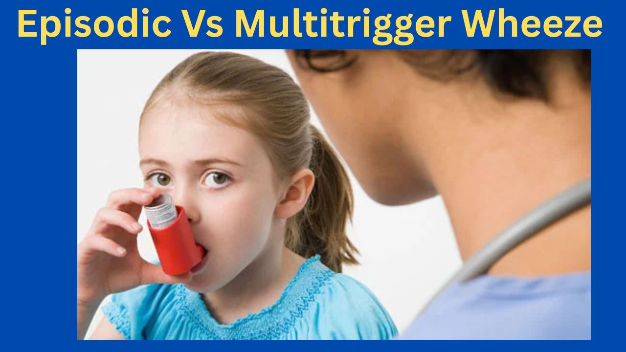
Episodic (Viral) Wheeze vs. Multiple Trigger Wheeze
A Clinically Oriented Review for the Practicing Pediatrician
Based on Nelson Textbook of Pediatrics (21st ed.) | Kendig’s Disorders of the Respiratory Tract in Children (9th ed.) | AAP & IAP-NAPCON Official Resources
1. Introduction
Wheezing in preschool children (0–5 years) is one of the most common reasons for pediatric consultation and hospital admission worldwide. It is now well established that ‘preschool wheeze’ is not a single disease but a heterogeneous group of phenotypes with distinct pathophysiology, natural history, and responses to therapy. The two most clinically useful and validated phenotypes—recognized in both the Nelson Textbook of Pediatrics and major international guidelines—are:
Episodic (Viral) Wheeze (EVW): wheezing episodes triggered exclusively by viral respiratory infections, with complete resolution between episodes.
Multiple Trigger Wheeze (MTW): wheezing triggered by multiple stimuli including viruses, aeroallergens, exercise, cold air, tobacco smoke, and emotional stimuli, with symptoms also occurring between discrete episodes.
This classification, initially proposed by Brand et al. and incorporated into the PRACTALL Consensus Report (2008) of the European Academy of Allergy and Clinical Immunology (EAACI) and the American Academy of Allergy, Asthma and Immunology (AAAAI), is now endorsed by the American Academy of Pediatrics (AAP) and the Indian Academy of Pediatrics (IAP) / National Asthma Consensus Group (NACG).
2. Epidemiology
According to Nelson Textbook of Pediatrics (21st edition, Chapter 169: Wheezing in Infants and Children), approximately 30–40% of all children will experience at least one wheezing episode in the first three years of life, yet fewer than one-third of these will develop persistent asthma. Data from the Tucson Children’s Respiratory Study (TCRS), cited prominently in Nelson, delineates three early wheezing trajectories:
Transient early wheezers: viral-triggered, remit by age 6; low atopic burden.
Non-atopic wheezers (EVW phenotype): episode-only wheeze; best aligned with EVW.
IgE-associated persistent wheezers (MTW/Asthma phenotype): atopic sensitization, family history, persistent into school age.
The IAP NAPCON 2019 Consensus Statement on Childhood Asthma notes that in South Asian children, including India and Nepal, the prevalence of preschool wheeze is significant, often complicated by high pollution exposure and early sensitization to house dust mite and cockroach allergens, features that shift the phenotype toward MTW.
3. Pathophysiology
3.1 Episodic (Viral) Wheeze
As described in Nelson (Chapter 169) and Kendig’s Disorders of the Respiratory Tract in Children (9th edition, Chapter 38), EVW is predominantly mediated by:
Rhinovirus (RV) and respiratory syncytial virus (RSV) — the principal triggers in children <3 years.
Neutrophilic airway inflammation: transient bronchial inflammation during the acute episode, with restoration of normal airway architecture between episodes. Unlike classical asthma, eosinophilic infiltration is typically absent or minimal.
Small airway mechanics: infants have a high ratio of airway resistance due to anatomically smaller caliber airways, making them more susceptible to luminal obstruction from viral-induced mucosal edema and secretions.
Immune dysregulation: reduced interferon-γ (IFN-γ) and impaired Th1 responses to RV have been demonstrated, contributing to prolonged viral shedding and exaggerated bronchospasm.
No persistent structural remodeling: between episodes, lung function is typically normal and there is no evidence of airway remodeling or eosinophilic inflammation.
3.2 Multiple Trigger Wheeze
MTW pathophysiology, as detailed in both Nelson and Kendig’s, resembles that of classic atopic asthma:
Eosinophilic airway inflammation: persistent even during asymptomatic intervals, with elevated fractional exhaled nitric oxide (FeNO).
Th2-skewed immune response: elevated IgE, IL-4, IL-5, IL-13; mast cell and eosinophil activation with allergen exposure.
Airway hyperresponsiveness (AHR): demonstrable on methacholine or exercise challenge, and persisting between symptomatic episodes.
Early sensitization: specific IgE to house dust mite (Dermatophagoides pteronyssinus), cockroach, Alternaria, or other regional allergens is frequently demonstrable by age 2–3 years.
Structural remodeling: subepithelial fibrosis and smooth muscle hypertrophy develop over time if left inadequately treated.
4. Clinical Features and Diagnosis
4.1 History
Nelson (21st ed., Chapter 169) and AAP Clinical Practice Guidelines for Asthma (2020 Update) recommend a detailed history focusing on:
Trigger identification: exclusive viral triggers (EVW) vs. multiple triggers including allergens, exercise, cold air, irritants (MTW).
Inter-episodic symptoms: nocturnal cough, exercise-induced wheeze, or persistent cough between viral episodes strongly suggests MTW.
Atopic comorbidities: personal history of eczema, allergic rhinitis; food allergy.
Family history: parental asthma/atopy increases the Asthma Predictive Index (API) score, supporting MTW/asthma phenotype.
Environmental history: tobacco smoke exposure, cooking fuel, pet ownership, damp housing — relevant especially per IAP guidelines for South Asian settings.
4.2 Asthma Predictive Index (API)
The modified API (mAPI), described in Nelson and endorsed by the AAP, is a validated tool to identify preschool wheezers likely to develop persistent asthma (MTW phenotype). A positive mAPI in a child with ≥3 wheezing episodes in the past year has a positive predictive value of ~80% for asthma at school age.
Digital clubbing, persistent hyperinflation, failure to thrive — suggest alternative diagnoses (cystic fibrosis, primary ciliary dyskinesia, structural airway anomalies).
Normal examination between episodes — expected in EVW; persistent wheeze or hyperinflation between episodes raises suspicion for MTW or alternative pathology.
4.4 Investigations
Kendig’s (9th ed., Chapter 38) and AAP Guidelines recommend the following investigations based on clinical context:
Spirometry (≥5–6 years): reversible airflow obstruction (post-bronchodilator FEV1 improvement ≥12%) supports MTW/asthma; may be normal in EVW.
Skin prick testing / Specific IgE: aeroallergen sensitization supports MTW phenotype; recommended in children with positive mAPI or recurrent MTW.
Complete blood count: peripheral eosinophilia (≥4%) is a minor API criterion.
Chest radiograph: to exclude structural anomalies, foreign body, or consolidation; not routinely needed for wheeze per AAP guidelines.
FeNO measurement: elevated (>25 ppb) supports eosinophilic airway inflammation (MTW/asthma); not universally available but referenced in Nelson and Kendig’s.
Bronchoscopy / BAL: reserved for diagnostically challenging cases; mentioned in Kendig’s for evaluation of structural/anatomic causes of wheeze.
5. Comparative Overview: EVW vs. MTW
Table 1 summarizes the key distinguishing features of the two preschool wheeze phenotypes.
Table 1. Episodic Viral Wheeze vs. Multiple Trigger Wheeze — Comparative Features
Per AAP Clinical Practice Guidelines (2020) and Nelson (Chapter 169), acute management is phenotype-independent and follows standard bronchodilator therapy:
Short-Acting Beta-2 Agonists (SABA): salbutamol (albuterol) 2.5–5 mg via nebulizer, or 2–4 puffs via spacer and face mask every 20 minutes for 3 doses in severe episodes. First-line therapy for all preschool wheeze.
Ipratropium bromide: may be added for moderate-to-severe exacerbations; reduces hospitalization when combined with salbutamol.
Systemic corticosteroids: oral prednisolone (1–2 mg/kg/day, max 40 mg, for 3–5 days) for moderate-to-severe exacerbations. Per the AAP, short courses do not significantly affect adrenal function or growth in children.
Supplemental oxygen: titrate to maintain SpO2 ≥94% (AAP target); SpO2 ≥95% per IAP-NAPCON 2019.
Hospitalization criteria: SpO2 <92% on room air, severe respiratory distress (HR >60/min in infants), inability to maintain oral feeds, poor response to initial bronchodilators.
7.2 Preventive/Controller Therapy
This is where the phenotype distinction critically guides management:
7.2.1 Episodic (Viral) Wheeze
Per Nelson, Kendig’s, and AAP Guidelines:
Continuous ICS: NOT routinely recommended for EVW. Multiple RCTs (including the PEAK and MIST trials cited in Nelson) show no significant reduction in episode frequency or severity with continuous low-dose ICS in non-atopic preschool wheezers.
Intermittent/episodic ICS: high-dose ICS at the onset of a viral URTI (e.g., budesonide 400 mcg/day or fluticasone 200 mcg/day for 7–10 days) may reduce episode severity in selected children, though evidence remains inconsistent across trials.
Montelukast: episodic use at onset of wheeze shows modest benefit in some studies (Bisgaard et al., NEJM, cited in Nelson); may be considered for children with 3 or more episodes per year.
Bronchodilator reliever therapy: salbutamol as needed during episodes. Continuous reliever use between episodes is not indicated in pure EVW.
Avoidance: passive smoking cessation, hand hygiene, daycare modifications to reduce viral exposure.
7.2.2 Multiple Trigger Wheeze
Per Nelson, Kendig’s, AAP (2020), and IAP-NAPCON (2019):
Low-dose ICS: first-line preventer therapy. Budesonide 100–200 mcg/day or fluticasone propionate 100 mcg/day (BDP-equivalent). Initiate when diagnosis of MTW/persistent asthma is established.
Montelukast: may be used as an alternative to ICS in mild MTW or as add-on therapy in moderate MTW. IAP-NAPCON recognizes its role given high house dust mite sensitization in the South Asian context.
Medium-dose ICS: step up to 200–400 mcg/day (budesonide equivalent) if low-dose ICS fails to achieve symptom control after 6–8 weeks.
LABA addition: for children ≥5 years with inadequate control on medium-dose ICS, salmeterol or formoterol can be added. Not approved or recommended for children <4 years as monotherapy.
Allergen avoidance: mattress/pillow encasements, HEPA filtration, pet removal — strongly recommended by AAP and IAP for sensitized children with MTW.
Allergen Immunotherapy (AIT): subcutaneous or sublingual AIT for house dust mite-sensitized children with MTW/asthma is recommended in international guidelines and endorsed in IAP-NAPCON for appropriate candidates ≥5 years.
Omalizumab: anti-IgE therapy; approved for moderate-to-severe persistent allergic asthma in children ≥6 years; referenced in Nelson and AAP guidelines for refractory MTW/asthma with high IgE and allergen sensitization.
7.3 Step-Therapy Summary
Table 2. Stepwise Treatment Approach for EVW and MTW
Step
EVW Management
MTW Management
Acute
SABA (salbutamol) via spacer/nebulizer; oral prednisolone for moderate-severe
SABA; oral/systemic corticosteroids; consider early ICS step-up
Preventer
Not routinely indicated; trial ICS only if frequent/severe episodes (≥3/year)
Low-dose ICS (e.g., budesonide 100–200 mcg/day) as first-line preventer
Step-up
Episodic ICS at onset of URTI (intermittent therapy); montelukast episodic use
Increase ICS dose; add montelukast or LABA (≥5 yr); consider specialist referral
Monitoring
Symptom diary; reassess trigger pattern at each visit
Nebulizers are not superior to pMDI+spacer for acute bronchodilation and carry infection transmission risk in healthcare settings. Both AAP and IAP recommend prioritizing spacer-based delivery.
8. Monitoring and Follow-Up
Nelson, AAP (2020 Expert Panel Report 3 Update), and IAP-NAPCON recommend the following monitoring framework:
Review diagnosis every 3–6 months: re-evaluate whether phenotype has shifted from EVW to MTW as the child grows.
Assess symptom control using validated tools: \Childhood Asthma Control Test (C-ACT) for children ≥4 years; parent-report tools for younger children.
Spirometry when developmentally feasible (≥5 years): monitor FEV1, FVC, and FEV1/FVC ratio at each visit.
Reassess trigger profile at each visit: new aeroallergen sensitization, school exposures, change in environment.
Monitor growth: height and weight percentile; ICS at low doses does not significantly affect final adult height per Nelson; monitor with medium-to-high doses.
Adherence and inhaler technique: check at every visit; poor technique is the most common cause of apparent treatment failure per AAP.
Consider step-down: if well-controlled for ≥3 months, cautiously step down therapy, reassessing trigger pattern.
9. Prognosis and Natural History
The TCRS and birth cohort studies cited in Nelson provide the most robust data on prognosis:
EVW (Transient wheeze): ~60% of preschool wheezers remit by 6 years of age. These children, corresponding to the EVW phenotype, generally have normal lung function at school age. The absence of atopic sensitization, normal lung function between episodes, and non-positive API predict favorable outcome.
MTW (Persistent/Asthma phenotype): ~40% of preschool wheezers continue to wheeze at school age. Risk factors for persistence include: positive mAPI, maternal asthma, early sensitization to aeroallergens, frequent episodes in the first 3 years, male sex, and exposure to high-dose indoor allergens.
Lung function trajectory: Lung function deficits, if present at age 6 years in the MTW group, tend to track into adult life and are associated with increased risk of COPD in adulthood (“early origins of adult lung disease” concept, cited in Nelson and Kendig’s).
South Asian context (IAP): earlier sensitization to perennial allergens (HDM, cockroach), higher pollution burden, and lower vitamin D levels may confer worse outcomes in the MTW phenotype in Indian children, as noted in IAP-NAPCON 2019.
10. Special Clinical Situations
10.1 The “Overlap” Child
Many children present with features of both EVW and MTW, especially between ages 2–4 years. Nelson recommends using the mAPI as a practical decision aid in such cases. If the mAPI is positive, treat as MTW (initiate regular ICS); if negative, manage as EVW (episodic/as-needed therapy).
10.2 Very Young Infants (<12 months)
Wheezing in infants under 12 months is most commonly due to bronchiolitis (RSV) and should not be classified as EVW or MTW. Per AAP Clinical Practice Guideline for the Diagnosis, Management, and Prevention of Bronchiolitis (2014, reaffirmed 2020), bronchodilators are not recommended for infants with bronchiolitis. ICS and systemic steroids are similarly not recommended in this age group for acute bronchiolitis.
10.3 COVID-19 and Respiratory Viruses
The AAP has issued guidance noting that SARS-CoV-2 infection in young children may trigger wheezing episodes similar to other viral URTI triggers in EVW. Standard asthma action plans should include COVID-19 as a potential EVW trigger; ICS should not be stopped during COVID-19 illness in MTW/asthma patients.
10.4 Vaccination
Both AAP and IAP recommend annual influenza vaccination for all children with recurrent wheezing (EVW or MTW), as influenza is a significant trigger for severe exacerbations. Pneumococcal vaccination per national immunization schedules is also recommended.
11. Parent and Caregiver Education
AAP and IAP emphasize that education is a cornerstone of management:
Provide written Asthma Action Plan (AAP template available at healthychildren.org) for all children with recurrent wheeze.
Educate on symptom recognition: early signs of exacerbation (nocturnal cough, reduced exercise tolerance, increased rescue inhaler use).
Inhaler technique training at every visit; video demonstrations and teach-back methods are recommended by AAP.
Environmental control counseling: tobacco smoke, allergen avoidance, mold reduction, pet dander management.
Address caregiver anxiety: explain phenotype, natural history, and that EVW does not inevitably become asthma.
Emphasize adherence to preventive therapy in MTW: parents often reduce ICS doses prematurely when symptoms improve.
12. Key Clinical Takeaways
Phenotype matters: Distinguish EVW from MTW at every clinical encounter; this distinction drives preventive therapy decisions.
mAPI guides therapy: A positive mAPI in a high-frequency preschool wheezer indicates MTW/asthma phenotype and justifies early ICS therapy.
ICS is not universal: Continuous ICS is not recommended for pure EVW; reserve for MTW or EVW with frequent/severe episodes.
Trigger profile shapes management: Allergen sensitization testing is indicated when MTW is suspected; AIT may be indicated in sensitized children ≥5 years.
Phenotypes are dynamic: Reassess at every visit; EVW may evolve to MTW as atopic sensitization develops.
Guideline resources: Use AAP (healthychildren.org, aappublications.org) and IAP-NAPCON (iapindia.org) official resources for updated local guidance.
Exclude mimics: Always consider structural, infectious, and congenital causes of recurrent wheeze, especially in children <12 months or with atypical features.
References
Primary Textbook References:
Kliegman RM, St. Geme JW, Blum NJ, et al. Nelson Textbook of Pediatrics, 21st Edition. Philadelphia: Elsevier; 2020. Chapter 169: Wheezing in Infants and Young Children; Chapter 170: Asthma.
Wilmott RW, Deterding R, Li A, et al. Kendig’s Disorders of the Respiratory Tract in Children, 9th Edition. Philadelphia: Elsevier; 2019. Chapter 38: Wheezing in Infancy and Early Childhood; Chapter 39: Asthma in the Pediatric Patient.
AAP Official Resources:
American Academy of Pediatrics. Clinical Practice Guideline for the Diagnosis, Evaluation, and Management of Childhood Asthma. Pediatrics. 2020;145(3):e20193432. Available at: https://publications.aap.org
American Academy of Pediatrics. Clinical Practice Guideline: The Diagnosis, Management, and Prevention of Bronchiolitis. Pediatrics. 2014;134(5):e1474-e1502. Reaffirmed 2020. Available at: https://publications.aap.org
American Academy of Pediatrics. Asthma Action Plan templates and parent education resources. HealthyChildren.org. Available at: https://www.healthychildren.org
IAP Official Resources:
Indian Academy of Pediatrics, National Asthma Consensus Group (NAPCON). IAP-NAPCON Consensus Statement on Childhood Asthma 2019. Indian Pediatrics. 2020;57(1):42–58. Available at: https://www.indianpediatrics.net
Indian Academy of Pediatrics. IAP Standard Treatment Guidelines: Bronchial Asthma in Children. 2022. Available at: https://www.iapindia.org
Landmark Studies and Consensus Documents (cited in Nelson/Kendig’s):
Brand PL, Baraldi E, Bisgaard H, et al. Definition, assessment and treatment of wheezing disorders in preschool children: an evidence-based approach. European Respiratory Journal. 2008;32(4):1096–1110. [PRACTALL Consensus Report, cited in Nelson 21e and Kendig’s 9e]
Martinez FD, Wright AL, Taussig LM, et al. Asthma and wheezing in the first six years of life: The Group Health Medical Associates. New England Journal of Medicine. 1995;332(3):133–138. [Tucson Children’s Respiratory Study, cited in Nelson 21e]
National Asthma Education and Prevention Program (NAEPP). Expert Panel Report 3 (EPR-3): Guidelines for the Diagnosis and Management of Asthma. National Heart, Lung, and Blood Institute (NHLBI). 2007 (Updated 2020). Available at: https://www.nhlbi.nih.gov
Global Initiative for Asthma (GINA). Difficult-to-Treat and Severe Asthma in Adolescent and Adult Patients: A GINA Pocket Guide. 2023. [Referenced in Nelson and Kendig’s for management framework]
Malnutrition: Complications, Assessment, and Prevention
Malnutrition is a serious public health concern affecting individuals of all ages, particularly children in low-resource settings. It can lead to both acute and chronic complications, impacting survival, growth, and overall health.
Acute Complications of Malnutrition
A helpful mnemonic for remembering the main acute complications is “Shieldeded”:
Sugar deficiency / Hypoglycemia – Low blood sugar levels can lead to lethargy, seizures, and even coma.
Hypothermia – Impaired thermoregulation increases vulnerability to cold stress.
Infection – Reduced immunity predisposes to frequent and severe infections.
Electrolyte disorder – Commonly includes imbalances in sodium, potassium, and magnesium.
Dehydration – Often due to diarrhea or inadequate fluid intake.
Deficiency of vitamins and minerals – Leads to a range of specific deficiency syndromes (e.g., anemia, rickets, night blindness).
Next we will discuss 10 essential steps in the management of malnutrition (Severe Acute Malnutrition – SAM) based on standard WHO guidelines.
Anthropometry – Measurement of weight, height, mid-upper arm circumference (MUAC), and growth charts.
Biochemical markers – Blood tests to assess nutrient levels and detect deficiencies.
Clinical evaluation – Physical examination for signs of malnutrition.
Dietary evaluation – Analysis of food intake patterns and adequacy.
Epidemiological assessment – Community-based data to identify at-risk populations.
Prevention of Malnutrition: GOBIFFF Strategy
The GOBIFFF approach is widely promoted for prevention:
G – Growth monitoring
O – Oral rehydration solution (ORS) use
B – Breastfeeding promotion
I – Immunization coverage
F – Family planning
F – Female education
F – Feeding improvement (appropriate complementary feeding)
Follow-up in Malnutrition
Monitoring recovery is crucial for preventing relapse:
Initial follow-up: At 2 weeks, 1 month, and 3 months after starting treatment.
Long-term follow-up: Every 3 months thereafter until the Z-score is greater than –1.
Assessment of Physical Growth Schedule
Monthly for children under 1 year
Every 2 months for ages 1–2 years
Every 3 months for ages 3–5 years
Conclusion
Malnutrition remains preventable through early detection, community education, and targeted interventions. A combination of clinical vigilance and public health measures can ensure healthier growth and development in children worldwide.
Gastrointestinal (GI) Bleeding in Children: High-Yield Overview
Table of Contents(toc)
Gastrointestinal (GI) Bleeding in Children: High-Yield Overview
GI bleeding in children is classified into upper and lower sources. Understanding the common causes and their relative prevalence helps in timely diagnosis and management.
Upper GI Bleeding(More Common)
Esophagitis, Gastritis, Duodenitis – 30–40%
Most frequent causes; often associated with infections, NSAIDs, or stress.
Gastroesophageal Reflux Disease (GERD) – 20–30%
Chronic reflux can lead to mucosal damage and bleeding.
Peptic Ulcer Disease – 10–20%
Associated with H. pylori, stress, or NSAIDs.
Esophageal Varices – 5–10%
Seen in children with chronic liver disease or portal hypertension.
Mallory-Weiss Tear – ~5%
Mucosal tear due to forceful vomiting.
Coagulopathies / Bleeding Disorders – 2–5%
Underlying bleeding diathesis may present with GI hemorrhage.
Foreign Body Ingestion (with mucosal injury) – <5%
Particularly in toddlers; bleeding due to mucosal erosion or ulceration.
Lower GI Bleeding
Anal Fissures – 30–40%
Most common cause in infants and toddlers; associated with hard stools.
Infectious Colitis / Gastroenteritis – 20–25%
Caused by bacterial or viral pathogens, often with diarrhea.
Juvenile Polyps – 10–15%
Benign but can cause painless rectal bleeding in young children.
Meckel’s Diverticulum – 5–10%
Congenital anomaly; may bleed due to ectopic gastric mucosa.
Inflammatory Bowel Disease (IBD) – 5–10%
Includes Crohn’s and ulcerative colitis; chronic inflammation leads to bleeding.
Intussusception – 2–5%
Often presents with “currant jelly” stools and abdominal pain.
Henoch-Schönlein Purpura (HSP) – 1–5%
Small vessel vasculitis; GI involvement can cause bleeding and pain.
Here is a quick-reference table summarizing the common causes of GI bleeding in children, categorized by location and including approximate prevalence:
Vitamins are types of micronutrients needed for proper growth and maintainance of health. Vitamins are found in the foods we eat daily.
How much vitamin does a person require?
Vitamins are required in very small quantity for our body. The growing a children require more amount of vitamins and minerals than adults. Adults are also required to eat sufficient amount of vitamins for maintenance of their body. Vitamins are also required to boost the immune system of our body. This helps our body to fight against different kind of communicable and non communicable disease like infection of bacteria, viruses and fungus as well as cancer and heart diseses.
Where do we get nutrients and vitamins from?
Generally if a person is eating healthy and nutritious balanced diet and he or she is not suffering from any disease then there is high chance that the person is already taking sufficient amount of nutrition and nutrients in his or her diet. But in some cases and if the person is suffering from disease or not able to eat properly then there might be nutrition defeciency.
Why do children need nutrients supplement?
Children are special population, nutrition during childhood refers to their health growth and long term health benefit. If a child suffers from nutrition deficiency in childhood the person might suffer is who life due to this. Nutrition supplement during childhood also means that the child will be healthy his whole life. Growth and development failure during his or her childhood means he/she will have problem with his/her whole life.
Does my child need nutrition supplement?
The answer to this question is not all the children may need nutrition supplement. But still there is possibility that your child might be having nutrition deficiency. If the nutrients are supplied in safe doors then there is no side effect of it. So basically there is no harm in supplementing nutrients to your child but this may benefit your child instead. Supplementing nutrition will treat the nutrition deficiency while not harming your baby. For the same reason you should provide supplement nutrition to your baby rather than not doing it. There is more benefit of providing supplement. There is high chance that you are baby might have nutrition deficiency so it should be advised to treat the nutrition deficiency.
What are the nutrition supplement that I can give to my baby?
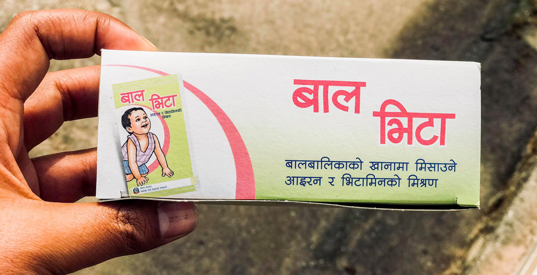
There are multiple supplementary options available for our baby. There is a famous nutrition supplement which contains 16 types of micro nutrients all in one called Balvita supplied by government of nepal for free. Special nutrients supplement required for children is vitamin A.
What is Balvita?
Balvita is a type of nutritional supplement that is designed to provide essential vitamins and minerals to support the growth and development of children. Balvita is typically recommended for children who may have nutritional deficiencies or who require additional support for healthy growth and development.
Baal Vita (also spelled Balvita) is a multiple micronutrient powder (MNP) provided to young children, primarily in Nepal, to combat malnutrition, prevent anemia, and promote healthy physical and cognitive development.
It is also called multi micronutrient powder as well.
How to feed balvita to my baby?
In Nepal, “Balvita” (also called Baal Vita or micronutrient powder) is usually given to babies from 6 months onward along with complementary foods. It is meant to add vitamins and minerals to homemade food. (Dr Chaitanya Joshi, MD)
Here’s the recommended way to feed it:
Wash your hands and use a clean bowl/spoon.
Prepare soft semi-solid food your baby already eats, such as:
lito
jaulo/khichdi
mashed rice
mashed potato or vegetables
Open one full sachet from the pointed edge.
Mix the entire sachet thoroughly into a small serving of food that the baby can finish in one sitting.
Feed immediately with a spoon.
Important precautions:
Do not mix Balvita into very hot food because heat can reduce vitamin effectiveness.
Do not mix it into watery liquids or a feeding bottle.
Use the whole sachet at once; do not divide it for multiple meals.
Continue breastfeeding along with complementary feeding.
If your baby develops vomiting, rash, diarrhea, or feeding problems, consult a pediatrician.
The Nepal nutrition program commonly provides about 60 sachets every 6 months for children aged 6–24 months.
Deworming
According to WHO the country in which there is more than 20 % of ppulation suffering from worm infestation then the country should deworm the population twice a year.
Neapal also has hugh prevalence of worm infestation and among children and the pregnant women the prevalence of infestatio nis more than one third.
It has been shown that one tablet of albendazole 400 mg or mebendazole 500 mg can kill almost all of the intestinal worms.
Nepal has decided to distribute albendazole for purpose of deworming. This tablet can kill various types of worms including Ancylostoma canium, Ancylostoma duodenale (Nectar americanus), Ascaris lumbricoids (intestinal worm), Enteribiasis (pinworm), Oesophagostomum bifurcum etc.
Mechanism of action of albendazole:
The active metabolite of albendazole is albendazole sulfoxide. It causes selective degenration if the cytoplasmic degeneration of the microtubules in intestinal and tegmental cells of intestinal helminths and larvae; glycogen is depleted, glucose uptake and cholinesterase secretion is imparired, and desecratory substance is accumulated intracellularly. ATP production decreases. This causes energy depletion and immobilization and subsequent worm death. Thus dead worm is passed down in stool.
Dose of albendazole:
Childres one year to less than 2 years -> 200 mg (1/2 tablet twice yearly
Children 2 years to 5 years -> one tablet of 400 mg twice yearly
Pregnant woman after first trimester -> one tablet of albendazole 400 mg
What is vitamin A?
Vitamin A is a micronutrient found mostly in green leafy vegetables and animal source foods like meat, eggs and dairy products.
What does vitamin a do in our body:
High dose vitamin A supplementation is done to prevent and treat many conditions among children and adults.
The dose of vitamin A in different population is summerized below
Children 6 to under 12 months of age – one oral vitamin A dose of 100,000 IU two times per year
Children 12 to 59 months of age – one oral vitamin A dose of 200,000 IU two times per year
Women immediately following childbirth, or as soon as possible up to six weeks post-partum, can be given one oral dose of vitamin A 200,000 IU
To treat complications related to deficiency- xerophthalmia etc three doses one upon diagnosis, one the following day and third dose one month later (Note: one dose: 100,000 IU for children aged 6-<12 months; 200,000 IU for children more than 12 months)
For measles: one upon diagnosis and another the following day(Note: one dose: 100,000 IU for children aged 6-<12 months; 200,000 IU for children more than 12 months)
Prolonged diarrhea: more than two weeks: one dose immidiately after diagnosis (Note: one dose: 100,000 IU for children aged 6-<12 months; 200,000 IU for children more than 12 months)
Severe malnutrition: one dose immidiately after diagnosis (Note: one dose: 100,000 IU for children aged 6-<12 months; 200,000 IU for children more than 12 months)
For pregnant women with nightblindness: 25000 IU once a week for four weeks (4 doses)
Viral hepatitis in children is a primary inflammation of the liver caused by at least five specific hepatotropic viruses (A, B, C, D, and E). While often milder in pediatric patients than in adults, it can progress to acute liver failure or chronic liver disease, particularly with types B and C.
I. Classification and Etiological Profile of Primary Viral Hepatitis
The five main viruses are distinguished by their genomic structure and mode of transmission:
Hepatitis A Virus (HAV): An RNA virus identical to enteroviruses; transmitted primarily via the feco-oral route through contaminated food or water. It never causes chronic infection.
Hepatitis B Virus (HBV): A DNA virus (Dane particle) transmitted parenterally, sexually, or vertically (perinatal). It is the most common cause of acute and chronic hepatitis worldwide.
Hepatitis C Virus (HCV): An RNA virus primarily transmitted via percutaneous blood exposure (IV drug use, transfusions before 1991) and vertically (5-6% risk).
Hepatitis D Virus (HDV): A defective RNA virus that requires co-infection or superinfection with HBV to replicate, as it uses the HBV lipoprotein envelope.
Hepatitis E Virus (HEV): An RNA virus transmitted enterally (water-borne), similar to HAV; it is a major cause of high mortality in pregnant women.
II. High-Yield Incubation Periods and Transmission Routes
Virus
Incubation Period
Main Route of Transmission
HAV
28–42 days
Feco-oral (“The vowels go through the bowels”)
HBV
60–150 days
Parenteral, Sexual, Vertical (Perinatal)
HCV
30–60 days
Parenteral (Blood exposure), Vertical
HDV
60–80 days
Parenteral (Requires HBV co-infection)
HEV
25–60 days
Feco-oral (Often water-borne epidemics)
Exam Point: “The Window Period” In HBV infection, the “window period” occurs when HBsAg has disappeared but Anti-HBs has not yet appeared. During this time, Anti-HBc IgM is the only marker of acute infection.
III. Detailed Clinical Presentation and Extrahepatic Features
Symptoms in children are often non-specific and vary by age:
Prodromal Phase:Fever, malaise, anorexia, nausea, vomiting, and right upper quadrant abdominal pain.
Icteric Phase: Subside of fever and anorexia, followed by jaundice (1–3 days after prodrome), dark urine, and pale stools.
Physical Findings: Tender hepatomegaly is common; splenomegaly occurs in 30% of cases.
HBV Extrahepatic Manifestations: High-yield exam points include serum sickness-like syndrome, polyarteritis nodosa (PAN), and membranous glomerulonephritis.
Chronic Hepatitis: Defined as continuing inflammation for \(\ge\)3–6 months; markers include persistently raised transaminases.
IV. Interpretation of Serological Markers (Crucial for MD Exams)
Diagnostic confirmation relies heavily on serology:
HAV: Diagnosis by Anti-HAV IgM (acute); Anti-HAV IgG indicates past infection and lifelong immunity.
HBV Complex Serology:
HBsAg: Indicates current infection (acute or chronic).
Anti-HBs: Indicates immunity (either via vaccine or recovered infection).
Anti-HBc IgM: Indicates acute/recent infection (useful in the window period).
Anti-HBc IgG: Indicates past or chronic infection.
HBeAg: Correlates with high viral replication and high infectivity.
Anti-HBe: Indicates lower infectivity.
HCV: Screen with Anti-HCV; confirm with HCV RNA PCR (detectable 1–2 weeks post-exposure).
V. Management and Pediatric Treatment Protocols
Treatment is primarily supportive for acute cases, but chronic cases require targeted therapy:
Supportive Care: Bed rest during jaundice, high carbohydrate diet, and avoidance of fats and hepatotoxic drugs (e.g., paracetamol, chlorpromazine).
Chronic HBV: Preferred treatments include Entecavir (\(\ge\)2 years) or Tenofovir (\(\ge\)12 years). Interferon-alfa is also an option for children 1–18 years.
Chronic HCV: Revolutionized by Direct-Acting Antivirals (DAAs). Recommended for all children \(\ge\)3 years. Regimens like Sofosbuvir/Ledipasvir (Harvoni) or Glecaprevir/Pibrentasvir (Mavyret) are highly effective.
Fulminant Hepatitis: Requires ICU admission, management of cerebral edema (Mannitol), and evaluation for liver transplantation.
VI. Prevention and Post-Exposure Prophylaxis (PEP)
Hepatitis A Vaccine: Two-dose series starting at 12 months.
Hepatitis B Vaccine: Routine three-dose series at birth, 1–2 months, and 6 months.
Perinatal HBV PEP: If a mother is HBsAg-positive, the neonate must receive HBIG (0.5 mL) and the first HBV vaccine dose within 12 hours of birth at separate sites.
Hygiene: Improving water supply and personal hygiene is the mainstay for preventing enteral (A and E) types.
High-Yield Laboratory “Rule of Thumb”: In acute viral hepatitis, ALT is typically > AST. If AST is twice as high as ALT, consider other etiologies like alcoholic hepatitis or hemolysis. Very high levels (>1000 U/L) are characteristic of acute viral or toxic injury.
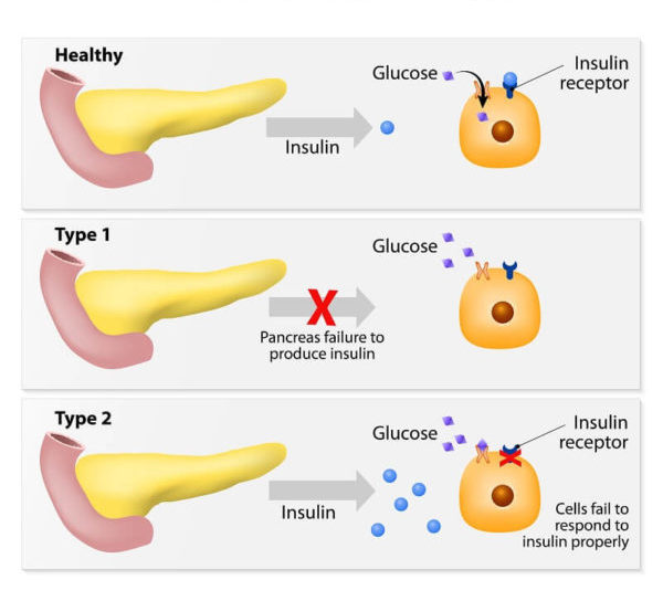
Diabetes is a chronic medical condition characterized by high levels of blood sugar (glucose). This occurs either because the body doesn’t produce enough insulin (a hormone that regulates blood sugar) or because the cells don’t respond properly to the insulin that is produced. Insulin is necessary for the body to effectively use glucose as a source of energy.
History for diabetes mellitus type 1 and 2:
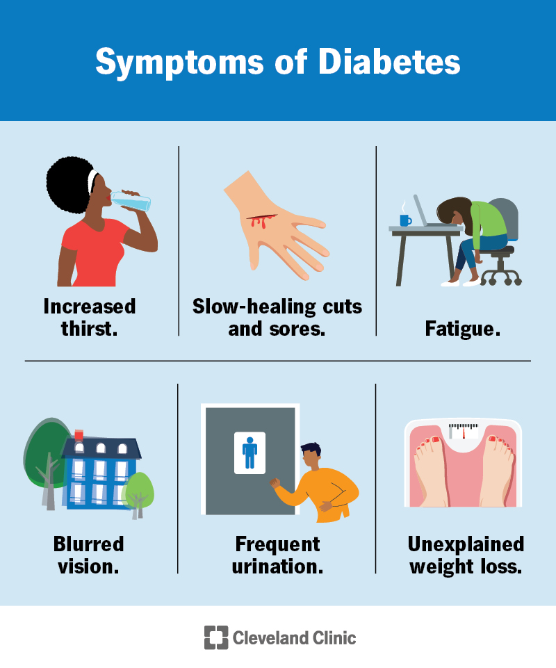
Symptoms of hyperglycemia
Thirst, dry mouth
Polyuria
Nocturia
Tiredness, fatigue, lethargy
Noticeable change in weight (usually weight loss)
Blurring of vision
Pruritus vulvae, balanitis (genital candidiasis)
Nausea; headache
Hyperphagia; predilection for sweet foods
Mood change, irritability, difficulty in concentrating, apathy
Family history
Physical examination for diabetes mellitus type 1 and 2
BMI
Retinal examination
Orthostatic blood pressure
Foot examination
Peripheral pulses
Insulin injection sites
Peripheral neuropathy
Type 1 Vs Type 2 Diabetes mellitus DM
Type 1
Type 2
Onset
Sudden
Gradual
Age at onset
Any (mostly young)
Mostly in adults
Body habitus
Thin or normal
Often obese
Ketoacidosis
Common
Rare
Autoantibodies
Usually +
Absent
Endogeneous insulin
Low or absent
Normal, decreased or increased
Concordance in identical twins
~ 50%
~90%
Prevalence
Less prevalent
More prevalent (~90-95% of US diabetics)
Biochemical
C-peptide disappears
C-peptide persists
Mechanism and pathophysiology of Diabetes mellitus
Increased bloog glucose sugar level definition is called when
Two-hour glucose during OGTT <140 mg/dL (7.8 mmol/L)
Categories of increased risk for diabetes:
•Impaired fasting glucose(IFG)
FPG between 100 and 125 mg/dL (5.6 to 6.9 mmol/L).
•Impaired glucose tolerance(IGT)
Two-hour – 75 g OGTT between 140 and 199 mg/L (7.8 to 11.0 mmol/L).
•A1C – Persons with 5.7 to 6.4 percent (39 to 46 mmol/mol
Diagnostic criteria of Diabetes mellitus
1. A1C ≥6.5 percent OR 2. Fasting Plasma Glucose ≥126 mg/dL (7.0 mmol/L) (Fasting is defined as no caloric intake for at least eight hours.) OR 3. Two-hour plasma glucose ≥200 mg/dL (11.1 mmol/L) during an OGTT. (The test should be performed using a glucose load containing the equivalent of 75-gram anhydrous glucose dissolved in water.) OR 4. In a patient with classic symptoms of hyperglycemia or hyperglycemic crisis, a random plasma glucose ≥200 mg/dL (11.1 mmol/L).
1. A1C ≥6.5 percent
OR
2. Fasting Plasma Glucose ≥126 mg/dL (7.0 mmol/L)
(Fasting is defined as no caloric intake for at least eight hours.)
OR
3. Two-hour plasma glucose ≥200 mg/dL (11.1 mmol/L) during an OGTT.
(The test should be performed using a glucose load containing the equivalent of 75-gram anhydrous glucose dissolved in water.)
OR
4. In a patient with classic symptoms of hyperglycemia or hyperglycemic crisis, a random plasma glucose ≥200 mg/dL (11.1 mmol/L).
Advice to patients with Impaired glucose tolerance
Have an increased risk both of progression to type 2 diabetes and of developing macrovascular disease
Advice lifestyle modification reduces the risk of progression in IGT
Monitor annually by measurement of fasting blood glucose
Other cardiovascular risk factors treate aggressively