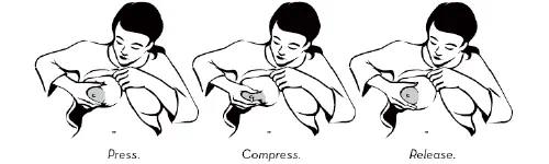
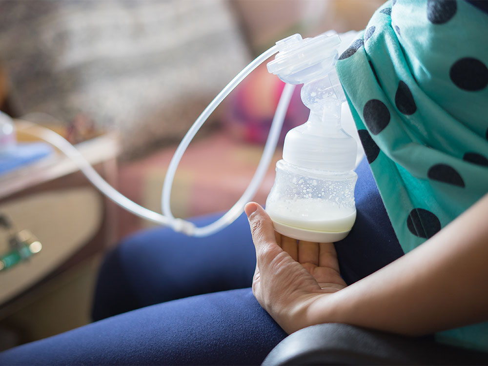
Proper storage of expressed breast milk (EBM) helps preserve its nutritional and immunological properties for later feeding when the mother is away.
2. Containers for Storage
Use clean, sterilized glass or BPA-free plastic containers with tight-fitting lids.
Breast milk storage bags (food-grade, pre-sterilized) are convenient for freezing.
Avoid ordinary plastic bottles or disposable liners.
3. Labeling
Each container should be labeled with:
Date and time of expression
Baby’s name (if used in hospital or daycare)
4. Storage Guidelines
Location
Temperature
Duration
Remarks
Room temperature
Up to 25°C (77°F)
4–6 hours
Keep in a cool, clean area away from direct sunlight
Refrigerator (back portion)
2–4°C (35–40°F)
Up to 72 hours (3 days)
Do not store in refrigerator door due to temperature fluctuation
Freezer (separate door)
–18°C or lower
Up to 3–6 months
Keep in small portions; leave space for expansion
Deep freezer
–20°C
Up to 6–12 months
Best for long-term storage
Insulated cooler box with ice packs
~15°C
Up to 24 hours
For temporary transport or travel
5. Thawing and Warming
Thaw in refrigerator overnight or by placing the container in warm water (<37°C).
Do not microwave or boil — destroys antibodies and nutrients.
Swirl gently (do not shake) to mix separated fat layers.
Once thawed, use within 24 hours and do not refreeze.
6. Hygiene
Wash hands before expressing or handling milk.
Use clean pump parts and containers for each session.
Avoid touching the inside of lids or bottles.
7. Key Points
Always use oldest milk first (“first in, first out”).
Discard leftover milk from feeding bottle after use.
Observe for odor or curdling — discard if spoiled.
Summary:
Safe breast milk storage requires hygienic handling, correct temperature, and appropriate duration. Properly stored milk retains most of its nutritional, immunological, and protective properties, ensuring safe feeding for infants when direct breastfeeding isn’t possible.
An enlarged placenta isn’t usually a reason to panic. The word is big (placentomegaly), but most of the time it doesn’t cause problems.
Some conditions play a role. A larger placenta may be linked to hypertension, anemia, or diabetes — but your doctor will monitor these and the health of your baby.
Your baby’s growth matters most. Even if your placenta measures big, steady fetal development is the important goal, so keep up your regular appointments to be sure everything’s right on track.
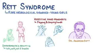
Rett syndrome is a neurodevelopmental disorder that primarily affects girls, characterized by normal early development followed by regression of acquired skills, especially speech and purposeful hand movements, with onset typically between 6–18 months of age.
Etiology
Genetic cause: Mutation in MECP2 gene (methyl-CpG-binding protein 2) on the X chromosome (Xq28)
Inheritance: Usually sporadic (de novo); rarely familial
Pathophysiology: Dysfunction of MECP2 protein → abnormal brain maturation and synaptic development
Epidemiology
Affects 1 in 10,000–15,000 female births
Lethal in males (most do not survive infancy unless mosaic or XXY)
Clinical Features
Phases of Disease
Early Onset (6–18 months)
Normal development initially
Gradual loss of interest in surroundings
Loss of purposeful hand skills
Deceleration of head growth (acquired microcephaly)
Rapid Destructive Phase (1–4 years)
Loss of speech and purposeful hand use
Stereotyped hand movements: hand-wringing, washing, clapping, or mouthing
Gait ataxia, truncal apraxia
Autistic-like behavior
Plateau Phase (2–10 years)
Some improvement in social interaction and eye contact
Hypothyroidism is a clinical state resulting from deficiency of thyroid hormone production or action, leading to a generalized slowing of metabolic processes.
It may be:
Congenital (Neonatal) – present at birth.
Acquired (Childhood) – develops later due to autoimmune, iatrogenic, or other causes.
2. Classification
A. Based on Level of Defect
Type
Site of Defect
TSH
T4/T3
Primary
Thyroid gland
↑
↓
Secondary
Pituitary
↓/N
↓
Tertiary
Hypothalamus
↓/N
↓
Peripheral (Resistance)
Target tissue
N/↑
N/↑
B. Based on Onset
Congenital hypothyroidism (CH)
Acquired hypothyroidism
3. Epidemiology
CH: ~1 in 2,000–4,000 live births.
More common in females.
Acquired form common in older children/adolescents, often autoimmune (Hashimoto’s).
thyroid gland
4. Etiology
A. Congenital Hypothyroidism
Thyroid dysgenesis (80–85%)
Agenesis, ectopy, or hypoplasia.
Usually sporadic.
Dyshormonogenesis (10–15%)
Inborn errors of thyroid hormone synthesis (autosomal recessive).
Leishmaniasis — MD Pediatrics Note (Based on Nelson Textbook of Pediatrics)
Table of Contents(toc)
Introduction
Leishmaniasis is a spectrum of protozoal diseases caused by Leishmania species, transmitted by the bite of infected female phlebotomine sandflies.
cutaneous leishmaniasis
Disease manifestations depend on the species involved and the host immune response.
Major clinical forms:
Visceral leishmaniasis (VL / kala-azar)
Cutaneous leishmaniasis (CL)
Mucocutaneous leishmaniasis (MCL)
Etiology and Classification
Form
Causative Species
Geographic Distribution
Visceral
L. donovani, L. infantum (chagasi)
South Asia, East Africa, Latin America
Cutaneous
L. tropica, L. major, L. mexicana, L. braziliensis
Middle East, Africa, Americas
Mucocutaneous
L. braziliensis complex
Central & South America
Epidemiology
Endemic in >80 countries; affects poor, rural populations.
Vectors:Phlebotomus (Old World), Lutzomyia (New World).
Reservoirs: Humans (L. donovani), dogs, rodents.
Transmission: Sandfly bite, rarely congenital or via transfusion.
Phlebotomus
Pathogenesis
Inoculation of promastigotes → engulfed by macrophages → transform into amastigotes → intracellular multiplication → spread to RES (liver, spleen, bone marrow).
Disease severity depends on cell-mediated immunity (CMI).
Category: Inherited bone marrow failure syndrome (IBMFS) Inheritance: Autosomal recessive (rarely X-linked) Gene defects: >22 genes identified (FANCA, FANCC, FANCG most common) → defective DNA interstrand crosslink repair.
fanconi anemia notes
1. Pathophysiology
Defect in DNA repair (Fanconi/BRCA pathway) → chromosomal breakage and hypersensitivity to DNA cross-linking agents (e.g., mitomycin C, diepoxybutane).
Progressive bone marrow failure (due to stem cell depletion) and genomic instability → predisposition to malignancies.
Multisystem developmental abnormalities due to impaired cell proliferation during embryogenesis.
2. Epidemiology
Incidence: ~1 in 100,000–250,000 live births.
Carrier frequency: ~1 in 200.
Median age of diagnosis: 7–9 years.
~90% develop marrow failure by age 40.
3. Clinical Features
A. Hematologic
Pancytopenia (usually first manifests with thrombocytopenia or macrocytic anemia).
हामी प्रायः रगत परीक्षणका लागि हातको नसाबाट (vein) रगत निकालिन्छ भन्ने कुरा जान्दछौं। तर कहिलेकाहीँ स्वास्थ्यकर्मीले नाडीबाट (artery) पनि रगत निकाल्छन्। यो सामान्य रगत परीक्षणभन्दा फरक र विशिष्ट उद्देश्यका लागि गरिन्छ।
ABG sampling technique why and when
नाडीबाट रगत निकाल्नुको मुख्य कारण — “Arterial Blood Gas (ABG)” परीक्षण
नाडीबाट रगत निकाल्ने मुख्य उद्देश्य Arterial Blood Gas (ABG) test हो।
यो परीक्षणले शरीरमा रहेका अक्सिजन (O₂), कार्बन डाइअक्साइड (CO₂) र रगतको अम्ल–क्षार (pH) सन्तुलन कस्तो छ भन्ने देखाउँछ।
यो जानकारी फोक्सो र मुटुको कार्य कस्तो छ भन्ने बुझ्न अत्यन्त जरुरी हुन्छ।
यो परीक्षण कहिले गरिन्छ ?
जब बिरामीलाई अक्सिजन कमी (hypoxia) को शंका हुन्छ।
सास फेर्न गाह्रो भएको अवस्थामा (जस्तै– दमा, COPD, pneumonia, ARDS)।
भेन्टिलेटरमा राखिएका बिरामीहरूमा, अक्सिजनको मात्रा ठिक छ कि छैन भनेर हेर्न।
ABG गर्नुअघि प्रायः Allen’s test गरिन्छ, जसले हातको रक्तप्रवाह सुरक्षित छ कि छैन भन्ने पक्का गर्छ।
कसरी निकालिन्छ ?
बिरामीलाई आराम दिन्छ।
छालालाई सफा गरिन्छ (antiseptic)।
नाडीको धड्कन भेटाएर सुई प्रयोग गरी सिधै नाडीभित्र सुई प्रवेश गरिन्छ।
रगत सिधै syringe मा स्वचालित रूपमा भरिन्छ, किनकि नाडीको दबाब (pressure) बढी हुन्छ।
त्यसपछि तुरुन्तै syringe लाई बर्फमा राखी ल्याबमा पठाइन्छ ताकि ग्यासहरू नबदलिऊन्।
नसाबाट होइन, नाडीबाट किन ?
नसाको रगतले शरीरको अक्सिजन र कार्बन डाइअक्साइडको सन्तुलन सही रूपमा देखाउँदैन, किनभने त्यो पहिले नै ऊतकहरूबाट फर्किएको हुन्छ।
तर नाडीको रगत भने फोक्सोबाट निस्किएको ताजा अक्सिजनयुक्त रगत हो, जसले शरीरको साँच्चिकै ग्यास स्थिति जनाउँछ।
त्यसैले फोक्सो, सासफेर्ने प्रणाली वा अक्सिजन थेरापी मूल्याङ्कन गर्न नाडीबाट रगत आवश्यक पर्छ।
के जोखिम हुन्छ ?
सामान्यतया सुरक्षित भए पनि केही साइड इफेक्ट हुन सक्छन् —
नाडीमा दबाबको कारण दुखाइ वा निलो दाग (bruise)
कहिलेकाहीँ रगत बग्ने वा clot बन्ने समस्या
धेरै पटक सुई लगाउँदा नाडीको क्षति वा हात सुन्निनु
त्यसैले यो परीक्षण प्रशिक्षित स्वास्थ्यकर्मी (जस्तै चिकित्सक वा नर्स) ले मात्र गर्नुपर्छ।
सारांशमा
नाडीबाट रगत निकाल्नु साधारण परीक्षण होइन, तर अत्यन्त महत्त्वपूर्ण चिकित्सकीय प्रक्रिया हो जसले शरीरको अक्सिजन, कार्बन डाइअक्साइड र अम्ल–क्षार सन्तुलनबारे सटीक जानकारी दिन्छ।
यसले चिकित्सकलाई बिरामीको सासफेर्ने स्थिति बुझ्न, भेन्टिलेटर मिलाउन, र उपचारको प्रभाव मूल्याङ्कन गर्न मद्दत गर्छ।
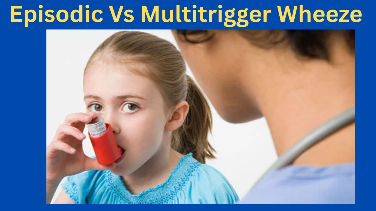
Episodic (Viral) Wheeze vs. Multiple Trigger Wheeze
A Clinically Oriented Review for the Practicing Pediatrician
Based on Nelson Textbook of Pediatrics (21st ed.) | Kendig’s Disorders of the Respiratory Tract in Children (9th ed.) | AAP & IAP-NAPCON Official Resources
1. Introduction
Wheezing in preschool children (0–5 years) is one of the most common reasons for pediatric consultation and hospital admission worldwide. It is now well established that ‘preschool wheeze’ is not a single disease but a heterogeneous group of phenotypes with distinct pathophysiology, natural history, and responses to therapy. The two most clinically useful and validated phenotypes—recognized in both the Nelson Textbook of Pediatrics and major international guidelines—are:
Episodic (Viral) Wheeze (EVW): wheezing episodes triggered exclusively by viral respiratory infections, with complete resolution between episodes.
Multiple Trigger Wheeze (MTW): wheezing triggered by multiple stimuli including viruses, aeroallergens, exercise, cold air, tobacco smoke, and emotional stimuli, with symptoms also occurring between discrete episodes.
This classification, initially proposed by Brand et al. and incorporated into the PRACTALL Consensus Report (2008) of the European Academy of Allergy and Clinical Immunology (EAACI) and the American Academy of Allergy, Asthma and Immunology (AAAAI), is now endorsed by the American Academy of Pediatrics (AAP) and the Indian Academy of Pediatrics (IAP) / National Asthma Consensus Group (NACG).
2. Epidemiology
According to Nelson Textbook of Pediatrics (21st edition, Chapter 169: Wheezing in Infants and Children), approximately 30–40% of all children will experience at least one wheezing episode in the first three years of life, yet fewer than one-third of these will develop persistent asthma. Data from the Tucson Children’s Respiratory Study (TCRS), cited prominently in Nelson, delineates three early wheezing trajectories:
Transient early wheezers: viral-triggered, remit by age 6; low atopic burden.
Non-atopic wheezers (EVW phenotype): episode-only wheeze; best aligned with EVW.
IgE-associated persistent wheezers (MTW/Asthma phenotype): atopic sensitization, family history, persistent into school age.
The IAP NAPCON 2019 Consensus Statement on Childhood Asthma notes that in South Asian children, including India and Nepal, the prevalence of preschool wheeze is significant, often complicated by high pollution exposure and early sensitization to house dust mite and cockroach allergens, features that shift the phenotype toward MTW.
3. Pathophysiology
3.1 Episodic (Viral) Wheeze
As described in Nelson (Chapter 169) and Kendig’s Disorders of the Respiratory Tract in Children (9th edition, Chapter 38), EVW is predominantly mediated by:
Rhinovirus (RV) and respiratory syncytial virus (RSV) — the principal triggers in children <3 years.
Neutrophilic airway inflammation: transient bronchial inflammation during the acute episode, with restoration of normal airway architecture between episodes. Unlike classical asthma, eosinophilic infiltration is typically absent or minimal.
Small airway mechanics: infants have a high ratio of airway resistance due to anatomically smaller caliber airways, making them more susceptible to luminal obstruction from viral-induced mucosal edema and secretions.
Immune dysregulation: reduced interferon-γ (IFN-γ) and impaired Th1 responses to RV have been demonstrated, contributing to prolonged viral shedding and exaggerated bronchospasm.
No persistent structural remodeling: between episodes, lung function is typically normal and there is no evidence of airway remodeling or eosinophilic inflammation.
3.2 Multiple Trigger Wheeze
MTW pathophysiology, as detailed in both Nelson and Kendig’s, resembles that of classic atopic asthma:
Eosinophilic airway inflammation: persistent even during asymptomatic intervals, with elevated fractional exhaled nitric oxide (FeNO).
Th2-skewed immune response: elevated IgE, IL-4, IL-5, IL-13; mast cell and eosinophil activation with allergen exposure.
Airway hyperresponsiveness (AHR): demonstrable on methacholine or exercise challenge, and persisting between symptomatic episodes.
Early sensitization: specific IgE to house dust mite (Dermatophagoides pteronyssinus), cockroach, Alternaria, or other regional allergens is frequently demonstrable by age 2–3 years.
Structural remodeling: subepithelial fibrosis and smooth muscle hypertrophy develop over time if left inadequately treated.
4. Clinical Features and Diagnosis
4.1 History
Nelson (21st ed., Chapter 169) and AAP Clinical Practice Guidelines for Asthma (2020 Update) recommend a detailed history focusing on:
Trigger identification: exclusive viral triggers (EVW) vs. multiple triggers including allergens, exercise, cold air, irritants (MTW).
Inter-episodic symptoms: nocturnal cough, exercise-induced wheeze, or persistent cough between viral episodes strongly suggests MTW.
Atopic comorbidities: personal history of eczema, allergic rhinitis; food allergy.
Family history: parental asthma/atopy increases the Asthma Predictive Index (API) score, supporting MTW/asthma phenotype.
Environmental history: tobacco smoke exposure, cooking fuel, pet ownership, damp housing — relevant especially per IAP guidelines for South Asian settings.
4.2 Asthma Predictive Index (API)
The modified API (mAPI), described in Nelson and endorsed by the AAP, is a validated tool to identify preschool wheezers likely to develop persistent asthma (MTW phenotype). A positive mAPI in a child with ≥3 wheezing episodes in the past year has a positive predictive value of ~80% for asthma at school age.
Digital clubbing, persistent hyperinflation, failure to thrive — suggest alternative diagnoses (cystic fibrosis, primary ciliary dyskinesia, structural airway anomalies).
Normal examination between episodes — expected in EVW; persistent wheeze or hyperinflation between episodes raises suspicion for MTW or alternative pathology.
4.4 Investigations
Kendig’s (9th ed., Chapter 38) and AAP Guidelines recommend the following investigations based on clinical context:
Spirometry (≥5–6 years): reversible airflow obstruction (post-bronchodilator FEV1 improvement ≥12%) supports MTW/asthma; may be normal in EVW.
Skin prick testing / Specific IgE: aeroallergen sensitization supports MTW phenotype; recommended in children with positive mAPI or recurrent MTW.
Complete blood count: peripheral eosinophilia (≥4%) is a minor API criterion.
Chest radiograph: to exclude structural anomalies, foreign body, or consolidation; not routinely needed for wheeze per AAP guidelines.
FeNO measurement: elevated (>25 ppb) supports eosinophilic airway inflammation (MTW/asthma); not universally available but referenced in Nelson and Kendig’s.
Bronchoscopy / BAL: reserved for diagnostically challenging cases; mentioned in Kendig’s for evaluation of structural/anatomic causes of wheeze.
5. Comparative Overview: EVW vs. MTW
Table 1 summarizes the key distinguishing features of the two preschool wheeze phenotypes.
Table 1. Episodic Viral Wheeze vs. Multiple Trigger Wheeze — Comparative Features
Per AAP Clinical Practice Guidelines (2020) and Nelson (Chapter 169), acute management is phenotype-independent and follows standard bronchodilator therapy:
Short-Acting Beta-2 Agonists (SABA): salbutamol (albuterol) 2.5–5 mg via nebulizer, or 2–4 puffs via spacer and face mask every 20 minutes for 3 doses in severe episodes. First-line therapy for all preschool wheeze.
Ipratropium bromide: may be added for moderate-to-severe exacerbations; reduces hospitalization when combined with salbutamol.
Systemic corticosteroids: oral prednisolone (1–2 mg/kg/day, max 40 mg, for 3–5 days) for moderate-to-severe exacerbations. Per the AAP, short courses do not significantly affect adrenal function or growth in children.
Supplemental oxygen: titrate to maintain SpO2 ≥94% (AAP target); SpO2 ≥95% per IAP-NAPCON 2019.
Hospitalization criteria: SpO2 <92% on room air, severe respiratory distress (HR >60/min in infants), inability to maintain oral feeds, poor response to initial bronchodilators.
7.2 Preventive/Controller Therapy
This is where the phenotype distinction critically guides management:
7.2.1 Episodic (Viral) Wheeze
Per Nelson, Kendig’s, and AAP Guidelines:
Continuous ICS: NOT routinely recommended for EVW. Multiple RCTs (including the PEAK and MIST trials cited in Nelson) show no significant reduction in episode frequency or severity with continuous low-dose ICS in non-atopic preschool wheezers.
Intermittent/episodic ICS: high-dose ICS at the onset of a viral URTI (e.g., budesonide 400 mcg/day or fluticasone 200 mcg/day for 7–10 days) may reduce episode severity in selected children, though evidence remains inconsistent across trials.
Montelukast: episodic use at onset of wheeze shows modest benefit in some studies (Bisgaard et al., NEJM, cited in Nelson); may be considered for children with 3 or more episodes per year.
Bronchodilator reliever therapy: salbutamol as needed during episodes. Continuous reliever use between episodes is not indicated in pure EVW.
Avoidance: passive smoking cessation, hand hygiene, daycare modifications to reduce viral exposure.
7.2.2 Multiple Trigger Wheeze
Per Nelson, Kendig’s, AAP (2020), and IAP-NAPCON (2019):
Low-dose ICS: first-line preventer therapy. Budesonide 100–200 mcg/day or fluticasone propionate 100 mcg/day (BDP-equivalent). Initiate when diagnosis of MTW/persistent asthma is established.
Montelukast: may be used as an alternative to ICS in mild MTW or as add-on therapy in moderate MTW. IAP-NAPCON recognizes its role given high house dust mite sensitization in the South Asian context.
Medium-dose ICS: step up to 200–400 mcg/day (budesonide equivalent) if low-dose ICS fails to achieve symptom control after 6–8 weeks.
LABA addition: for children ≥5 years with inadequate control on medium-dose ICS, salmeterol or formoterol can be added. Not approved or recommended for children <4 years as monotherapy.
Allergen avoidance: mattress/pillow encasements, HEPA filtration, pet removal — strongly recommended by AAP and IAP for sensitized children with MTW.
Allergen Immunotherapy (AIT): subcutaneous or sublingual AIT for house dust mite-sensitized children with MTW/asthma is recommended in international guidelines and endorsed in IAP-NAPCON for appropriate candidates ≥5 years.
Omalizumab: anti-IgE therapy; approved for moderate-to-severe persistent allergic asthma in children ≥6 years; referenced in Nelson and AAP guidelines for refractory MTW/asthma with high IgE and allergen sensitization.
7.3 Step-Therapy Summary
Table 2. Stepwise Treatment Approach for EVW and MTW
Step
EVW Management
MTW Management
Acute
SABA (salbutamol) via spacer/nebulizer; oral prednisolone for moderate-severe
SABA; oral/systemic corticosteroids; consider early ICS step-up
Preventer
Not routinely indicated; trial ICS only if frequent/severe episodes (≥3/year)
Low-dose ICS (e.g., budesonide 100–200 mcg/day) as first-line preventer
Step-up
Episodic ICS at onset of URTI (intermittent therapy); montelukast episodic use
Increase ICS dose; add montelukast or LABA (≥5 yr); consider specialist referral
Monitoring
Symptom diary; reassess trigger pattern at each visit
Nebulizers are not superior to pMDI+spacer for acute bronchodilation and carry infection transmission risk in healthcare settings. Both AAP and IAP recommend prioritizing spacer-based delivery.
8. Monitoring and Follow-Up
Nelson, AAP (2020 Expert Panel Report 3 Update), and IAP-NAPCON recommend the following monitoring framework:
Review diagnosis every 3–6 months: re-evaluate whether phenotype has shifted from EVW to MTW as the child grows.
Assess symptom control using validated tools: \Childhood Asthma Control Test (C-ACT) for children ≥4 years; parent-report tools for younger children.
Spirometry when developmentally feasible (≥5 years): monitor FEV1, FVC, and FEV1/FVC ratio at each visit.
Reassess trigger profile at each visit: new aeroallergen sensitization, school exposures, change in environment.
Monitor growth: height and weight percentile; ICS at low doses does not significantly affect final adult height per Nelson; monitor with medium-to-high doses.
Adherence and inhaler technique: check at every visit; poor technique is the most common cause of apparent treatment failure per AAP.
Consider step-down: if well-controlled for ≥3 months, cautiously step down therapy, reassessing trigger pattern.
9. Prognosis and Natural History
The TCRS and birth cohort studies cited in Nelson provide the most robust data on prognosis:
EVW (Transient wheeze): ~60% of preschool wheezers remit by 6 years of age. These children, corresponding to the EVW phenotype, generally have normal lung function at school age. The absence of atopic sensitization, normal lung function between episodes, and non-positive API predict favorable outcome.
MTW (Persistent/Asthma phenotype): ~40% of preschool wheezers continue to wheeze at school age. Risk factors for persistence include: positive mAPI, maternal asthma, early sensitization to aeroallergens, frequent episodes in the first 3 years, male sex, and exposure to high-dose indoor allergens.
Lung function trajectory: Lung function deficits, if present at age 6 years in the MTW group, tend to track into adult life and are associated with increased risk of COPD in adulthood (“early origins of adult lung disease” concept, cited in Nelson and Kendig’s).
South Asian context (IAP): earlier sensitization to perennial allergens (HDM, cockroach), higher pollution burden, and lower vitamin D levels may confer worse outcomes in the MTW phenotype in Indian children, as noted in IAP-NAPCON 2019.
10. Special Clinical Situations
10.1 The “Overlap” Child
Many children present with features of both EVW and MTW, especially between ages 2–4 years. Nelson recommends using the mAPI as a practical decision aid in such cases. If the mAPI is positive, treat as MTW (initiate regular ICS); if negative, manage as EVW (episodic/as-needed therapy).
10.2 Very Young Infants (<12 months)
Wheezing in infants under 12 months is most commonly due to bronchiolitis (RSV) and should not be classified as EVW or MTW. Per AAP Clinical Practice Guideline for the Diagnosis, Management, and Prevention of Bronchiolitis (2014, reaffirmed 2020), bronchodilators are not recommended for infants with bronchiolitis. ICS and systemic steroids are similarly not recommended in this age group for acute bronchiolitis.
10.3 COVID-19 and Respiratory Viruses
The AAP has issued guidance noting that SARS-CoV-2 infection in young children may trigger wheezing episodes similar to other viral URTI triggers in EVW. Standard asthma action plans should include COVID-19 as a potential EVW trigger; ICS should not be stopped during COVID-19 illness in MTW/asthma patients.
10.4 Vaccination
Both AAP and IAP recommend annual influenza vaccination for all children with recurrent wheezing (EVW or MTW), as influenza is a significant trigger for severe exacerbations. Pneumococcal vaccination per national immunization schedules is also recommended.
11. Parent and Caregiver Education
AAP and IAP emphasize that education is a cornerstone of management:
Provide written Asthma Action Plan (AAP template available at healthychildren.org) for all children with recurrent wheeze.
Educate on symptom recognition: early signs of exacerbation (nocturnal cough, reduced exercise tolerance, increased rescue inhaler use).
Inhaler technique training at every visit; video demonstrations and teach-back methods are recommended by AAP.
Environmental control counseling: tobacco smoke, allergen avoidance, mold reduction, pet dander management.
Address caregiver anxiety: explain phenotype, natural history, and that EVW does not inevitably become asthma.
Emphasize adherence to preventive therapy in MTW: parents often reduce ICS doses prematurely when symptoms improve.
12. Key Clinical Takeaways
Phenotype matters: Distinguish EVW from MTW at every clinical encounter; this distinction drives preventive therapy decisions.
mAPI guides therapy: A positive mAPI in a high-frequency preschool wheezer indicates MTW/asthma phenotype and justifies early ICS therapy.
ICS is not universal: Continuous ICS is not recommended for pure EVW; reserve for MTW or EVW with frequent/severe episodes.
Trigger profile shapes management: Allergen sensitization testing is indicated when MTW is suspected; AIT may be indicated in sensitized children ≥5 years.
Phenotypes are dynamic: Reassess at every visit; EVW may evolve to MTW as atopic sensitization develops.
Guideline resources: Use AAP (healthychildren.org, aappublications.org) and IAP-NAPCON (iapindia.org) official resources for updated local guidance.
Exclude mimics: Always consider structural, infectious, and congenital causes of recurrent wheeze, especially in children <12 months or with atypical features.
References
Primary Textbook References:
Kliegman RM, St. Geme JW, Blum NJ, et al. Nelson Textbook of Pediatrics, 21st Edition. Philadelphia: Elsevier; 2020. Chapter 169: Wheezing in Infants and Young Children; Chapter 170: Asthma.
Wilmott RW, Deterding R, Li A, et al. Kendig’s Disorders of the Respiratory Tract in Children, 9th Edition. Philadelphia: Elsevier; 2019. Chapter 38: Wheezing in Infancy and Early Childhood; Chapter 39: Asthma in the Pediatric Patient.
AAP Official Resources:
American Academy of Pediatrics. Clinical Practice Guideline for the Diagnosis, Evaluation, and Management of Childhood Asthma. Pediatrics. 2020;145(3):e20193432. Available at: https://publications.aap.org
American Academy of Pediatrics. Clinical Practice Guideline: The Diagnosis, Management, and Prevention of Bronchiolitis. Pediatrics. 2014;134(5):e1474-e1502. Reaffirmed 2020. Available at: https://publications.aap.org
American Academy of Pediatrics. Asthma Action Plan templates and parent education resources. HealthyChildren.org. Available at: https://www.healthychildren.org
IAP Official Resources:
Indian Academy of Pediatrics, National Asthma Consensus Group (NAPCON). IAP-NAPCON Consensus Statement on Childhood Asthma 2019. Indian Pediatrics. 2020;57(1):42–58. Available at: https://www.indianpediatrics.net
Indian Academy of Pediatrics. IAP Standard Treatment Guidelines: Bronchial Asthma in Children. 2022. Available at: https://www.iapindia.org
Landmark Studies and Consensus Documents (cited in Nelson/Kendig’s):
Brand PL, Baraldi E, Bisgaard H, et al. Definition, assessment and treatment of wheezing disorders in preschool children: an evidence-based approach. European Respiratory Journal. 2008;32(4):1096–1110. [PRACTALL Consensus Report, cited in Nelson 21e and Kendig’s 9e]
Martinez FD, Wright AL, Taussig LM, et al. Asthma and wheezing in the first six years of life: The Group Health Medical Associates. New England Journal of Medicine. 1995;332(3):133–138. [Tucson Children’s Respiratory Study, cited in Nelson 21e]
National Asthma Education and Prevention Program (NAEPP). Expert Panel Report 3 (EPR-3): Guidelines for the Diagnosis and Management of Asthma. National Heart, Lung, and Blood Institute (NHLBI). 2007 (Updated 2020). Available at: https://www.nhlbi.nih.gov
Global Initiative for Asthma (GINA). Difficult-to-Treat and Severe Asthma in Adolescent and Adult Patients: A GINA Pocket Guide. 2023. [Referenced in Nelson and Kendig’s for management framework]


")