Blog

Remember these 3 types now: Do you know Breast milk types?

Types of Breast Milk: Colostrum, Foremilk, and Hindmilk


1. Colostrum


  • Definition: The first milk secreted during the initial 2–4 days after delivery.


  • Appearance: Thick, yellowish, sticky fluid.


  • Volume: Small in quantity (about 30–100 mL/day).


  • Composition:


    • High in proteins, especially immunoglobulin A (IgA) and lactoferrin.


    • Low in fat and lactose.


    • Rich in vitamins A, E, and K, and leukocytes.


  • Functions / Importance:


    • Provides passive immunity and protects against infections (especially GI and respiratory).


    • Acts as a natural laxative, promoting early meconium passage and decreasing risk of neonatal jaundice.


    • Helps in gut maturation and colonization by beneficial bacteria.


2. Foremilk


  • Definition: The milk secreted at the beginning of a feeding session.


  • Appearance: Thin, watery, bluish-white.


  • Composition:


    • High in lactose, water, and proteins.


    • Low in fat and calories.


  • Function:


    • Quenches the baby’s thirst.


    • Provides hydration and quick energy through lactose.


  • Clinical Relevance:


    • Excessive foremilk intake (without adequate hindmilk) may cause lactose overload → symptoms like colic, gas, and loose stools.


3. Hindmilk


  • Definition: The milk secreted towards the end of feeding.


  • Appearance: Creamy, thick, and whiter due to higher fat content.


  • Composition:


    • High in fat and calories.


    • Relatively low in lactose.


  • Function:


    • Provides satiety and helps in weight gain.


    • Supplies essential fatty acids for brain development and energy.


  • Practical Point:


    • Mothers should be advised to empty one breast fully before switching to the other to ensure the baby receives enough hindmilk.


Summary Table

TypeTime of SecretionAppearanceMajor ComponentFunction
Colostrum0–4 days postpartumYellow, thickProteins, IgAImmunity, meconium passage
ForemilkEarly in each feedWatery, bluishLactose, waterHydration, energy
HindmilkLater in each feedCreamy, whiteFatSatiety, growth

You must know these dangerous causes of Black Tongue!

Table of Contents(toc)

🦷 What Causes Black Tongue?

(Understanding the Causes, Symptoms, and Prevention)

A black tongue can look alarming — but in most cases, it’s harmless and temporary. Medically known as “black hairy tongue” (lingua villosa nigra), this condition occurs when tiny projections on the tongue (called papillae) become elongated and trap debris, bacteria, or yeast, causing discoloration.

Let’s explore the causes, symptoms, and what you can do about it.

black hairy tongue

🧬 What Actually Happens?

The surface of your tongue is covered with small bumps called filiform papillae. Normally, these shed continuously.
However, when this natural process slows down, dead cells accumulate, allowing bacteria, fungi, food pigments, or even tobacco to get trapped.
This buildup gives the tongue a black, brown, or dark furry appearance.


⚠️ Common Causes of Black Tongue

1. Poor Oral Hygiene

Not brushing or scraping the tongue regularly is the most common cause.
Dead cells and bacteria accumulate, leading to discoloration.

2. Smoking and Tobacco Use

Nicotine and tar in tobacco products stain the tongue and alter its normal surface, promoting bacterial growth.

3. Excessive Coffee or Tea

Dark beverages contain chromogens — substances that stain teeth and tongue alike.

4. Antibiotics or Medications

Certain drugs (like tetracyclines, bismuth-containing medicines such as Pepto-Bismol, or long-term antibiotics) can alter oral flora and cause dark discoloration.

5. Dry Mouth (Xerostomia)

Reduced saliva allows dead cells and bacteria to build up.
This is common in people who breathe through their mouth, use mouthwashes with peroxide, or take anticholinergic drugs.

6. Candida (Fungal Overgrowth)

Sometimes, fungal infection (oral thrush) coexists with black hairy tongue, worsening discoloration.

7. Dietary Factors

Soft diets with little chewing may fail to mechanically clean the tongue.
Similarly, diets low in roughage may promote papillae overgrowth.

8. Mouthwash Overuse

Ironically, some antiseptic or oxidizing mouthwashes (containing peroxide or menthol) can irritate the tongue and promote papillary elongation.

causes of black tongue


👅 Symptoms

  • Black, brown, green, or yellow discoloration on the tongue

  • Hair-like or furry surface texture

  • Bad breath (halitosis)

  • Altered taste or metallic taste

  • Gagging sensation in severe cases


🔍 Diagnosis

A doctor or dentist can usually diagnose black hairy tongue just by examining your mouth.
No special tests are needed unless a fungal or bacterial infection is suspected.


🩺 Treatment and Management

The good news — it’s reversible with proper oral hygiene!

✅ Steps to Treat and Prevent:

  1. Brush the tongue twice daily using a soft toothbrush or tongue scraper.

  2. Maintain good oral hygiene — regular brushing, flossing, and dental visits.

  3. Stop smoking or chewing tobacco.

  4. Reduce coffee, tea, and alcohol intake.

  5. Stay hydrated to prevent dry mouth.

  6. Avoid irritating mouthwashes — use mild, alcohol-free ones.

  7. If caused by medication, consult your doctor before stopping it — they may suggest an alternative.

  8. In persistent cases, antifungal or keratolytic treatments may be prescribed.


🚫 When to See a Doctor

Seek medical advice if:

  • The discoloration does not improve within 1–2 weeks of good oral hygiene

  • You have pain, swelling, or bleeding

  • You notice white patches, ulcers, or difficulty swallowing

These could indicate other conditions such as oral candidiasis, leukoplakia, or precancerous lesions that need medical attention.


Key Takeaway

A black tongue is usually harmless — it’s your mouth’s way of saying, “I need better cleaning!”

By maintaining simple oral hygiene habits and avoiding staining substances, you can keep your tongue healthy, pink, and fresh.

Causes of Anemia in Infants : The Shortcut and Mnemonics in Anemia Causes

Causes of Anemia in Infants (List Only):

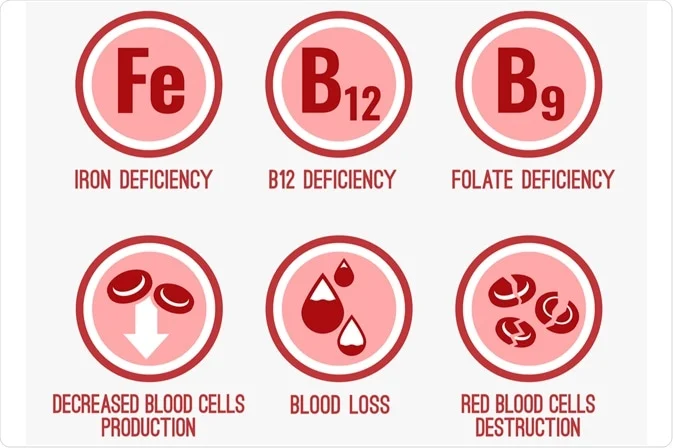
1. Nutritional causes

  • Iron deficiency
  • Vitamin B12 deficiency
  • Folate deficiency
  • Protein-energy malnutrition

2. Blood loss

  • Gastrointestinal bleeding (e.g., anal fissure, Meckel’s diverticulum
  • Birth trauma or internal hemorrhage
  • Iatrogenic blood loss (frequent phlebotomy in NICU)
  • Occult bleeding (cow’s milk protein-induced colitis)

3. Hemolytic causes

  • Hemolytic disease of the newborn (Rh or ABO incompatibility)
  • G6PD deficiency
  • Hereditary spherocytosis
  • Thalassemia
  • Sickle cell disease
  • Infections causing hemolysis (e.g., malaria, sepsis)

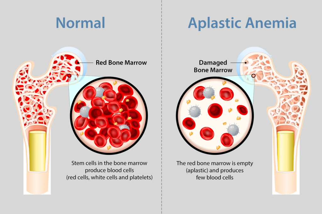
4. Bone marrow failure / decreased production

  • Aplastic anemia
  • Congenital pure red cell aplasia (Diamond–Blackfan anemia)
  • Transient erythroblastopenia of childhood
  • Bone marrow infiltration (leukemia, storage diseases)

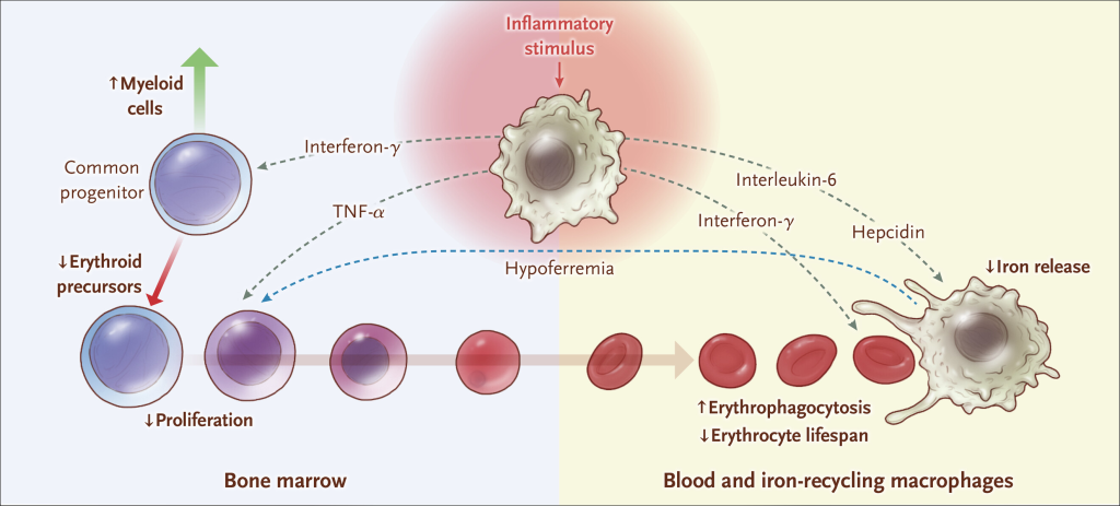
5. Chronic disease / inflammation

  • Anemia of chronic infection or inflammation
  • Renal failure (↓ erythropoietin)

6. Physiologic / other causes

  • Physiologic anemia of infancy
  • Prematurity (low iron stores, immature erythropoiesis)
  • Hypothyroidism
  • Chronic blood loss from parasites (hookworm in older infants)

How to store breast milk for future use?

1. Purpose

Proper storage of expressed breast milk (EBM) helps preserve its nutritional and immunological properties for later feeding when the mother is away.

2. Containers for Storage

  • Use clean, sterilized glass or BPA-free plastic containers with tight-fitting lids.
  • Breast milk storage bags (food-grade, pre-sterilized) are convenient for freezing.
  • Avoid ordinary plastic bottles or disposable liners.

3. Labeling

  • Each container should be labeled with:
  • Date and time of expression
  • Baby’s name (if used in hospital or daycare)

4. Storage Guidelines

LocationTemperatureDurationRemarks
Room temperatureUp to 25°C (77°F)4–6 hoursKeep in a cool, clean area away from direct sunlight
Refrigerator (back portion)2–4°C (35–40°F)Up to 72 hours (3 days)Do not store in refrigerator door due to temperature fluctuation
Freezer (separate door)–18°C or lowerUp to 3–6 monthsKeep in small portions; leave space for expansion
Deep freezer–20°CUp to 6–12 monthsBest for long-term storage
Insulated cooler box with ice packs~15°CUp to 24 hoursFor temporary transport or travel

5. Thawing and Warming

  • Thaw in refrigerator overnight or by placing the container in warm water (<37°C).
  • Do not microwave or boil — destroys antibodies and nutrients.
  • Swirl gently (do not shake) to mix separated fat layers.
  • Once thawed, use within 24 hours and do not refreeze.

6. Hygiene

  • Wash hands before expressing or handling milk.
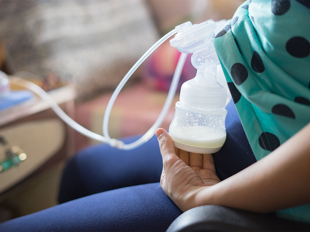
  • Use clean pump parts and containers for each session.
  • Avoid touching the inside of lids or bottles.

7. Key Points

  • Always use oldest milk first (“first in, first out”).
  • Discard leftover milk from feeding bottle after use.
  • Observe for odor or curdling — discard if spoiled.

Summary:

Safe breast milk storage requires hygienic handling, correct temperature, and appropriate duration. Properly stored milk retains most of its nutritional, immunological, and protective properties, ensuring safe feeding for infants when direct breastfeeding isn’t possible.

What causes Large and Thick placenta (placentomegaly)?

Placentomegaly — Causes

Table of Contents(toc)

Definition:

Placentomegaly refers to an abnormally thick or enlarged placenta, typically defined as:

  • >4 cm thick at 20 weeks gestation, or

  • >6 cm thick at term.

plaenta normal anterior image

I. Maternal Causes

  1. Diabetes mellitus (especially poorly controlled)

    → due to villous edema and increased fetal size.

  2. Maternal anemia (especially severe)

    → compensatory placental hypertrophy to improve oxygen transfer.

  3. Maternal infection

    • TORCH infections (Toxoplasmosis, Rubella, CMV, Herpes)

    • Syphilis, Malaria, Hepatitis, HIV

  4. Hypertension with superimposed infection or diabetes

  5. Rh isoimmunization (leading to fetal hydrops and placental edema)


II. Fetal Causes

  1. Fetal hydrops (immune or non-immune)

    • Most common fetal cause.

    • Due to excessive fluid accumulation → placental edema.

  2. Chromosomal abnormalities

    • Trisomy 13, 18, 21, Triploidy, Turner syndrome

  3. Fetal anemia (any cause, e.g., parvovirus B19 infection)

  4. Twin-to-twin transfusion syndrome (recipient twin side)

  5. Large-for-gestational-age (LGA) fetus

  • Often secondary to maternal diabetes.

a large placenta

III. Placental / Cord Causes

  1. Chorioangioma (benign vascular tumor of placenta)

  2. Molar pregnancy (partial mole)

  3. Chronic villitis or placentitis

  4. Placental edema due to venous obstruction (cord anomalies)


IV. Other / Miscellaneous Causes

  1. Congenital infections (CMV, syphilis, toxoplasmosis, parvovirus)

  2. Placental transfusion syndromes

  3. Maternal-fetal hemorrhage

  4. High altitude pregnancies (chronic hypoxia)


Mnemonic (for quick recall):

“BIG PLACENTA”

  • B – Beta-thalassemia / fetal anemia

  • I – Infections (TORCH, malaria, syphilis)

  • G – Gestational diabetes

  • P – Parvovirus / Polyhydramnios

  • L – Large baby (LGA)

  • A – Aneuploidy (Trisomy 13/18/21)

  • C – Chorioangioma

  • E – Erythroblastosis fetalis (Rh isoimmunization)

  • N – Nonimmune hydrops

  • T – Twin-to-twin transfusion

  • A – Anemia (maternal or fetal)

Key Takeaways:
  • An enlarged placenta isn’t usually a reason to panic. The word is big (placentomegaly), but most of the time it doesn’t cause problems.
  • Some conditions play a role. A larger placenta may be linked to hypertension, anemia, or diabetes — but your doctor will monitor these and the health of your baby.
  • Your baby’s growth matters most. Even if your placenta measures big, steady fetal development is the important goal, so keep up your regular appointments to be sure everything’s right on track.

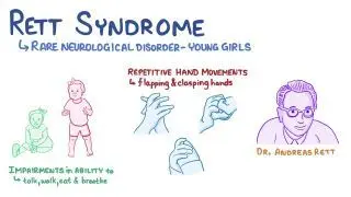
Rett Syndrome Notes for MD and DM level

Rett Syndrome

Definition

Rett syndrome is a neurodevelopmental disorder that primarily affects girls, characterized by normal early development followed by regression of acquired skills, especially speech and purposeful hand movements, with onset typically between 6–18 months of age.


Etiology

  • Genetic cause: Mutation in MECP2 gene (methyl-CpG-binding protein 2) on the X chromosome (Xq28)

  • Inheritance: Usually sporadic (de novo); rarely familial

  • Pathophysiology: Dysfunction of MECP2 protein → abnormal brain maturation and synaptic development


Epidemiology

  • Affects 1 in 10,000–15,000 female births

  • Lethal in males (most do not survive infancy unless mosaic or XXY)


Clinical Features

Phases of Disease

  1. Early Onset (6–18 months)

    • Normal development initially

    • Gradual loss of interest in surroundings

    • Loss of purposeful hand skills

    • Deceleration of head growth (acquired microcephaly)

  2. Rapid Destructive Phase (1–4 years)

    • Loss of speech and purposeful hand use

    • Stereotyped hand movements: hand-wringing, washing, clapping, or mouthing

    • Gait ataxia, truncal apraxia

    • Autistic-like behavior

  3. Plateau Phase (2–10 years)

    • Some improvement in social interaction and eye contact

    • Persistent motor problems and seizures

  4. Late Motor Deterioration (>10 years)

    • Progressive scoliosis, muscle wasting, rigidity, dystonia

    • Loss of ambulation in many cases


Other Features

  • Breathing abnormalities: hyperventilation, apnea during wakefulness

  • Seizures: common (up to 90%)

  • Bruxism, cold/purple extremities (autonomic dysfunction)

  • Sleep disturbances

  • Growth retardation


Investigations

  • Genetic testing: MECP2 mutation analysis (diagnostic)

  • EEG: slowing with epileptiform activity

  • MRI brain: may show nonspecific atrophy

  • Metabolic tests: normal (to rule out other causes)


Diagnosis

  • Clinical + confirmed MECP2 mutation

  • Diagnostic criteria include:

    • Regression after normal early development

    • Loss of purposeful hand skills and spoken language

    • Gait abnormalities

    • Stereotypic hand movements


Differential Diagnosis

  • Autism spectrum disorder

  • Angelman syndrome

  • Cerebral palsy (especially ataxic type)

  • Childhood disintegrative disorder


Management

  • No cure – supportive and multidisciplinary care

    • Physiotherapy & occupational therapy: maintain mobility

    • Speech therapy: communication support (eye-tracking devices)

    • Antiepileptics: for seizures

    • Nutritional support: adequate calories, manage feeding difficulties

    • Behavioral therapy: improve interaction

    • Orthopedic care: for scoliosis, contractures


Prognosis

  • Progressive but non-degenerative

  • Life expectancy: many survive into adulthood (40–50 years)

  • Main causes of death: sudden unexplained death, pneumonia, cardiac arrhythmias


Mnemonic (Key features) — “RETT”

  • R = Regression (speech, hand skills)

  • E = Episodic breathing abnormalities

  • T = Typical hand movements (wringing, washing)

  • T = Tiny head (acquired microcephaly)

Hypothyroidism in Neonates and Children

Table of Contents(toc)

1. Definition

Hypothyroidism is a clinical state resulting from deficiency of thyroid hormone production or action, leading to a generalized slowing of metabolic processes.

It may be:

  • Congenital (Neonatal) – present at birth.

  • Acquired (Childhood) – develops later due to autoimmune, iatrogenic, or other causes.


2. Classification

A. Based on Level of Defect

Type Site of Defect TSH T4/T3
Primary Thyroid gland ↑ ↓
Secondary Pituitary ↓/N ↓
Tertiary Hypothalamus ↓/N ↓
Peripheral (Resistance) Target tissue N/↑ N/↑

B. Based on Onset

  • Congenital hypothyroidism (CH)

  • Acquired hypothyroidism


3. Epidemiology

  • CH: ~1 in 2,000–4,000 live births.

  • More common in females.

  • Acquired form common in older children/adolescents, often autoimmune (Hashimoto’s).

thyroid gland


4. Etiology

A. Congenital Hypothyroidism

  1. Thyroid dysgenesis (80–85%)

    • Agenesis, ectopy, or hypoplasia.

    • Usually sporadic.

  2. Dyshormonogenesis (10–15%)

    • Inborn errors of thyroid hormone synthesis (autosomal recessive).

    • E.g. TPO, TG, Pendrin, NIS mutations.

  3. Central hypothyroidism (rare)

    • Pituitary/hypothalamic malformation, midline defects.

  4. Transient CH

    • Iodine excess/deficiency, maternal antibodies or antithyroid drugs.

  5. Thyroid hormone resistance – extremely rare.

B. Acquired Hypothyroidism

  • Autoimmune thyroiditis (Hashimoto’s) – most common.

  • Iodine deficiency/excess.

  • Post-irradiation or post-surgical.

  • Drugs: amiodarone, lithium, interferon-α.

  • Secondary causes: pituitary tumors, craniopharyngioma.


5. Pathophysiology

↓ Thyroid hormone → ↓ metabolic activity → impaired CNS myelination, growth retardation, delayed bone maturation.

  • In neonates: irreversible neurodevelopmental impairment if untreated.

  • In older children: growth failure and delayed puberty predominate.


6. Clinical Features

A. Neonatal / Congenital

Often asymptomatic at birth due to transplacental maternal T4.

Typical features (develop over weeks):

  • Prolonged jaundice

  • Lethargy, hypotonia

  • Feeding difficulty, constipation

  • Large fontanelles

  • Macroglossia

  • Umbilical hernia

  • Cold, dry skin

  • Hoarse cry

  • Poor growth

  • Delayed bone age

  • Delayed milestones (later)

B. Childhood / Acquired

  • Growth retardation, short stature

  • Weight gain with poor height velocity

  • Fatigue, cold intolerance

  • Constipation

  • Dry skin, coarse hair

  • Bradycardia

  • Delayed puberty / menstrual irregularities

  • Pseudoprecocious puberty (rare, due to high TRH → prolactin ↑)

  • Goiter (especially in Hashimoto’s)


7. Investigations

A. Screening

  • Neonatal screening: heel-prick sample at 48–72 hr.

    • Primary TSH (most programs).

    • Elevated TSH → confirm with serum free T4.

B. Diagnostic Tests

Test Interpretation
Serum TSH, Free T4 ↓T4 with ↑TSH → primary hypothyroidism
T3 less reliable in neonates
Thyroglobulin Low in agenesis, high in dyshormonogenesis
Imaging Thyroid scan (99mTc or I-123) – ectopy, agenesis, uptake defects
Ultrasound Gland location and size
Antibodies (TPO, Tg) Positive in autoimmune
Bone age X-ray Delayed
Additional: Pituitary MRI if central hypothyroidism suspected

8. Complications (if untreated)

  • Neurologic: irreversible intellectual disability, deaf-mutism, spasticity.

  • Growth: severe stunting, delayed bone age.

  • Metabolic: dyslipidemia.

  • Cardiac: bradycardia, pericardial effusion.


9. Management

A. Principles

  • Early, adequate, lifelong replacement.

  • Monitor and titrate carefully to maintain euthyroid state.

B. Drug

  • Levothyroxine (L-T4) – drug of choice.

    • Dose:

      • Neonates: 10–15 µg/kg/day.

      • Infants: 8–10 µg/kg/day.

      • Children: 4–6 µg/kg/day.

      • Adolescents: 2–4 µg/kg/day.

    • Given on empty stomach (preferably crushed with water or milk).

C. Monitoring

Age Frequency Parameters
0–6 mo Every 2 wk till T4 normal, then q1–2 mo T4, TSH
6–12 mo q2–3 mo 〃
1–3 yr q3–4 mo 〃
>3 yr q6–12 mo 〃

Target: Free T4 in upper half of normal range, TSH normal.

D. Developmental Follow-up

  • Neurodevelopmental assessment

  • Hearing evaluation

  • Growth chart monitoring


10. Prognosis

  • Normal IQ if therapy started within first 2 weeks of life.

  • Delay in treatment → irreversible intellectual deficit.

  • Acquired forms usually fully reversible with treatment.


11. Key Differentials

  • Pituitary insufficiency

  • Hypothyroxinemia of prematurity

  • Chronic systemic illness (euthyroid sick syndrome)

  • Constitutional growth delay


12. Summary Table

Feature Congenital Acquired
Onset Birth Childhood/adolescence
Cause Dysgenesis > dyshormonogenesis Hashimoto’s most common
Presentation Lethargy, constipation, macroglossia Growth failure, delayed puberty
TSH High High
T4 Low Low
Rx Levothyroxine Levothyroxine
Prognosis Excellent if early Excellent

References

  1. Nelson Textbook of Pediatrics, 22nd ed.

  2. Indian Academy of Pediatrics Guidelines (2021) — Screening and management of congenital hypothyroidism.

  3. Endocrine Society Clinical Practice Guideline (2020) – Congenital Hypothyroidism.

  4. Sperling MA, Pediatric Endocrinology, 5th ed.

Exit mobile version