Blog

What does Unequal blood pressure in the left and the right upper arms mean?

Unequal blood pressure in the left and right upper arms: Review

Table of Contents(toc)

Unequal blood pressure in the left and right upper arms can be caused by several underlying conditions. The difference is typically considered significant if it is greater than 10–15 mmHg. Common causes include:

1. Arterial Conditions:

Atherosclerosis: Plaque buildup in the arteries can lead to narrowing or obstruction, causing unequal blood flow and pressure.
Subclavian Artery Stenosis: Narrowing of the subclavian artery, usually on one side, leads to reduced blood pressure in the affected arm.
Aortic Dissection: A tear in the wall of the aorta can create a false channel, disrupting blood flow and causing pressure differences.

2. Anatomical Abnormalities:

Coarctation of the Aorta: A congenital condition causing narrowing of the aorta, which can lead to pressure differences.
Thoracic Outlet Syndrome: Compression of blood vessels (or nerves) as they pass through the thoracic outlet can affect blood pressure.

3. Inflammatory or Autoimmune Diseases:

Takayasu Arteritis: An inflammatory disease that can cause narrowing of large arteries, including the subclavian arteries.
Giant Cell Arteritis: Inflammation of blood vessels, typically in older adults, can affect blood flow.

4. Peripheral Vascular Disease (PVD):

Obstruction or narrowing of peripheral arteries can lead to unequal pressures.

5. Neurological or Vascular Compression:

Conditions like subclavian steal syndrome, where blood is “stolen” from the brain to supply the arm due to an obstruction, can cause differences.

6. Other Causes:

Blood Clots: In the subclavian or brachial arteries can reduce blood pressure in one arm.
Measurement Error: Incorrect cuff size, positioning, or technique can falsely create a pressure difference.

When to Seek Medical Attention:

If the difference in systolic blood pressure is greater than 20 mmHg, or if you have symptoms like dizziness, arm pain, or fainting, it’s essential to consult a healthcare provider. This may require further investigation using imaging techniques like Doppler ultrasound, CT angiography, or MRI.
Early detection and management of the underlying cause are crucial to preventing complications such as stroke or cardiovascular events.
Source: Uptodate , Medscape

MBBS third year medicine MCQs AIIMS (old Questions)

MBBS third year medicine AIIMS Multiple Choice Questions (MCQs)


Table of Contents(toc)

Q1. A patient who is being screened for vitamin B-12 deficiency undergoes a Schilling test. In this test, 1 mg of cobalamin is administered intramuscularly 1 hour after the administration of radiolabeled cobalamin. A normal Schilling test (excretion of 10% of the labeled cobalamin in a 24-hour urine specimen) would most likely be seen in which of the following situations?

  • (a) Intrinsic factor deficiency
  • (b) Chronic pancreatitis
  • (c) Surgically absent terminal ileum
  • (d) Celiac sprue

Q2. HIV infects cells bearing the CD4 receptor. Which co-receptor is required for HIV to enter the cell?

  • (a) CD5
  • (b) Chemokine receptor 5 (CCR5)
  • (c) Major histocompatibility complex receptor (MHC)
  • (d) CD3

Q3. Which of the following is a symptom of acute kidney injury?

  • (a) Increased urine output
  • (b) Decreased urine output
  • (c) Hypertension
  • (d) Proteinuria

Q4. Weight loss and malabsorption are commonly seen as features of which of the following conditions?

  • (a) Lactose intolerance
  • (b) Pernicious anemia
  • (c) Lymphocytic colitis
  • (d) Small bowel bacterial overgrowth

Q5. Dengue hemorrhagic fever is considered if all the following are present except:

  • (a) Systolic blood pressure < 90 mm Hg
  • (b) Fever (acute onset of 2–7 days)
  • (c) Hemorrhagic manifestations
  • (d) Platelet count ≤ 100,000/cu.mm

Q6. An 80-year-old male complains of a 3-day history of a painful rash extending over the left half of his forehead and down to his left eyelid. Physical examination reveals weeping vesicular lesions. What is the most likely diagnosis?

  • (a) Impetigo
  • (b) Adult chickenpox
  • (c) Shingles
  • (d) Herpes simplex

Q7. A 25-year-old patient presents with flank pain and fever. What is the most likely diagnosis?

  • (a) Acute pyelonephritis
  • (b) Renal tuberculosis
  • (c) Transitional cell carcinoma
  • (d) Pelvic inflammatory disease

Q8. According to Truelove and Witts’ classification, which of the following suggests severe ulcerative colitis?

  • (a) >10 bloody stools/day
  • (b) Pulse 84/min
  • (c) Hemoglobin 10.6 g/dL
  • (d) ESR 19 mm in 1 hour

Q9. Regarding pleural effusions, which of the following statements is true?

  • (a) A 500 mL pleural effusion is generally easy to detect
  • (b) A protein level of <30 g/L suggests an exudate
  • (c) In infections, a pleural fluid pH of 7.2 suggests the need for drainage
  • (d) Pleural effusions occur more commonly on the left

Q10. Which of the following is not a laboratory indicator of iron deficiency anemia?

  • (a) Microcytic red blood cells
  • (b) A low serum ferritin
  • (c) A low serum TIBC
  • (d) A low serum iron

Q11. Which of the following is a cause of bilateral spastic paraparesis?

  • (a) Vitamin B12 deficiency
  • (b) Cerebellar disease
  • (c) Peripheral neuropathy
  • (d) Stroke in the internal capsule

Q12. Which one of the following statements about paracetamol poisoning is not true?

  • (a) Fifteen tablets may be a fatal overdose
  • (b) Oral methionine may be a useful treatment
  • (c) Paracetamol levels should be checked 4 hours post-ingestion
  • (d) If liver function tests are normal at 4 hours post-ingestion, the liver has not been damaged

Q13. Hyponatremia is defined as a plasma sodium concentration of:

  • (a) <115 mmol/L
  • (b) <125 mmol/L
  • (c) <135 mmol/L
  • (d) <145 mmol/L

Q14. Which of the following is a cause of direct hyperbilirubinemia?

  • (a) Crigler-Najjar syndrome
  • (b) Gilbert syndrome
  • (c) G6PD deficiency
  • (d) Dubin-Johnson syndrome

Q15. Management of delirium includes:

  • (a) Neuroleptics
  • (b) Atypical neuroleptics
  • (c) Both can be used
  • (d) None of the above

Q16. Regarding the SPIKES protocol, which of the following is correct?

  • (a) It includes elements of breaking bad news
  • (b) It involves mentally preparing to interact with the patient and family
  • (c) It emphasizes providing information sensitively
  • (d) All of the above

Q17. Evidence-based medicine involves:

  • (a) Formulating the management question
  • (b) Searching literature and databases for research
  • (c) Applying gathered knowledge for the best possible outcome
  • (d) All of the above

Q18. Regarding the patient-physician relationship, which statement is true?

  • (a) Patients are individuals with problems that present as physical complaints
  • (b) Most patients are anxious and fearful
  • (c) A professional attitude with warmth and openness helps alleviate patient anxiety
  • (d) All of the above

Q19. Which of the following diseases is associated with gross hematuria?

  • (a) Subacute bacterial endocarditis
  • (b) Cystic kidney disease
  • (c) Mesangioproliferative glomerulonephritis
  • (d) IgA nephropathy

Q20. Which of the following is an appetite-regulating hormone?

  • (a) Amylin
  • (b) Ghrelin
  • (c) GLP-1
  • (d) Secretin

Here are the answer keys for the multiple-choice questions:

Answer Key:

  1. (b) Chronic pancreatitis
  2. (b) Chemokine receptor 5 (CCR5)
  3. (b) Decreased urine output
  4. (d) Small bowel bacterial overgrowth
  5. (a) Systolic Blood Pressure < 90 mm Hg
  6. (c) Shingles
  7. (a) Acute pyelonephritis
  8. (a) >10 bloody stools/day
  9. (c) In infections, a pleural fluid pH of 7.2 suggests the need for drainage
  10. (c) A low serum TIBC
  11. (d) Stroke in the internal capsule
  12. (d) If liver function tests are normal at 4 hours post-ingestion, the liver has not been damaged
  13. (c) <135 mmol/L
  14. (d) Dubin-Johnson syndrome
  15. (c) Both can be used
  16. (d) All of the above
  17. (d) All of the above
  18. (d) All of the above
  19. (d) IgA nephropathy
  20. (b) Ghrelin

Orthognathic Surgery: A Guide to Jaw Realignment and Recovery

Orthognathic Surgery: A Guide to Jaw Realignment and Recovery

Table of Contents(toc)

Orthognathic surgery, commonly known as jaw surgery, is a corrective procedure that addresses misalignment of the jaws and teeth. This surgical intervention is essential for patients with significant jaw irregularities that affect function, facial balance, and overall health. It is typically performed by an oral and maxillofacial surgeon in collaboration with an orthodontist.

For many patients, orthognathic surgery improves not only their facial aesthetics but also essential functions like chewing, speaking, and breathing. It is often recommended for individuals with severe malocclusions (bite issues) that cannot be corrected with orthodontics alone. The procedure involves precise planning, surgical repositioning of the jawbones, and a structured recovery period to ensure long-term stability and success.

Why is Orthognathic Surgery Needed?

Orthognathic surgery is performed for both medical and aesthetic reasons, including:

  • Correction of Jaw Misalignment: Treats conditions such as underbite, overbite, crossbite, and open bite.
  • Ensures proper alignment between the upper and lower jaws for improved function.
  • Facial Balance and Aesthetics: Helps create a symmetrical, proportionate facial appearance.
  • Addresses congenital or developmental jaw discrepancies.
  • Improved Chewing and Speech Function: Enhances bite efficiency, reducing strain on the teeth and jaw muscles.
  • Corrects speech difficulties caused by improper jaw positioning.
  • Treatment of Obstructive Sleep Apnea (OSA): Expands the airway to reduce breathing difficulties during sleep.
  • Helps patients who do not respond well to CPAP therapy.
  • Jaw Growth Abnormalities or Trauma Repair:
  • Corrects congenital jaw deformities or injuries that affect jaw function.

Types of Orthognathic Surgery

The type of jaw surgery depends on the specific issue being addressed:
1. Maxillary Osteotomy (Upper Jaw Surgery)

  • Performed on the maxilla (upper jaw) to correct:
  • Overgrowth or underdevelopment of the upper jaw.
  • Open bite, where the front teeth do not touch when the mouth is closed.
  • Crossbite, where the upper teeth sit inside the lower teeth.
  • Midface deficiencies that affect facial harmony.

2. Mandibular Osteotomy (Lower Jaw Surgery)

  • Performed on the mandible (lower jaw) to address:
  • Underbites, where the lower jaw protrudes beyond the upper jaw.
  • Overbites, where the lower jaw is too far back.
  • Jaw asymmetry that affects function and appearance.

3. Bimaxillary Osteotomy (Double Jaw Surgery)

  • Involves repositioning both the upper and lower jaws for:
  • Severe misalignment that affects facial balance.
  • Significant functional issues, such as difficulty chewing or breathing.
  • Cases where a single jaw surgery would not provide sufficient correction.

4. Genioplasty (Chin Surgery)

  • Involves reshaping or repositioning the chin to:
  • Improve facial symmetry and balance.
  • Correct a receding or overly prominent chin.
  • Enhance the results of other jaw surgeries.

The Surgical Process: What to Expect

1. Pre-Surgical Preparation

Orthodontic Treatment:

  • Most patients wear braces for 12-18 months before surgery to align their teeth properly.Imaging and Planning:
  • X-rays, 3D imaging, and digital models help guide precise surgical adjustments.Pre-Operative Evaluations:
  • Medical assessments ensure patients are fit for surgery.

2. The Surgery

Procedure Duration:

  • Performed under general anesthesia, typically lasting 3–6 hours, depending on complexity.

Surgical Technique:

  • Incisions are made inside the mouth to access and reposition the jawbones.
  • Bones are secured using titanium plates and screws for stability.

Hospital Stay:

  • Most patients stay in the hospital for 1–2 days post-surgery.

3. Post-Surgical Recovery

Initial Recovery (First Few Weeks):

  • Swelling, discomfort, and restricted jaw movement are common.
  • Liquid or soft-food diet is necessary until the jaw heals.
  • Intermediate Healing (6–12 Weeks):
  • Swelling gradually decreases.
  • Patients transition back to a normal diet under medical guidance.

Long-Term Recovery (Up to 12 Months):

  • Full bone healing and nerve recovery continue over several months.
  • Orthodontic treatment (braces) is often continued for 6–12 months post-surgery.

Benefits of Orthognathic Surgery

  • Improved jaw function, making it easier to chew, speak, and breathe.
  • Enhanced facial symmetry and aesthetics.
  • Reduction in jaw pain and TMJ (temporomandibular joint) issues.
  • Better airway function, reducing symptoms of sleep apnea.
  • Long-term dental health by preventing excessive tooth wear and bite strain.

Risks and Considerations

  • Swelling, bruising, and temporary discomfort are common post-surgery.
  • Risk of infection, bleeding, or delayed bone healing.
  • Temporary or permanent numbness due to nerve involvement.
  • In rare cases, additional procedures may be needed to refine jaw alignment.

Final Thoughts

Orthognathic surgery is a highly effective treatment for individuals with severe jaw misalignment and functional issues. While the process requires a commitment to preparation and recovery, the long-term benefits—including improved function, aesthetics, and overall well-being—make it a life-changing procedure for many patients.

If you’re considering jaw surgery, consult an experienced oral and maxillofacial surgeon to evaluate your specific needs and develop a personalized treatment plan.


Myocardial Infarction (MI): Causes, Symptoms, and Management

Myocardial Infarction (MI): Causes, Symptoms, and Management

Table of Contents(toc)

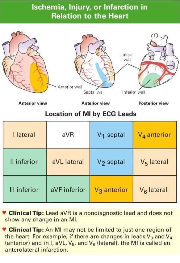
What is Myocardial Infarction?

Myocardial infarction (MI), commonly known as a heart attack, occurs
when blood flow to a part of the heart muscle is blocked. (Source)This is usually caused by a blood clot or atherosclerosis, leading to oxygen
deprivation and subsequent damage or necrosis of heart muscle tissue.

Symptoms of Myocardial Infarction

Symptoms of MI can vary but often include:

  • Chest
    Pain or Discomfort
  • Pain Radiating to Other Areas: Arms
    (especially the left arm), shoulders, neck, jaw, or back
  • Shortness of Breath
  • Nausea, Vomiting, or Abdominal Pain
  • Cold Sweats
  • Dizziness or Lightheadedness
  • Fatigue: More common in females

Management of Myocardial Infarction

Emergency Management (Acute Phase)

  • Immediate medical intervention is crucial to minimize heart muscle
    damage.
  • Aspirin (160–325 mg): Should be chewed unless
    contraindicated.
  • Oxygen Therapy: Administered if SpOâ‚‚ is below
    90%.
  • Sublingual Nitroglycerin (0.3–0.6 mg): Given unless
    contraindicated, such as in hypotension.
  • Morphine: Used for
    severe chest pain that does not respond to nitroglycerin.
  • ECG
    Monitoring: Essential for confirming diagnosis and identifying complications.

Hospital Management

  • Treatment focuses on restoring blood flow and preventing further
    complications.
  • Thrombolytic Therapy (Alteplase or Tenecteplase):
    Used when percutaneous coronary intervention (PCI) cannot be performed within 90
    minutes.
  • Percutaneous Coronary Intervention (PCI): The standard
    procedure for restoring blood flow.
  • Antiplatelet Therapy:
    Includes aspirin and P2Y12 inhibitors (Clopidogrel, Ticagrelor).
  • Anticoagulants: Heparin or low molecular weight heparin (e.g., Enoxaparin) is
    administered.
  • Beta-Blockers: Medications such as Metoprolol
    succinate, Carvedilol, or Bisoprolol help reduce heart workload.
  • ACE Inhibitors: Drugs like Lisinopril, Captopril, or Ramipril lower blood
    pressure and reduce strain on the heart.
  • ARBs (Angiotensin II
    Receptor Blockers): Used as an alternative to ACE inhibitors, including
    Losartan, Candesartan, Valsartan, and Telmisartan.
  • Statins
    (High-Intensity Therapy): Atorvastatin (40–80 mg) is recommended to lower
    cholesterol and prevent further blockages.

Long-Term Management (Post-MI Care)

Ongoing treatment helps prevent future heart attacks and supports
overall cardiovascular health.

Medications:

  • Antiplatelet Therapy: Dual antiplatelet
    therapy (DAPT) for 12 months.
  • Beta-Blockers
  • ACE
    Inhibitors or ARBs
  • Statins
  • Nitrates: Used for
    angina management.

Lifestyle Modifications:

  • Quit Smoking: Eliminating
    tobacco reduces heart disease risk.
  • Healthy Diet: Emphasizes
    fruits, vegetables, whole grains, and limits saturated fats, sodium, and sugar.
  • Regular Exercise: At least 30 minutes of physical activity, five
    days a week.
  • Weight Management: Maintaining a healthy body
    weight is essential.
  • Control of Chronic Conditions: Proper
    management of diabetes, hypertension, and cholesterol levels.

Cardiac Rehabilitation:

  • A structured rehabilitation program includes:
  • Supervised exercise programs.
  • Educational resources on heart
    health.
  • Psychological counseling for emotional well-being.

Psychological Support:

Addressing anxiety and
depression associated with MI recovery.

Monitoring and Follow-Up

  • Regular check-ups with a cardiologist are necessary to track
    progress and prevent complications.
  • Monitoring Vital Signs:
    Blood pressure, cholesterol, and blood glucose levels should be regularly
    assessed.
  • Routine ECGs and Echocardiograms: Used to evaluate
    heart function.

Prevention of Myocardial Infarction

  • Reducing risk factors plays a crucial role in preventing heart
    attacks.
  • Control of Hypertension, Diabetes, and High Cholesterol
  • Smoking Cessation
  • Limiting Alcohol Consumption
  • Stress Management: Includes relaxation techniques and
    psychotherapy.
  • Adherence to Medications: Ensuring compliance
    with prescribed treatments.

Potential Complications of Myocardial Infarction

  • If not properly managed, MI can lead to severe complications,
    including:
  • Arrhythmias (Irregular Heartbeat)
  • Heart Failure
  • Cardiogenic Shock
  • Pericarditis
    (Inflammation of the Heart’s Outer Layer)
  • Recurrent Myocardial
    Infarctions

Conclusion

Myocardial infarction is a medical emergency that requires immediate
and long-term management to reduce damage and prevent recurrence. Proper
treatment, lifestyle changes, and regular follow-ups significantly improve
survival and quality of life.

Congenital Glaucoma MCQs

Congenital Glaucoma MCQs 2025

Table of Contents(toc)


1. What is the most common cause of congenital glaucoma?


2. Which of the following is NOT a typical clinical feature of congenital glaucoma?


3. What is the first-line treatment for primary congenital glaucoma?


4. Which of the following findings is most suggestive of congenital glaucoma on slit-lamp examination?


5. At what intraocular pressure (IOP) is congenital glaucoma typically suspected in infants?


.question { margin-bottom: 20px; padding: 10px; border: 1px solid #ddd; border-radius: 5px; }
.options button { display: block; width: 100%; margin: 5px 0; padding: 10px; border: none; cursor: pointer; background-color: #f4f4f4; text-align: left; border-radius: 5px; }
.options button.correct { background-color: #c8e6c9; }
.options button.incorrect { background-color: #ffcccb; }
.explanation { display: none; margin-top: 10px; padding: 10px; background-color: #eef; border-left: 5px solid #007bff; border-radius: 5px; }

function checkAnswer(button, isCorrect, explanation) {
let optionsDiv = button.parentElement;
let buttons = optionsDiv.getElementsByTagName(“button”);
for (let btn of buttons) { btn.disabled = true; }
if (isCorrect) { button.classList.add(“correct”); }
else { button.classList.add(“incorrect”); }
let explanationPara = optionsDiv.nextElementSibling;
explanationPara.innerHTML = “Explanation: ” + explanation;
explanationPara.style.display = “block”;
}

MCQs based on hernias

MCQs based on hernias with explanation and answer

Table of contents (toc)


Here are some MCQs based on hernias:

1. Which is the most common type of hernia?

a) Femoral hernia
b) Umbilical hernia
c) Inguinal hernia
d) Epigastric hernia
Ans: c) Inguinal hernia

2. Which type of hernia is most common in females?

a) Femoral hernia
b) Umbilical hernia
c) Inguinal hernia
d) Obturator hernia
Ans: c) Inguinal hernia

3. A hernia that contains a portion of the intestine’s circumference is called?

a) Pantaloon hernia
b) Richter’s hernia
c) Northan’s hernia
d) Littre’s hernia
Ans: b) Richter’s hernia

4. Which hernia contains Meckel’s diverticulum?

a) Richter’s hernia
b) Littre’s hernia
c) Pantaloon hernia
d) Spigelian hernia
Ans: b) Littre’s hernia

5. A hernia that involves both direct and indirect hernias is called?

a) Richter’s hernia
b) Littre’s hernia
c) Pantaloon hernia
d) Amyand’s hernia
Ans: c) Pantaloon hernia

6. Which type of hernia occurs in the pouch of Douglas?

a) Richter’s hernia
b) Berger’s hernia
c) Spigelian hernia
d) Obturator hernia
Ans: b) Berger’s hernia

7. Hernia containing portion of intestine

a. Pantaloon hernia
b. Richter’s hernia
c. Northan’s hernia
d. Litter’s hernia
Ans: b. Richter’s hernia
Please refer to our lecture PowerPoint and video for details. 
Would you like to practice more questions? Contact us for more paid questions. 

Why is There a Measles Outbreak in Texas in 2025?

Why is There a Measles Outbreak in Texas?

Table of Contents(toc)


Measles is making a comeback in the United States, despite being rich and healthy country in the world and Texas is one of the states seeing a rise in cases. There are various factors causing this and its complex yet important to understand. 

We reviewed multiple sources and news agency along with health and givernment sources and came with explanations. 

Let’s break down why this is happening despite such precautions and what it means for public health.

1. Low Vaccination Rates

  • The CDC reports that vaccination rates have dropped in certain areas of the U.S., particularly in communities with vaccine hesitancy.
  • Texas has seen pockets of low vaccination rates due to misinformation, distrust in vaccines, and political factors.
  • Measles is highly contagious, and when vaccination rates fall below 95%, the disease can spread rapidly within a community.
  • The CDC recommends the MMR (measles, mumps, and rubella) vaccine at 1 year and again at 4-6 years old for maximum protection.

2. Measles is Highly Contagious

  • Measles is one of the most contagious diseases, with a basic reproduction number (R0) between 12-18, which means one person can infect up to 18 others.
  • It spreads through respiratory droplets when an infected person coughs, sneezes, or even talks.
  • The virus can remain in the air or on surfaces for up to 2 hours, making it highly transmissible.
  • A person with measles is contagious from 4 days before to 4 days after the rash appears.

3. Lack of Herd Immunity

  • Herd immunity occurs when a sufficient percentage of the population is immune to a disease (either through vaccination or previous infection), preventing its spread.
  • With vaccination rates in Texas dipping, herd immunity has been compromised, making it easier for the virus to spread.
  • Children, the elderly, pregnant women, and those with weakened immune systems are most at risk when herd immunity is inadequate.

4. International Travel and Imported Cases

  • The WHO has warned that global travel can increase the risk of importing diseases like measles.
  • Outbreaks in countries with lower vaccination coverage, such as parts of Europe and Africa, can lead to cases arriving in the U.S.
  • Once imported, the disease can spread if local vaccination rates are low and individuals are unprotected.

5. Outbreaks in Schools and Communities

  • Children are especially vulnerable to measles, and outbreaks often start in schools where students are in close contact.
  • Texas has seen clusters of measles cases tied to schools and daycare centers.
  • The virus can easily spread in closed environments, especially among children who have not received the MMR vaccine.
  • Some schools in Texas have lower-than-expected vaccination rates, which increases the risk of outbreaks.

6. The Role of Misinformation

  • Misinformation surrounding vaccines is a significant factor in declining vaccination rates.
  • Despite multiple studies debunking the myth that the MMR vaccine causes autism, this false claim still contributes to vaccine hesitancy.
  • Social media and anti-vaccine groups have amplified these fears, influencing parents’ decisions to delay or refuse vaccination.

7. Vaccine Effectiveness and Disease Spread

  • The MMR vaccine is highly effective, with an efficacy rate of around 97% after two doses.
  • However, even small reductions in vaccination coverage can lead to outbreaks. Measles can spread rapidly in under-vaccinated communities, as seen in recent Texas outbreaks.
  • According to the CDC, outbreaks of measles are often a wake-up call that vaccination levels need to be improved.

8. Measles Symptoms and Complications

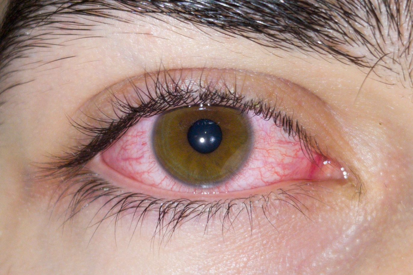
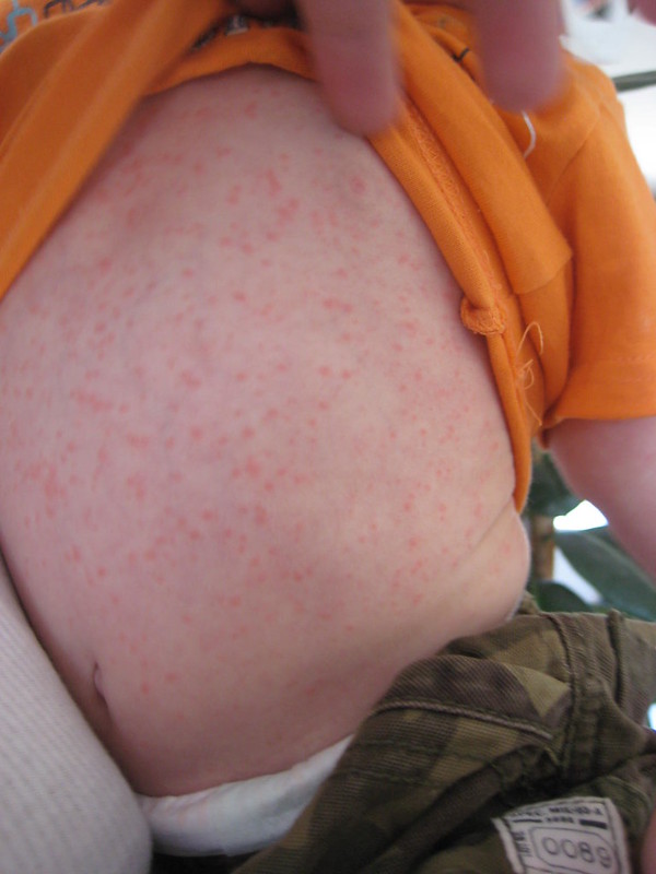
  • Measles starts with high fever, cough, runny nose, and red eyes, followed by a characteristic red, blotchy rash.
  • Complications from measles can be severe, including pneumonia, encephalitis (swelling of the brain), and even death.
  • Children under 5 years old, pregnant women, and individuals with weakened immune systems are particularly vulnerable to these complications.

9. Why Texas?

  • Texas has faced challenges in vaccine accessibility and education, especially in rural and underserved areas.
  • In some regions, the perception that vaccine-preventable diseases are no longer a threat has contributed to lower immunization rates.
  • Texas has also been a focus of outbreaks due to its large population, diverse communities, and geographic proximity to other countries.

What’s Being Done?

  • Health authorities are intensifying efforts to increase vaccination rates through public awareness campaigns, especially in areas with low coverage.
  • The Texas Department of State Health Services (DSHS) is working closely with local health departments to monitor and control outbreaks.
  • Local health officials are urging parents to ensure their children are up-to-date with their vaccinations, including the MMR shot.
  • The CDC continues to provide guidance on outbreak control measures, including quarantine recommendations for exposed individuals.

Conclusion

Measles outbreaks in Texas are a result of a combination of factors, including low vaccination rates, misinformation, and the highly contagious nature of the disease. Public health experts, including those from the CDC, WHO, and local health departments, emphasize that vaccination is the best way to protect children and communities from measles. With measles outbreaks on the rise, it’s more important than ever for parents to ensure their children are vaccinated.

Protect your family. Protect your community. Vaccinate against measles.

For more information on measles, refer to the CDC, WHO, Medscape, and UpToDate for comprehensive guidance on prevention and control.

Why is texas good or bad for nursing

Can Cancer be diagnosed by urine test? If yes how?

Cancer Cells May Be Found in Urine?

Table of Contents(toc)


Cancer cells can sometimes be seen in urine, depending on the type and location of the cancer. This is particularly true for cancers affecting the urinary tract. The detection of cancer cells in urine is typically done through a urine cytology test, where a sample of urine is examined under a microscope.

Types of Cancers Where Cancer Cells May Be Found in Urine:

  1. Bladder Cancer: One of the most common cancers detected through urine cytology.
  2. Ureteral and Renal Pelvis Cancer: Cancer of the upper urinary tract may shed cells into the urine.
  3. Prostate Cancer: In rare cases, prostate cancer cells may be found in urine, particularly after a prostate massage.
  4. Kidney Cancer: Uncommon, but cells from advanced renal cancers may occasionally appear in urine.
  5. Urethral Cancer: Cancer of the urethra may shed cancer cells into urine.

Urine Cytology Test:

  • Used to identify abnormal or cancerous cells in urine.
  • Best for detecting high-grade cancers like urothelial carcinoma.
  • May not be as sensitive for detecting low-grade cancers.

Limitations:

  • Not all cancers shed cells into the urine, so a negative urine cytology does not rule out cancer.
  • If cancer is suspected but urine cytology is negative, further tests like cystoscopy, biopsy, or imaging studies (e.g., CT or MRI) may be required.

Cytocentrifugation and staining are commonly used techniques to prepare urine samples for cytological examination to detect cancer cells. These methods enhance the visualization of cells and improve the accuracy of detecting abnormal or cancerous cells in urine. Here’s how it works:

Cytocentrifugation:

  • Purpose: Concentrates cells from a urine sample onto a single slide, making it easier to examine under a microscope.
  • Process:
    1. The urine sample is centrifuged to collect cells.
    2. The collected cells are deposited onto a glass slide using a cytocentrifuge.
    3. The slide is then air-dried or fixed for staining.
  • Benefit: Reduces background debris and improves the clarity of cellular features.

Staining:

  • After cytocentrifugation, the prepared slide is stained to highlight the morphology of cells.
  • Common staining techniques include:
    1. Papanicolaou (Pap) Stain:
      • Most commonly used for urine cytology.
      • Highlights nuclear and cytoplasmic details to identify malignant changes.
    2. Hematoxylin and Eosin (H&E):
      • Provides excellent contrast for cellular structures.
    3. Special Stains:
      • Immunohistochemistry (IHC) may be added for specific cancer markers (e.g., p53, Ki-67) if needed.
    4. Romanowsky Stains (e.g., Giemsa or Wright’s stain):
      • Occasionally used for detecting inflammatory or atypical cells.

Diagnostic Utility:

  • These techniques are particularly effective for detecting urothelial carcinoma, the most common cancer of the urinary tract.
  • Cytocentrifugation and staining improve sensitivity in detecting abnormal cells, especially in early or high-grade cancers.
  • If the results are unclear or suspicious, further diagnostic procedures, such as cystoscopy or biopsy, may be required.

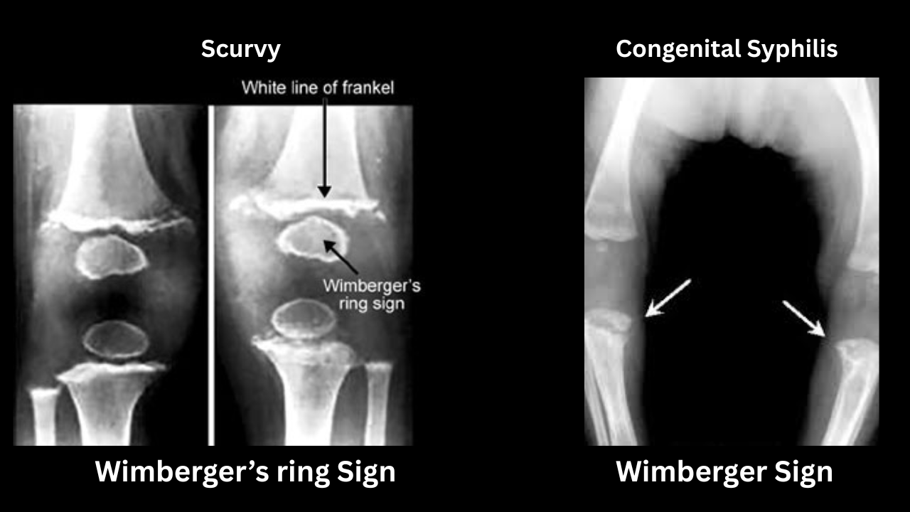
Wimberger’s Sign vs Wimberger Ring Sign A comparison

Wimberger’s Sign

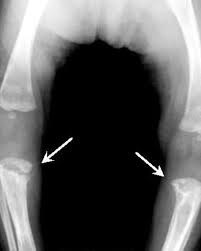
Wimberger’s sign refers to localized bone destruction or demineralization of the medial aspect of the proximal tibial metaphysis. It is often considered a radiological finding suggestive of congenital syphilis in infants. This condition occurs due to the involvement of rapidly growing bones in syphilitic periostitis or osteitis.

Key Points:

  • Cause: Congenital syphilis (usually secondary to spirochetal infection transmitted from the mother to the fetus).
  • Appearance: Bilateral, symmetrical defects in the medial portion of the proximal tibial metaphysis.
  • Differential Diagnosis: Other conditions, such as rickets or scurvy, may cause bone deformities, but the symmetry and specific localization are more characteristic of congenital syphilis.

Wimberger Ring Sign

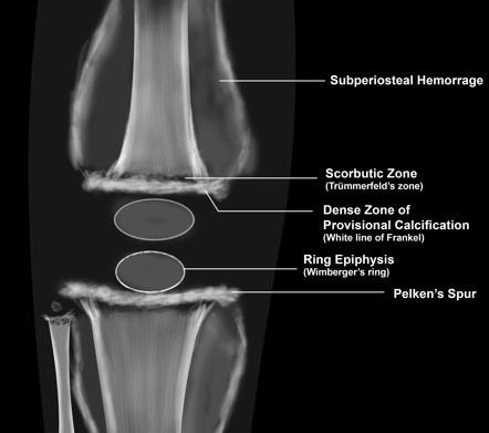
The Wimberger ring sign refers to a radiological finding characterized by a dense, sclerotic ring around the epiphysis of long bones, typically seen in cases of scurvy. This condition occurs due to subperiosteal hemorrhages and impaired collagen synthesis caused by vitamin C deficiency.

Key Features of the Wimberger Ring Sign:

  1. Appearance: A sharply defined, dense, sclerotic ring surrounding the epiphysis.
  2. Cause: Reflects disorganized bone growth due to vitamin C deficiency.
  3. Associated Findings in Scurvy:
    • Subperiosteal hemorrhages.
    • Fragility fractures.
    • “Corner sign” (a small metaphyseal fracture).
    • “Trümmerfeld zone” or scorbutic zone (an area of lucency in the metaphysis).
    • Generalized osteopenia.

Clinical Context:

  • Seen in: Malnourished children or adults with prolonged vitamin C deficiency.
  • Symptoms: Bone pain, weakness, irritability, bleeding gums, and anemia.

Comparison Between Wimberger’s Sign and Wimberger Ring Sign

FeatureWimberger’s Sign (Congenital Syphilis)Wimberger Ring Sign (Scurvy)
DefinitionDestruction or demineralization of the medial proximal tibial metaphysisSclerotic ring surrounding the epiphysis of long bones
CauseCongenital syphilis (due to Treponema pallidum infection)Scurvy (Vitamin C deficiency)
PathophysiologyInflammatory osteitis and periostitis caused by congenital syphilisImpaired collagen synthesis leading to defective bone formation and subperiosteal hemorrhage
Radiological AppearanceBilateral, symmetrical bony defects in the medial proximal tibial metaphysisDense white ring around the epiphysis due to disorganized bone deposition
Commonly Affected BonesProximal tibia (pathognomonic), but also femur and other long bonesLong bones, especially around the knees, wrists, and ankles
Associated Radiological SignsPeriostitis, saber shins, metaphyseal destructionSubperiosteal hemorrhages, “Trümmerfeld zone” (lucent metaphysis), corner sign
Clinical SymptomsPseudoparalysis (Parrot’s pseudoparalysis), bone pain, fever, irritabilityBone pain, swollen joints, bleeding gums, anemia, irritability
Age Group AffectedInfants (congenital)Infants and adults with severe vitamin C deficiency
TreatmentPenicillin (antibiotics for syphilis)Vitamin C supplementation

Key Differences:

  • Wimberger’s Sign is a destructive lesion in congenital syphilis, whereas Wimberger Ring Sign is a sclerotic epiphyseal ring due to scurvy.
  • Syphilis affects metaphyses, while scurvy primarily affects the epiphysis.
  • Syphilis is an infectious disease, while scurvy is a nutritional deficiency.

What kind of questions are asked to physician assistants in USA?

Here are model MCQs for Physician Assistants just like in USA

Table of Contents(toc)


Clinical Topics

  1. Which of the following is the most common cause of community-acquired pneumonia?
    a) Streptococcus pneumoniae
    b) Mycoplasma pneumoniae
    c) Haemophilus influenzae
    d) Legionella pneumophila
    Answer: a) Streptococcus pneumoniae
    Explanation: Streptococcus pneumoniae is the leading cause of community-acquired pneumonia in adults and children.

  2. What is the most effective treatment for type 2 diabetes?
    a) Insulin therapy
    b) Metformin
    c) Sulfonylureas
    d) Thiazolidinediones
    Answer: b) Metformin
    Explanation: Metformin is the first-line pharmacological treatment for type 2 diabetes due to its efficacy and safety profile.

  3. What is the most common cause of acute pancreatitis?
    a) Alcohol consumption
    b) Gallstones
    c) Trauma
    d) Viral infection
    Answer: b) Gallstones
    Explanation: Gallstones obstruct the pancreatic duct, leading to inflammation and acute pancreatitis.

  4. What is the best initial test for suspected deep vein thrombosis (DVT)?
    a) D-dimer assay
    b) Venous ultrasound
    c) CT angiography
    d) Magnetic resonance imaging (MRI)
    Answer: b) Venous ultrasound
    Explanation: Venous ultrasound is the gold standard for diagnosing DVT due to its accuracy and non-invasive nature.

  5. Which of the following is a common side effect of beta-blocker therapy?
    a) Hypertension
    b) Bradycardia
    c) Hyperglycemia
    d) Hyponatremia
    Answer: b) Bradycardia
    Explanation: Beta-blockers slow heart rate by blocking beta-adrenergic receptors, which can lead to bradycardia.

  6. Which of the following is a risk factor for the development of osteoporosis?
    a) Obesity
    b) Physical activity
    c) High calcium intake
    d) Menopause
    Answer: d) Menopause
    Explanation: Estrogen deficiency post-menopause accelerates bone loss, increasing the risk of osteoporosis.

  7. Which of the following is the most common cause of chronic kidney disease?
    a) Diabetes mellitus
    b) Hypertension
    c) Glomerulonephritis
    d) Polycystic kidney disease
    Answer: a) Diabetes mellitus
    Explanation: Diabetic nephropathy is the leading cause of chronic kidney disease due to prolonged hyperglycemia and microvascular damage.

  8. What is the most common cause of viral gastroenteritis?
    a) Norovirus
    b) Rotavirus
    c) Adenovirus
    d) Astrovirus
    Answer: a) Norovirus
    Explanation: Norovirus is highly contagious and the leading cause of acute gastroenteritis outbreaks worldwide.

  9. What is the recommended first-line treatment for hypertension in most patients?
    a) Beta-blockers
    b) Calcium channel blockers
    c) ACE inhibitors
    d) Diuretics
    Answer: c) ACE inhibitors
    Explanation: ACE inhibitors reduce blood pressure by inhibiting angiotensin-converting enzyme, decreasing vasoconstriction.

  10. Which of the following is the most common type of skin cancer?
    a) Basal cell carcinoma
    b) Squamous cell carcinoma
    c) Melanoma
    d) Kaposi sarcoma
    Answer: a) Basal cell carcinoma
    Explanation: Basal cell carcinoma is the most prevalent type of skin cancer, typically caused by prolonged UV exposure.

Clinical Topics (Continued)

  1. Which of the following is the most common cause of a urinary tract infection (UTI)?
    a) Escherichia coli (E. coli)
    b) Streptococcus agalactiae
    c) Staphylococcus aureus
    d) Klebsiella pneumoniae
    Answer: a) Escherichia coli
    Explanation: E. coli accounts for the majority of UTIs, primarily due to its presence in the gastrointestinal tract and its ability to ascend the urinary tract.

  2. What is the most effective treatment for major depressive disorder (MDD)?
    a) Cognitive-behavioral therapy (CBT)
    b) Selective serotonin reuptake inhibitors (SSRIs)
    c) Tricyclic antidepressants (TCAs)
    d) Benzodiazepines
    Answer: b) Selective serotonin reuptake inhibitors (SSRIs)
    Explanation: SSRIs are considered first-line treatment due to their efficacy, tolerability, and safety profile compared to older antidepressants.

  3. What is the most common cause of bronchiolitis in infants?
    a) Respiratory syncytial virus (RSV)
    b) Rhinovirus
    c) Influenza virus
    d) Parainfluenza virus
    Answer: a) Respiratory syncytial virus (RSV)
    Explanation: RSV is the leading cause of bronchiolitis, a lower respiratory tract infection common in children under 2 years of age.

  4. What is the most common cause of chronic obstructive pulmonary disease (COPD)?
    a) Smoking
    b) Air pollution
    c) Alpha-1 antitrypsin deficiency
    d) Asthma
    Answer: a) Smoking
    Explanation: Long-term smoking damages the airways and lung tissue, making it the primary cause of COPD.

  5. Which of the following is a common side effect of statin therapy?
    a) Hypotension
    b) Hyperkalemia
    c) Myalgia
    d) Hypoglycemia
    Answer: c) Myalgia
    Explanation: Muscle pain (myalgia) is a frequent side effect of statins due to their effect on muscle metabolism.

  6. What is the most common cause of acute diarrhea in children?
    a) Rotavirus
    b) Norovirus
    c) E. coli
    d) Salmonella
    Answer: a) Rotavirus
    Explanation: Rotavirus is the leading cause of severe diarrhea in young children, often leading to dehydration.

  7. What is the recommended treatment for first-degree atrioventricular (AV) block?
    a) Pacemaker implantation
    b) Calcium channel blockers
    c) Beta-blockers
    d) No treatment is necessary
    Answer: d) No treatment is necessary
    Explanation: First-degree AV block is typically asymptomatic and does not require intervention unless it progresses.

  8. Which of the following is a risk factor for the development of Alzheimer’s disease?
    a) High educational attainment
    b) Regular exercise
    c) Family history of Alzheimer’s disease
    d) Low-fat diet
    Answer: c) Family history of Alzheimer’s disease
    Explanation: A family history of Alzheimer’s increases the risk due to genetic predisposition, particularly the APOE-e4 gene.

  9. What is the most common cause of acute otitis media (AOM) in children?
    a) Streptococcus pneumoniae
    b) Haemophilus influenzae
    c) Moraxella catarrhalis
    d) Staphylococcus aureus
    Answer: a) Streptococcus pneumoniae
    Explanation: Streptococcus pneumoniae is the leading bacterial cause of AOM, often following an upper respiratory infection.

  10. What is the recommended treatment for a first-time uncomplicated urinary tract infection (UTI) in non-pregnant women?
    a) Amoxicillin-clavulanate
    b) Trimethoprim-sulfamethoxazole (TMP-SMX)
    c) Ciprofloxacin
    d) Nitrofurantoin
    Answer: d) Nitrofurantoin
    Explanation: Nitrofurantoin is preferred due to its effectiveness and low risk of resistance in uncomplicated UTIs.

Clinical Topics (Continued)

  1. Which of the following is the most common type of stroke?
    a) Ischemic stroke
    b) Hemorrhagic stroke
    c) Transient ischemic attack (TIA)
    d) Subarachnoid hemorrhage
    Answer: a) Ischemic stroke
    Explanation: Ischemic strokes account for approximately 87% of all strokes and occur due to blockage of a cerebral artery, often from a thrombus or embolism.

  2. Which of the following medications is used as first-line therapy for anaphylaxis?
    a) Diphenhydramine
    b) Epinephrine
    c) Methylprednisolone
    d) Ranitidine
    Answer: b) Epinephrine
    Explanation: Epinephrine is the primary treatment for anaphylaxis as it rapidly reverses airway constriction, hypotension, and other life-threatening symptoms.

  3. Which of the following conditions is associated with a “thumb sign” on lateral neck X-ray?
    a) Epiglottitis
    b) Croup
    c) Retropharyngeal abscess
    d) Peritonsillar abscess
    Answer: a) Epiglottitis
    Explanation: Epiglottitis causes swelling of the epiglottis, leading to the characteristic “thumb sign” on lateral neck radiographs.

  4. Which of the following is a common cause of secondary hypertension?
    a) Essential hypertension
    b) Chronic kidney disease
    c) Obesity
    d) High salt intake
    Answer: b) Chronic kidney disease
    Explanation: Secondary hypertension results from an underlying condition, with chronic kidney disease being a major contributor due to fluid and electrolyte imbalances.

  5. Which of the following is a hallmark feature of nephrotic syndrome?
    a) Hematuria
    b) Proteinuria >3.5 g/day
    c) Hypertension
    d) Hypokalemia
    Answer: b) Proteinuria >3.5 g/day
    Explanation: Nephrotic syndrome is characterized by massive proteinuria (>3.5 g/day), hypoalbuminemia, and edema due to increased glomerular permeability.

  6. What is the most common cause of acute myocardial infarction (MI)?
    a) Coronary artery embolism
    b) Coronary artery spasm
    c) Atherosclerotic plaque rupture
    d) Hypertension
    Answer: c) Atherosclerotic plaque rupture
    Explanation: MI typically occurs when an atherosclerotic plaque ruptures, leading to thrombus formation and occlusion of the coronary artery.

  7. Which of the following is the best initial imaging study for suspected appendicitis?
    a) MRI
    b) Abdominal ultrasound
    c) CT scan
    d) X-ray
    Answer: b) Abdominal ultrasound
    Explanation: Ultrasound is the preferred initial imaging study, especially in children and pregnant women, due to its accuracy and lack of radiation. CT scan is used when ultrasound findings are inconclusive.

  8. Which of the following is the primary mechanism of action of aspirin in cardiovascular disease prevention?
    a) Beta-adrenergic blockade
    b) Inhibition of cyclooxygenase-1 (COX-1)
    c) Vasodilation of coronary arteries
    d) Activation of fibrinolysis
    Answer: b) Inhibition of cyclooxygenase-1 (COX-1)
    Explanation: Aspirin irreversibly inhibits COX-1, reducing thromboxane A2 production and preventing platelet aggregation.

  9. Which of the following is a common complication of untreated peptic ulcer disease?
    a) Gastroesophageal reflux disease (GERD)
    b) Perforation
    c) Irritable bowel syndrome (IBS)
    d) Diverticulitis
    Answer: b) Perforation
    Explanation: Peptic ulcers can erode the stomach or duodenal wall, leading to perforation, peritonitis, and potential sepsis.

  10. Which of the following is the most common cause of lower gastrointestinal bleeding in adults?
    a) Diverticulosis
    b) Hemorrhoids
    c) Colorectal cancer
    d) Peptic ulcer disease
    Answer: a) Diverticulosis
    Explanation: Diverticulosis is a major cause of painless lower GI bleeding due to rupture of small arteries near diverticula in the colon.

Clinical Topics (Continued)

  1. Which of the following is the most common bacterial cause of urinary tract infections (UTIs)?
    a) Klebsiella pneumoniae
    b) Escherichia coli
    c) Proteus mirabilis
    d) Staphylococcus saprophyticus
    Answer: b) Escherichia coli
    Explanation: E. coli is responsible for the majority of UTIs due to its ability to adhere to the urinary tract via fimbriae.

  2. Which of the following is the most common cause of Cushing’s syndrome?
    a) Pituitary adenoma
    b) Adrenal adenoma
    c) Ectopic ACTH secretion
    d) Prolonged corticosteroid use
    Answer: d) Prolonged corticosteroid use
    Explanation: Exogenous corticosteroid use is the leading cause of Cushing’s syndrome, leading to symptoms such as central obesity, moon facies, and muscle weakness.

  3. Which of the following ECG changes is most commonly associated with hyperkalemia?
    a) ST elevation
    b) T wave flattening
    c) Peaked T waves
    d) U waves
    Answer: c) Peaked T waves
    Explanation: Hyperkalemia typically presents with tall, peaked T waves due to altered cardiac repolarization. Severe cases may progress to arrhythmias.

  4. Which of the following is the best initial test for suspected hyperthyroidism?
    a) T3 level
    b) T4 level
    c) TSH level
    d) Thyroid ultrasound
    Answer: c) TSH level
    Explanation: TSH is the most sensitive test for thyroid dysfunction. In hyperthyroidism, TSH is suppressed due to negative feedback from elevated thyroid hormones.

  5. Which of the following organisms is the most common cause of infective endocarditis in intravenous drug users?
    a) Streptococcus viridans
    b) Enterococcus faecalis
    c) Staphylococcus aureus
    d) Pseudomonas aeruginosa
    Answer: c) Staphylococcus aureus
    Explanation: S. aureus is the most common cause of infective endocarditis in IV drug users due to direct bloodstream access and its high virulence.

  6. Which of the following is the most common type of shock in trauma patients?
    a) Cardiogenic shock
    b) Hypovolemic shock
    c) Distributive shock
    d) Obstructive shock
    Answer: b) Hypovolemic shock
    Explanation: Trauma often leads to significant blood loss, resulting in hypovolemic shock due to reduced intravascular volume and inadequate tissue perfusion.

  7. Which of the following is the gold standard diagnostic test for pulmonary embolism?
    a) D-dimer
    b) Ventilation-perfusion (V/Q) scan
    c) CT pulmonary angiography
    d) Chest X-ray
    Answer: c) CT pulmonary angiography
    Explanation: CT pulmonary angiography is the most accurate and widely used test for diagnosing pulmonary embolism, as it directly visualizes emboli in the pulmonary arteries.

  8. Which of the following is the primary treatment for acute angle-closure glaucoma?
    a) Pilocarpine
    b) Acetazolamide
    c) Latanoprost
    d) Timolol
    Answer: b) Acetazolamide
    Explanation: Acetazolamide, a carbonic anhydrase inhibitor, reduces aqueous humor production and is used as an emergency treatment to lower intraocular pressure in acute angle-closure glaucoma.

  9. Which of the following signs is most characteristic of pericarditis?
    a) Pulsus paradoxus
    b) Kussmaul’s sign
    c) Pericardial friction rub
    d) Water hammer pulse
    Answer: c) Pericardial friction rub
    Explanation: A pericardial friction rub is a hallmark sign of pericarditis and is best heard with the patient leaning forward.

  10. Which of the following is a major risk factor for hepatocellular carcinoma?
    a) Alcoholic hepatitis
    b) Hepatitis B or C infection
    c) Gallstones
    d) Wilson’s disease
    Answer: b) Hepatitis B or C infection
    Explanation: Chronic hepatitis B and C infections significantly increase the risk of hepatocellular carcinoma due to ongoing liver inflammation and fibrosis.

Exit mobile version